

Abstract
Procedures are described for measuring sucrose in plant extracts or freeze-dried tissue in the range between 10−7 and 10−14 moles. The method is based on the destruction of pre-existing glucose and fructose, followed by the hydrolysis of sucrose and reduction of NADP+ by a series of coupled enzymic reactions. Depending on the sensitivity required, the NADPH is determined directly with a spectrophotometer or a fluorometer, or is amplified as much as 30,000 times before fluorometric assay. The procedures suggested for the macro level are simpler than current methods, and those suggested for microanalysis are several orders of magnitude more sensitive.
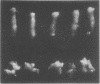
With this technique, single palisade parenchyma cells and single spongy parenchyma cells of Vicia faba leaflets were each found to contain about 2.2 pmoles of sucrose.
Full text
PDF
Images in this article
Selected References
These references are in PubMed. This may not be the complete list of references from this article.
- Chick W. L., Like A. A. Ultramicro method for determination of glycogen and glucose by enzymic labeling with adenosine triphosphate-gamma-32P. Anal Biochem. 1969 Nov;32(2):340–347. doi: 10.1016/0003-2697(69)90095-5. [DOI] [PubMed] [Google Scholar]
- Conrad H. E., Sr, Varboncouer E., James M. E. Qualitative and quantitative analysis of reducing carbohydrates by radiochromatography on ion-exchange papers. Anal Biochem. 1973 Feb;51(2):486–500. doi: 10.1016/0003-2697(73)90505-8. [DOI] [PubMed] [Google Scholar]
- Geiger D. R., Giaquinta R. T., Sovonick S. A., Fellows R. J. Solute distribution in sugar beet leaves in relation to Phloem loading and translocation. Plant Physiol. 1973 Dec;52(6):585–589. doi: 10.1104/pp.52.6.585. [DOI] [PMC free article] [PubMed] [Google Scholar]
- Lowry O. H., Carter J. G. Stabilizing the alkali-generated fluorescent derivatives of NAD and NADP. Anal Biochem. 1974 Jun;59(2):639–642. doi: 10.1016/0003-2697(74)90319-4. [DOI] [PubMed] [Google Scholar]
- Lust W. D., Passonneau J. V., Crites S. K. The measurement of glycogen in tissues by amylo-alpha-1,4-alpha-1,6-glucosidase after the destruction of preexisting glucose. Anal Biochem. 1975 Sep;68(1):328–331. doi: 10.1016/0003-2697(75)90712-5. [DOI] [PubMed] [Google Scholar]
- Morrison W. H., Lou M. F., Hamilton P. B. The determination of hexoses and pentoses by anion-exchange chromatography: a method of high sensitivity. Anal Biochem. 1976 Apr;71(2):415–425. doi: 10.1016/s0003-2697(76)80007-3. [DOI] [PubMed] [Google Scholar]
- Outlaw W. H., Fisher D. B., Christy A. L. Compartmentation in Vicia faba Leaves: II. Kinetics of C-Sucrose Redistribution among Individual Tissues following Pulse Labeling. Plant Physiol. 1975 Apr;55(4):704–711. doi: 10.1104/pp.55.4.704. [DOI] [PMC free article] [PubMed] [Google Scholar]
- Outlaw W. H., Fisher D. B. Compartmentation in Vicia faba Leaves: I. Kinetics of C in the Tissues following Pulse Labeling. Plant Physiol. 1975 Apr;55(4):699–703. doi: 10.1104/pp.55.4.699. [DOI] [PMC free article] [PubMed] [Google Scholar]
- Outlaw W. H., Schmuck C. L., Tolbert N. E. Photosynthetic Carbon Metabolism in the Palisade Parenchyma and Spongy Parenchyma of Vicia faba L. Plant Physiol. 1976 Aug;58(2):186–189. doi: 10.1104/pp.58.2.186. [DOI] [PMC free article] [PubMed] [Google Scholar]
- Van Handel E. Direct microdetermination of sucrose. Anal Biochem. 1968 Feb;22(2):280–283. doi: 10.1016/0003-2697(68)90317-5. [DOI] [PubMed] [Google Scholar]
- Wolosiuk R. A., Pontis H. G. The role of sucrose and sucrose synthetase in carbohydrate plant metabolism. Mol Cell Biochem. 1974 Sep 30;4(2):115–123. doi: 10.1007/BF01770292. [DOI] [PubMed] [Google Scholar]