

Abstract
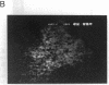
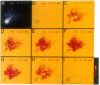
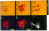
Real-time visualization of intracellular calcium concentration ([Ca2+]i) changes in mammalian Purkinje cells in vitro, utilizing the dye Fura-2, indicates that calcium action potentials are generated in the dendritic tree and follow a particular activation sequence. During spontaneous oscillations or after direct current injection, dendritic spikes are initiated as slow and graded plateau potentials at the level of the tertiary or spiny branchlets of the dendrite. As the plateau potentials become sufficiently high to reach the firing threshold for full dendritic spike generation, calcium entry is observed at the more proximal branches of the dendritic tree. These action potentials are then conducted orthodromically toward the soma and may invade other branches in the arbor antidromically. Simultaneous recording of the intracellular electrical activity and the Fura-2 fluorescent signal indicates that the intracellular calcium transients are accompanied by a very rapid increase in intracellular calcium concentration. This increase in [Ca2+]i exhibits an almost equally fast return to baseline after the termination of the action potential, indicating the presence of very efficient calcium sequestering and extruding mechanisms in the dendrites. Iontophoretic application of glutamate at the dendritic level provided a further demonstration of the spatial separation of plateau potentials from dendritic spikes and gives further insights into the details of dendritic integration in this neuron.
Full text
PDF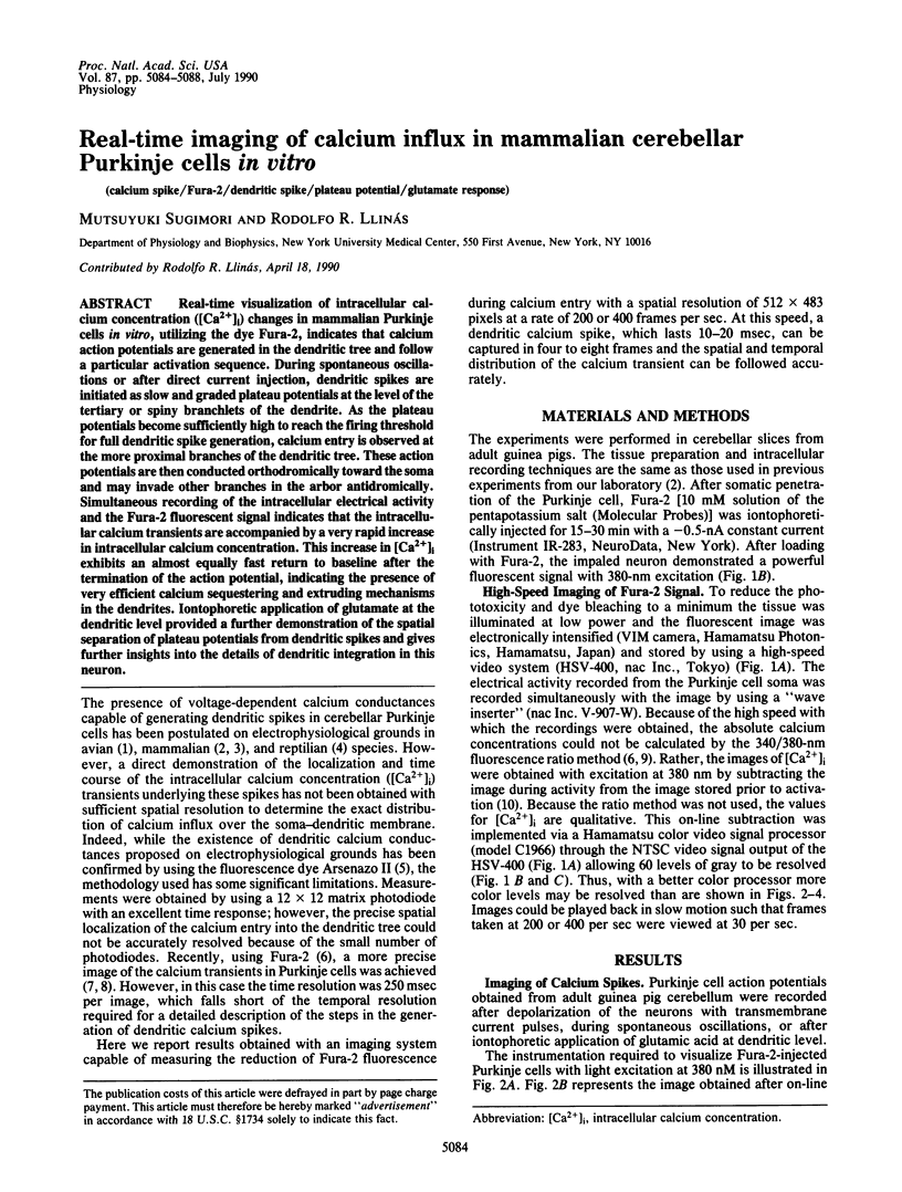
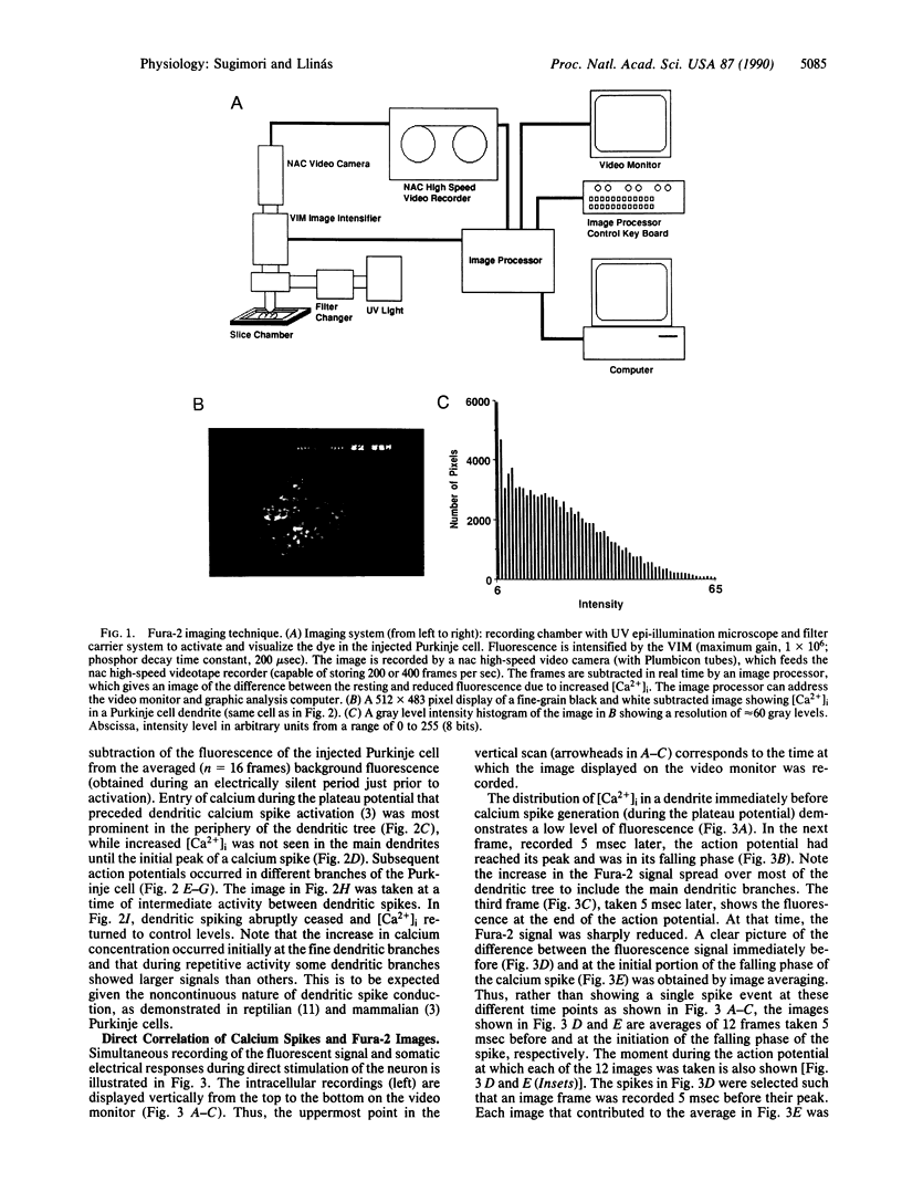
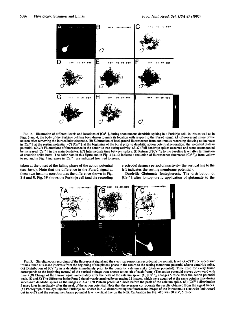
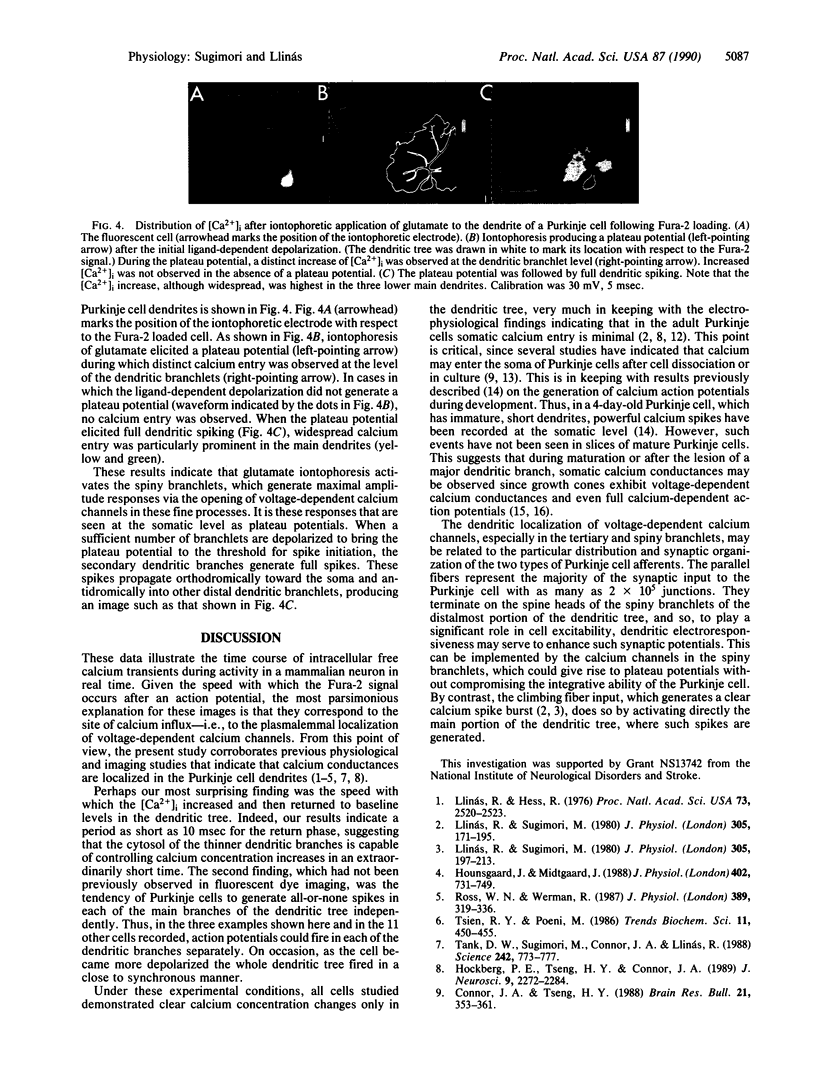

Images in this article

Selected References
These references are in PubMed. This may not be the complete list of references from this article.
- Connor J. A., Kater S. B., Cohan C., Fink L. Ca2+ dynamics in neuronal growth cones: regulation and changing patterns of Ca2+ entry. Cell Calcium. 1990 Feb-Mar;11(2-3):233–239. doi: 10.1016/0143-4160(90)90074-5. [DOI] [PubMed] [Google Scholar]
- Connor J. A., Tseng H. Y. Measurement of intracellular Ca2+ in cerebellar Purkinje neurons in culture: resting distribution and response to glutamate. Brain Res Bull. 1988 Sep;21(3):353–361. doi: 10.1016/0361-9230(88)90147-5. [DOI] [PubMed] [Google Scholar]
- Gruol D. L., Franklin C. L. Morphological and physiological differentiation of Purkinje neurons in cultures of rat cerebellum. J Neurosci. 1987 May;7(5):1271–1293. doi: 10.1523/JNEUROSCI.07-05-01271.1987. [DOI] [PMC free article] [PubMed] [Google Scholar]
- Hockberger P. E., Tseng H. Y., Connor J. A. Fura-2 measurements of cultured rat Purkinje neurons show dendritic localization of Ca2+ influx. J Neurosci. 1989 Jul;9(7):2272–2284. doi: 10.1523/JNEUROSCI.09-07-02272.1989. [DOI] [PMC free article] [PubMed] [Google Scholar]
- Hounsgaard J., Midtgaard J. Intrinsic determinants of firing pattern in Purkinje cells of the turtle cerebellum in vitro. J Physiol. 1988 Aug;402:731–749. doi: 10.1113/jphysiol.1988.sp017231. [DOI] [PMC free article] [PubMed] [Google Scholar]
- Llinas R., Nicholson C. Electrophysiological properties of dendrites and somata in alligator Purkinje cells. J Neurophysiol. 1971 Jul;34(4):532–551. doi: 10.1152/jn.1971.34.4.532. [DOI] [PubMed] [Google Scholar]
- Llinás R., Hess R. Tetrodotoxin-resistant dendritic spikes in avian Purkinje cells. Proc Natl Acad Sci U S A. 1976 Jul;73(7):2520–2523. doi: 10.1073/pnas.73.7.2520. [DOI] [PMC free article] [PubMed] [Google Scholar]
- Llinás R., Sugimori M. Calcium conductances in Purkinje cell dendrites: their role in development and integration. Prog Brain Res. 1979;51:323–334. doi: 10.1016/S0079-6123(08)61312-6. [DOI] [PubMed] [Google Scholar]
- Llinás R., Sugimori M. Electrophysiological properties of in vitro Purkinje cell dendrites in mammalian cerebellar slices. J Physiol. 1980 Aug;305:197–213. doi: 10.1113/jphysiol.1980.sp013358. [DOI] [PMC free article] [PubMed] [Google Scholar]
- Llinás R., Sugimori M. Electrophysiological properties of in vitro Purkinje cell somata in mammalian cerebellar slices. J Physiol. 1980 Aug;305:171–195. doi: 10.1113/jphysiol.1980.sp013357. [DOI] [PMC free article] [PubMed] [Google Scholar]
- MacVicar B. A., Llinás R. R. Barium action potentials in regenerating axons of the lamprey spinal cord. J Neurosci Res. 1985;13(1-2):323–335. doi: 10.1002/jnr.490130121. [DOI] [PubMed] [Google Scholar]
- Ross W. N., Werman R. Mapping calcium transients in the dendrites of Purkinje cells from the guinea-pig cerebellum in vitro. J Physiol. 1987 Aug;389:319–336. doi: 10.1113/jphysiol.1987.sp016659. [DOI] [PMC free article] [PubMed] [Google Scholar]
- Tank D. W., Sugimori M., Connor J. A., Llinás R. R. Spatially resolved calcium dynamics of mammalian Purkinje cells in cerebellar slice. Science. 1988 Nov 4;242(4879):773–777. doi: 10.1126/science.2847315. [DOI] [PubMed] [Google Scholar]