

Summary:
We present a case of a woman, 79 years old, followed by Psychiatry for depressive episodes after breast cancer removal. She was operated on for ductal breast carcinoma in 1983. Afterward she was submitted to adjuvant radiotherapy. She came to our attention for a chronic skin ulcer that developed into the radio-treated area about 4 years ago. We performed a skin biopsy and programed adipose tissue grafts to promote wound healing. The result of the biopsy was unexpected: dermal localization of not differentiated breast carcinoma. She is currently under systemic chemotherapy treatment. The key message is to always perform a skin biopsy of a chronic skin ulcer developed after breast cancer removal before planning surgical reconstruction.
CASE PRESENTATION
We present a case of a woman, 79 years old, followed by Psychiatry for depressive episodes after breast cancer removal. She was operated on for ductal breast carcinoma in 1983. Afterward she was submitted to adjuvant radiotherapy. She came to our attention for a chronic skin ulcer that developed into the radio-treated area about 4 years ago (Fig. 1). We programed adipose tissue grafts of the area to improve the thickness and quality of the skin. We performed a skin biopsy because of the persistence of the ulcer, despite several medical treatments. The result of the biopsy was unexpected: dermal localization of not differentiated breast carcinoma, compatible with primitive breast cancer. The patient is alive and is under systemic chemotherapy treatment, because of metastasis of the lung, seen at positron emission tomography examination, and is under strict oncologist follow-up.
Fig. 1.
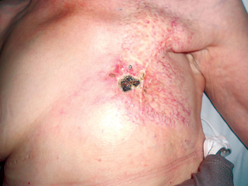
Chronic skin ulcer developed into the radio-treated area.
DISCUSSION
Ulceration and necrosis of the skin are significant aspects of chronic radiation dermatitis. Because of biological mechanisms caused by ionizing radiation, vascularization of ulcerated areas of skin is commonly very poor and refractory to conservative treatment.1 These chronic wounds may be treated with advanced dressings: contaminated wounds may be treated by silver-based dressings, whereas a wound with abundant exudate needs absorbent dressings.1 Severe ulceration and/or necrosis requires surgical management, which includes methods from surgical removal of the necrotic tissues and adipose tissue grafts with “regenerative purpose” to advanced reconstructions with skin flaps.1 Hyperbaric oxygen therapy could be a support to promote wound healing.2 The ever-present possibility of recurrence or persistence of the primary malignant neoplasm within the irradiated tissue must be always suspected and excluded by multiple biopsies before planning an eventual cancer removal and subsequent reconstruction.3,4 In case of recurrent malignancy, although the long-term prognosis may be poor, a multidisciplinary approach involving oncologists and general, thoracic and plastic surgeons could improve the quality of life of patients.
Footnotes
Disclosure: The authors have no financial interest to declare in relation to the content of this article. The Article Processing Charge was paid for by the authors.
REFERENCES
- 1.Spałek M. Chronic radiation-induced dermatitis: challenges and solutions. Clin Cosmet Investig Dermatol. 2016;9:473–482.. [DOI] [PMC free article] [PubMed] [Google Scholar]
- 2.Enomoto M, Yagishita K, Okuma K, et al. Hyperbaric oxygen therapy for a refractory skin ulcer after radical mastectomy and radiation therapy: a case report. J Med Case Rep. 2017;11:5. [DOI] [PMC free article] [PubMed] [Google Scholar]
- 3.Sironi I, Crespi AM, Magnoni E, et al. [Late radiation-induced injuries: breast carcinoma in post-actinic ulceration of the thoracic-breast region with infected osteoradionecrosis. A case report]. G Chir. 1996;17:37–42.. [PubMed] [Google Scholar]
- 4.Loveland-Jones CE, Wang F, Bankhead RR, et al. Squamous cell carcinoma of the nipple following radiation therapy for ductal carcinoma in situ: a case report. J Med Case Rep. 2010;4:186. [DOI] [PMC free article] [PubMed] [Google Scholar]