Abstract
The ability to culture different cell types is essential for answering many questions in developmental and regenerative biology. Studies in marine organisms, in particular echinoderms, have been limited by the lack of well-described cellular culture systems. Here we describe a cell culture system, for normal or regenerating holothurian cells, that allows cell characterization by immunohistochemistry and scanning electron microscopy. These cell cultures can now be used to perform multiple types of experiments in order to explore the cellular, biochemical, and genomic aspects of echinoderm regenerative properties.
Keywords: Echinoderm, Sea cucumber, Primary cell culture, Regeneration, Gastrointestinal tract, Immunohistochemistry, Scanning electron microscopy
1 Introduction
Echinoderms stand out among the group of animals known for their remarkable regenerative properties. Members from different classes of the Echinodermata can regenerate, both as adults or larvae, a large number of tissues and organs. Well-known examples include the regeneration of spines by sea urchins, arm regeneration by sea stars and brittle stars, and the regeneration of viscera by holothurians. The latter has been exploited in our laboratory using the species Holothuria glaberrima as a model system to study not only intestinal regeneration following evisceration but also nervous system regeneration following radial nerve transection [1–4].
Investigators using novel model systems need to develop many of the tools for modern molecular biology analyses, tools that are readily available for most classical model organisms. Echinoderm studies, in particular, are limited by the lack of a well-described technique for culturing cells or tissues from adult organisms. In fact, the culture of primary cells from marine invertebrates has been one of the limiting factors not only to study regeneration in echinoderms but also for the study of different processes in several animal groups. Initial attempts have been done previously where investigators have been able to grow explants and cells from crinoid arms [5, 6] and different tissues of asteroids [7, 8]. Other investigators, using cells from regenerating tissues of the sea cucumber Apostichopus japonicus, established cell cultures where different cell activities could be identified [9]. We have built upon these previous studies and have established a culture system that is amenable for the maintenance of dissociated cells of holothurians. Cells in these cultures can be identified using the same markers applied to in vivo tissues, namely immunohistochemical and morphological analyses.
Tissue cultures provide a unique tool where many cellular events and processes can be characterized. Moreover, these cultures will provide a tool for the development of techniques and methodology such as gene transfections, single cell transcriptomics, and lineages studies, among others, that are hitherto unavailable to study regeneration in adult echinoderms.
2 Materials
Prepare all solutions using ultrapure water and analytical-grade reagents. Diligently follow all waste disposal regulations when disposing waste materials. All cell culture procedures must be carried out under sterile conditions in a laminar flow hood.
2.1 Sea Cucumber Collection and Evisceration
Potassium chloride (KCl): 0.35 M solution in distilled water. Add 250 mL of distilled water in a 500 mL beaker. Weight 6.5 g KCl and transfer to the beaker. Mix using a magnetic stirrer and store at room temperature (RT).
2.2 Sea Cucumber Disinfection and Gut Dissection
Anesthetic solution: 1,1,1-Trichloro-2-methyl-2-propanol hemihydrate (chlorobutanol, Sigma), 0.2 % in natural sea water (SW). Add 950 mL of filtered SW in a glass beaker. Weigh 2 g chlorobutanol and transfer to the beaker. Stir for about 45 min until solubilized. Make up to 1 L with SW. Store at 4 °C.
Sodium hypochlorite: 10 % solution in water.
Ethanol: 95 % solution in water.
Antibiotic/antifungal solution (see Note 1): Penicillin/Streptomycin (300 U/mL and 300 μg/mL, respectively), neomycin (150 μg/mL), and amphotericin B (7.5 μg/mL) in filtered SW. Add 1.5 mL of the concentrated penicillin/streptomycin solution (10,000 U penicillin and 10 mg/mL streptomycin), 750 μL of the concentrated neomycin solution (10 mg/mL), and 150 μL of the amphotericin B stock solution (2.5 mg/mL) to 45 mL of SW in a 50 mL tube. Adjust the pH to 7.7. Complete to 50 mL with SW and sterilize by filtration (0.22 μm filters). Store at 4 °C.
2.3 Gut Rudiment Dissociation and Cell Culture
Calcium and Magnesium Free Artificial Sea Water Solution (CMFSS) [10]: Weight 0.8 g potassium chloride (KCl), 25.5 g sodium chloride (NaCl), 3.0 g sodium phosphate dibasic (Na2HPO4), 3.0 g glucose, and 2.86 g (4-(2-hydroxyethyl)-1-piperazineethanesulfonic acid) HEPES and transfer to a 1 L beaker. Add 950 mL of ultrapure water and mix by magnetic stirring. Adjust the pH to 7.7 and complete to 1 L with ultrapure water. Sterilize by filtration (0.22 μm filters) and store at RT (see Note 2).
Collagenase Type IA (Sigma): 0.125 % in CMFSS [9] supplemented with antibiotics (200 U/mL penicillin, 200 μg/mL streptomycin, 100 μg/mL neomycin) and antifungal (5 μg/mL amphotericin B). Add 45 mL of CMFSS in a 50 mL tube. Add 1 mL of the concentrated penicillin/streptomycin solution (10,000 U penicillin and 10 mg/mL streptomycin), 500 μL of the concentrated neomycin solution (10 mg/mL neomycin), and 100 μL of the amphotericin B stock solution (2.5 mg/mL) in the 50 mL tube. Weight 62.5 mg of collagenase type IA and transfer to the 50 mL tube. Adjust the pH to 7.7. Complete to 50 mL with CMFSS and sterilize by filtration (0.22 μm filters). Aliquot 5 mL of the solution in 15 mL tubes and store at −20 °C until use.
L-15 Medium (Leibovitz, Sigma): conditioned for marine species adding salts to the original composition [11, 12]. Weight 6.9 g L-15 powder, 6.25 g NaCl, 3.12 g glucose, 1. 58 g magnesium sulfate (MgSO4), 172 mg KCl, 96 mg sodium bicarbonate (NaHCO3), 1.33 g magnesium chloride (MgCl2), 150 mg L-glutamine, 745 mg calcium chloride dihydrate (CaCl2·2H2O) and transfer to a 500 mL beaker. Complete to 500 mL with ultrapure water. Mix by magnetic stirring. Sterilize by filtration (0.22 μm filters). Store at 4 °C in a bottle wrapped in aluminum foil.
Cell culture medium: L-15 medium conditioned for marine species supplemented with antibiotics (100 U/mL penicillin, 100 μg/mL streptomycin, 50 μg/mL gentamicin), antifungal (2.5 μg/mL amphotericin B), 1x MEM nonessential amino acids, 1 mM sodium pyruvate, 1.75 mg/mL α-tocopherol acetate, 1 μg/mL hydrocortisone, 1 x ITS (Insulin, Transferrin, and sodium Selenite), 2 % Fetal Bovine Serum, 10 % cell-free coelomic fluid (see Note 3). Add 500 μL of penicillin/streptomycin stock solution (10,000 U penicillin and 10 mg/mL streptomycin), 250 μL of gentamicin stock solution (10 mg/mL), 50 μL of amphotericin B stock solution (2.5 mg/mL), 500 μL of MEM nonessential amino acid stock solution (100 x), 500 μL of sodium pyruvate stock solution (100 mM), 50 μL of α-tocopherol-acetate stock solution (17.5 mg/mL), 100 μL of hydrocortisone stock solution (500 μg/mL), 500 μL of the ITS (Insulin, Transferrin, and sodium Selenite) stock solution (100×), 1 mL of fetal bovine serum (FBS, Sigma), and 5 mL of sea cucumber cell-free coelomic fluid in a 50 mL tube. Transfer L-15 medium conditioned for marine species to the 50 mL tube to complete 45 mL. Mix gently by inversion of the tube. Adjust the pH to 7.7–7.8 using sodium hydroxide (NaOH) 1 N. Complete to 50 mL adding L-15 medium. Sterilize by filtration (0.22 μm filters). Store at 4 °C in a bottle wrapped in aluminum foil (see Note 4).
Sea cucumber cell-free coelomic fluid: The method that we apply to obtain the cell-free coelomic fluid was described by Zhao et al. [13] with some modifications. Briefly, we wash the animals in clean SW to remove particles attached to the animal skin. Then, a person holds the animal on a sterile dissecting pan while another person cut its oral side (where the tentacles are present) using a sterile razor blade. The animal is raised and squeezed so that the coelomic fluid squirts into a sterile beaker which is on ice. Transfer the coelomic fluid to 50 mL tubes and centrifuge at 2,000 × g × 10 min at 4 °C to remove the cells. Transfer the supernatant to clean tubes. Sterilize by filtration (0.22 μm filter). Aliquot the supernatant in 5 mL aliquots and store at −20 °C. A new aliquot of coelomic fluid must be used every time culture medium is prepared (see Note 5).
Trypan blue dye (Sigma): 0.4 % solution in CMFSS. Sterilize by filtration (0.22 μm membranes). Store at RT.
2.4 Cell Identification by Indirect Immuno-histochemistry
Lab-Tek™ II Chamber Slide System, 8 wells per slide (0.7 cm2/well) (Thermo Fisher Scientific). Each pack of glass slides includes a slide separator system to remove the polysty-rene chamber.
Poly-L-lysine (Sigma): 0.01 % solution in water.
Paraformaldehyde (PFA, Sigma): 4 % in filtered SW (see Note 6).
Blocking solution: Normal goat serum (Sigma) diluted 1:50 in PBS containing 0.01 % sodium azide (NaN3).
Triton X-100 (Sigma): 0.1 % solution in PBS.
Phosphate Buffered Saline (PBS): 0.1 M. Weight 8 g NaCl and 13.4 g sodium phosphate dibasic heptahydrate (NaH2PO4·7 H2O) and transfer to a 1 L beaker. Add 800 mL to the beaker and mix using a magnetic stirrer. Adjust the pH to 7.4. Complete to 1 L with ultrapure water.
RIA buffer: Weight 7.06 g potassium phosphate dibasic (K2HPO4), 1.29 g potassium phosphate monobasic (KH2PO4), 9 g NaCl, 5 g bovine serum albumin (BSA), 0.1 g sodium azide (NaN3) and transfer to a 1 L beaker. Add 800 mL to the beaker and mix using a magnetic stirrer. Adjust the pH to 7.4. Complete to 1 L with ultrapure water. Aliquot in bottles wrapped in aluminum foil and store at −20 °C.
Monoclonal antibody Meso 1 [14]: diluted 1:20 in RIA buffer. This antibody was developed in mouse in our laboratory and specifically labels the mesothelial cells from the mesentery and the gut rudiment of holothurians.
Monoclonal anti-β tubulin antibody (Sigma, clone 2-28-33): diluted 1: 500 in RIA buffer. This antibody was prepared from sea urchin sperm axonemes. It labels neuron-like cells and nerve bundles in the mesothelium and connective tissue of normal and regenerating mesenteries in H. glaberrima [15]. Aliquot 10 μL of the anti-β tubulin antibody in microtubes. Store at −20 °C. Immediately before use add 990 μL of the RIA buffer to a microtube to obtain a stock solution (1:100). Transfer 100 μL of the stock solution (1:100) to a new tube and add 400 μL of the RIA buffer to obtain a working solution of 1:500 in RIA buffer.
Secondary antibody: Goat anti-mouse conjugated with the fluorophore Cy3 (GAM-Cy3, Jackson Immuno Research Laboratories) diluted 1:1,000 in RIA buffer. Aliquot 10 μL of the GAM-Cy3 solution in microtubes. Store at −20 °C. Immediately before use, add 990 μL of the RIA buffer to a microtube to obtain a stock solution (1:100). Transfer 100 μL of the stock solution (1:100) to a new tube and add 900 μL of the RIA buffer to obtain a working solution of 1:1,000 in RIA buffer.
Phalloidin-Fluorescein Isothiocyanate (FITC) labeled (Sigma): diluted 1:1,000 solution in RIA buffer. Prepare the stock solution (1:100) and the working solution (1:1,000) as indicated for the GAM-Cy3 antibody.
Mounting media: buffered glycerol solution containing 2 μg/mL 4′,6-diamidino-2-phenylindole (DAPI) (Sigma). To make a 5 mg/mL DAPI stock solution (14.3 mM), dissolve the contents of one vial (10 mg) in 2 mL of ultrapure water. Then prepare a 1 mg/mL DAPI working solution adding 200 μL of stock solution to 800 μL of ultrapure water. To prepare the mounting media add 100 μL of the DAPI working solution (1 mg/mL) to 24.9 mL of 0.1 M PBS in a 50 mL tube. Finally add 25 mL of glycerol to the same 50 mL tube and mix. Wrap in aluminum foil and store at 4 °C.
2.5 Cell Identification by Scanning Electron Microscopy
Round German glass coverslips (Electron Microscopy Sciences): 8 mm in diameter, 0.16–0.19 mm in thickness.
24-well Cell Culture Plate (BD Falcon): tissue culture-treated polystyrene, flat-bottom with lid.
Glutaraldehyde (Sigma) (see Notes 6 and 7): 2 % solution in 85 % natural SW. Mix 85 mL of natural SW with 15 mL of distilled water to obtain 100 mL of 85 % natural SW. To prepare 100 mL of the 2 % glutaraldehyde solution, mix 96 mL of 85 % natural SW with 4 mL of 50 % glutaraldehyde. The final osmolality is 1.1 Osm.
Osmium Tetroxide (OsO4, Electron Microscopy Sciences): 1 % in natural SW with the contribution of glucose. Add 0.37 g of glucose to 6 mL of natural SW. To produce 8 mL of 1 % OsO4 mix the 6 mL of natural SW containing glucose with 2 mL of 4 % OsO4 (see Notes 6 and 7).
Ethanol (Sigma): 25, 40, 50, 60, 70, 80, 90, 100 % solutions in distilled water.
Hexamethyldisilazane (HMDS, Electron Microscopy Sciences): diluted in ethanol at 25 %, 50 %, 75 %, and pure (100 %).
Carbon adhesive tabs (Electron Microscopy Sciences): 12 mm in diameter.
Aluminum mounts (Electron Microscopy Sciences): stubs of 12.2 mm in diameter.
3 Methods
3.1 Sea Cucumber Collection and Evisceration
Collect sea cucumbers of the species Holothuria glaberrima (see Note 8) and maintain them in the laboratory in sea water aquaria with constant oxygenation.
Eviscerate the sea cucumbers by injecting 0.35 M KCl (3–5 mL) into the coelomic cavity. During evisceration sea cucumbers expel most of their viscera (gut, hemal vessels, gonads, and part of the respiratory trees) through the cloaca in approximately 5 min. After evisceration, return the animals to the aquaria and allow them to regenerate for 5 days.
3.2 Sea Cucumber Disinfection and Gut Dissection
Immerse ten of the sea cucumbers which have regenerated their gastrointestinal tract for 5 days in 500 mL beakers containing 250 mL of anesthetic solution (prewarmed to RT) for 45 min at RT.
Decontaminate the sea cucumber surface immersing the animals one at a time in a 10 % sodium hypochlorite solution for 1 min, 95 % ethanol for 5 min, and a quick rinse in sterile ultrapure water.
Put the animals, ventral side up, on an autoclaved vinyl pad in a dissecting pan. The ventral side can be identified by the presence of the ambulacral feet. Cut with scissors along the longitudinal line that separates the dorsal and ventral axis. In Holothuria glaberrima this line rests at the boundary between the ventral side and the side without ambulacral feet (dorsal). To decrease the risk of contamination, make the cut from about 1 cm from the mouth (surrounded by a crown of tentacles) to approximately 1 cm from the cloaca. Once the body wall is opened, hold down the sea cucumber using pushpins. Dissect the gut rudiments under the stereoscopic microscope using fine tweezers and spring scissors. The gut rudiment may be recognized at day 5 of regeneration because in H. glaberrima it appears as a pink thickening at the free end of the mesenterium.
Collect the gut rudiments of ten sea cucumbers in 10 mL of the antibiotic/antifungal solution in a 15 mL tube and keep on ice.
Wash the gut rudiments with fresh antibiotic/antifungal solution twice for 30 min at 4 °C.
3.3 Gut Rudiment Dissociation and Cell Culture
Cut the gut rudiments into small pieces of approximately 1 mm3 on a sterile glass petri dish using a scalpel with #12 blades in the laminar flow hood. Transfer the pieces to 5 mL of 0.125 % collagenase in a 15 mL tube using a sterile dissecting needle.
Incubate the pieces in a water bath with constant agitation (15 rpm) (see Note 9) for approximately 2 h at 28 °C. Check for the presence of remaining tissue pieces after 1 h of incubation, the length of the dissociation will depend on the amount of tissue.
Centrifuge the cell suspension at low speed (300 × g, 4 °C) for 1 min to form a pellet with the tissue that remains nondissociated. Transfer the supernatant containing the cell suspension to a new 15 mL tube and discard the pellet (nondissociated tissue pieces).
Pass the cell suspension through a sterile cell strainer (mesh size 70 μm) placed on a 50 mL tube to remove large cell aggregates.
Centrifuge the cell suspension at 900 × g for 10 min at 4 °C, discard the supernatant containing the collagenase solution. Resuspend the cells in 5 mL of CMFSS supplemented with antibiotics and centrifuge again (900 × g,10 min, 4 °C) to wash the cells and to eliminate the cell debris.
Resuspend the cells in 1 mL of CMFSS (see Note 10). Perform trypan blue exclusion test to estimate the number of cells in suspension and their viability. Transfer 20 μL of the cell suspension to a microcentrifuge tube and add 20 μL trypan blue dye (0.4 % in CMFSS) (see Note 11). Mix and load approximately 10 μL of the cell suspension by capillary action in each side of a hemocytometer and count the cells under an inverted microscope. Dead cells will stain blue.
Count the number of live and dead cells in each of the four corner squares of the hemocytometer. To estimate the cell concentration in a milliliter of CMFSS follow the formula: (total live cells/4) x Dilution Factor (DF) x 104. As an example, if you counted on average 80 cells in the four squares of the hemocytometer and used the DF mentioned in 6, then the formula will be: 80 × 2 × 104 = 1.6 × 10⁶ cells/mL of CMFSS. The cell viability is determined by: (total live cells)/(total live cells + total dead cells). Following the dissociation protocol mentioned previously, the cell viability should be >80 %.
Seed the cells on 8-well glass slides treated with poly-L-lysine (see Note 12) at a cell density of 6 × 10⁴ cells/cm2, diluting the cell suspension in the supplemented cell culture medium. Add 300 μL of culture medium per well.
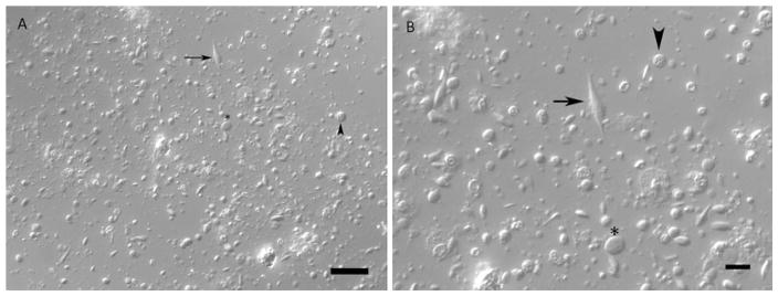
Incubate the cells in a modular incubator chamber (Billups-Rothenberg Inc) at RT. Change the culture medium every 3–5 days to reduce the risk of contamination (see Note 4). Check your cultures daily under an inverted microscope to check the cell morphology and possible contamination. Figure 1 shows an example of a 5 day cell culture observed by phase contrast microscopy.
Fig. 1.
Cells from regenerating gut rudiments (5 days post-evisceration) of the sea cucumber Holothuria glaberrima after 5 days in culture observed by phase contrast microscopy. Various cell sizes and morphologies can be observed, among them are spherical (arrowhead), oval-(asterisk), and spindle-shaped structures (arrow). Scale: (a) = 30 μm, and (b) = 10 μm
3.4 Cell Identification by Indirect Immuno-histochemistry
After culturing cells on glass slides for the previously deter-mined length of time, discard the culture medium and wash carefully the cultures with 300 μL of filter sterilized SW (see Note 13) per well. Add 300–500 μL of 4 % paraformaldehyde solution for 24 h (see Note 14) at 4 °C to fix the cells. After fixation rinse the cultures twice with filtered SW for 5 min each. At this point you can store the cultures in SW with 0.01 % sodium azide (NaN3) at 4 °C.
Add 300 μL of normal goat serum (diluted 1:50) in each well and incubate for 1 h at RT to block the nonspecific binding of the primary antibodies.
Discard the normal goat serum. Add 300 μL of 0.1 % triton X-100 in 0.1 M of Phosphate Buffered Saline (PBS) for 10 min at RT to each well (see Note 15). Wash twice carefully with 0. 1 M PBS for 10 min each.
Incubate the cells overnight (ON) at 4 °C with the primary antibody(ies) at the appropriate dilution in RIA buffer (see Note 16). The primary antibody may be monoclonal (generally raised in mice) or polyclonal (generally raised in rabbits) or a mix of both. As an example, we have used the monoclonal antibody Meso-1 [14] or the monoclonal antibody β-tubulin. For the 8-well glass slides with a surface area of 0.7 cm2/well, we use at least 100 μL of the primary antibody solution per well. Keep in mind that this solution must completely cover the cells.
After incubation with the primary antibody, wash cultures thrice carefully with 0.1 M PBS for 10 min each.
Incubate the cultures with the secondary antibody, conjugated with a fluorophore, and diluted in RIA buffer for 1 h at RT in the dark. In this case, we have used the goat anti-mouse antibody conjugated with the fluorophore Cy3 (GAM-Cy3) diluted 1:1,000 in RIA buffer. To label SLSs (see Note 17) incubate the samples with phalloidin-FITC. The samples can be simultaneously labeled with GAM-Cy3 and phalloidin-FITC. Transfer 100 μL of the GAM-Cy3 and 100 μL of the phalloidin-FITC stock solutions (1:100) in a microtube and add 800 μL to obtain a working solution (diluted 1:1,000) of secondary antibody and phalloidin. Use at least 100 μL of the working solution per well.
After incubation with the secondary antibody and phalloidin-FITC, wash cultures thrice carefully with 0.1 M PBS for 10 min each.
Carefully remove the polystyrene medium chamber using the slide separator included in each pack of the glass slides. Mount each slide adding mounting media and a 24 × 55 mm cover-glass (VWR), sealing the borders with nail polish and drying under a blower. The DAPI is used to stain the cell nuclei.
Examine the slides using a microscope equipped with the appropriate filters (see Note 18).
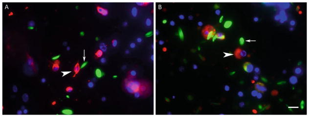
Figure 2 shows a 5 day primary cell culture from a regenerating gut of H. glaberrima displaying cells immunoreactive for β-tubulin (Fig. 2a) and Meso-1 (Fig. 2b).
Fig. 2.
Anti-β-tubulin and Meso-1 immunoreactivity of primary cell cultures from regenerating gut rudiments (5 days post-evisceration) of the sea cucumber Holothuria glaberrima after 5 days in culture. (a) Anti-β-tubulin labels (red) spherical and oval-shaped cells some of which exhibit cell projections (arrowhead). (b) Meso-1 labels (red) spherical cells (arrowhead). Phalloidin labels (green) spindle-like structures (arrow) that lack nuclei in (a) and (b). Cell nuclei are labeled with DAPI (blue). Scale = 10 μm
3.5 Cell Identification by Scanning Electron Microscopy (See Note 7)
1. Proceed to gut rudiment dissociation as indicated in Subheading 3.2 from steps 1–7. Then seed the cells on round glass coverslips treated with poly-L-lysine (see Note 12) at a cell density of 1 × 10⁵ cells/cm2. The 8 mm round coverslips fit inside the 24 well plates. Dilute the cell suspension in the supplemented cell culture medium. Add 500 μL of culture medium per well (see Note 19) and incubate the cells as in 3.3.9.
After culturing cells on glass slides for the previously determined length of time, discard the culture medium and wash carefully the cultures with 500 μL of filtered SW (see Note 13) per well. Add 300–500 μL of 2 % glutaraldehyde in filtered SW. Incubate for 2 h at 4 °C. Discard the glutaraldehyde and incubate in 500 μL of filtered SW ON at 4 °C.
Post-fix the cells adding 300–500 μL of 1 % osmium tetroxide (OsO4) in SW (see Note 13) for 2 h at RT. Wash specimens in sterile distilled water with two rapid washes.
Dehydrate the cultured cells in graded ethanol (25, 40, 50, 60, and 70 %) for 10 min each; (80, 90 %) twice for 10 min each, and 100 % ethanol thrice for 10 min each.
Chemically dry the samples using Hexamethyldisilazane (HMDS, Electron Microscopy Sciences) (see Note 20) incubating successively with 25, 50, and 75 % HMDS in 100 % ethanol for 10 min each. Finally, wash thrice the samples with 100 % HMDS for 10 min each. Let the third wash in HMDS to evaporate ON in a clean protected place at RT.
Put a carbon adhesive tab on each stub (Electron Microscopy Sciences, 12.2 mm in diameter) with fine tweezers and proceed to mount the sample. This is done carefully picking the glass coverslips from the 24 well plates with fine tweezers one at a time and placing them on the adhesive tab with the poly-L-lysine (cell) side facing up. If you are not sure, check the coverslips under the stereoscopic microscope.
Coat the samples with a layer of 25 nm of gold and then examine them with a scanning electron microscope (JEOL JSM 6480 LV) using variable magnifications.
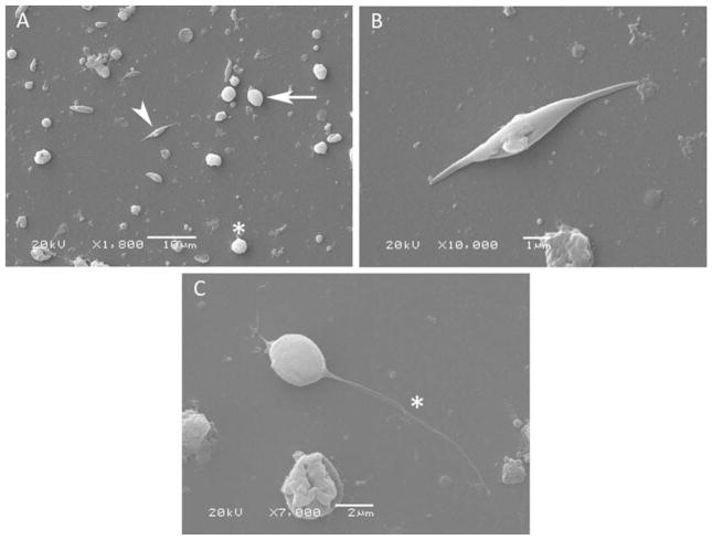
Figure 3 shows a 5 day primary cell culture from a regenerating gut of H. glaberrima displaying cells with different morphologies by Scanning Electron Microscopy.
Fig. 3.
Cell morphology of cultured cells from regenerating gut rudiments (5 days post-evisceration) of the sea cucumber Holothuria glaberrima after 5 days in culture shown by Scanning Electron Microscopy. Spherical (asterisk), ovoid-shaped (arrow), and spindle-shaped (arrowhead) structures are observed in (a). Higher magnification of spindle-like structures (b) and an ovoid-shaped cell with a cell projection (asterisk) (c) are shown. Scale: (a) = 10 μm, (b) = 1 μm, and (c) = 2 μm
Acknowledgments
This project was supported by NIH (Grant 1SC1GM084770-01, 1R03NS065275-01), NSF (IOS-0842870), and the University of Puerto Rico. We would like to thank to the Material Characterization Center of the University of Puerto Rico for the use of the Scanning Electron Microscopy Facility. We gratefully acknowledge Dr. Cristiano Di Benedetto (University of Milan) for his help concerning the scanning electron microscopy protocol.
Footnotes
The antibiotic/antifungal solution concentration is three times higher compared to that in cell culture medium and is used to disinfect the dissected tissues. Diluted antibiotic and antifungal solutions are heat labile, thus, it is recommended to store the SW supplemented with antibiotics/antifungal at 4 °C until use.
There are different formulations to prepare calcium and magnesium free artificial sea water. The formulation presented here is the one reported by Van der Merwe et al. [10].
Cell-free coelomic fluid is used as a growth factor source in our cell culture protocol.
Antibiotics and antifungal lose their activity in culture medium in approximately 3–5 days. Additionally, supplements such as L-glutamine also lose their activity in a few days in culture. Thus, it is recommended to add the supplements to the L-15 medium the same day or the day before the cells are cultured, and to change the culture medium every 3–5 days. The L-15 supplemented medium can be stored up to 2 weeks at 4 °C. Some of the supplements used to enrich our culture medium are used as reported by Odintsova et al. [9].
We obtain the coelomic fluid from sea cucumbers from the species Holothuria mexicana, that, due to their size contain a higher volume of coelomic fluid per animal when compared to Holothuria glaberrima.
Prepare and manipulate the paraformaldehyde, glutaraldehyde, and osmium tetroxide (OsO4) solutions carefully under the fume or chemical hood since these solutions and their vapors are highly hazardous by inhalation or skin contact. Use personal protective equipment: chemical goggles, disposable nitrile gloves, disposable laboratory coat, and closed-toed shoes. Before manipulation of these solutions read the corresponding Material Safety Data Sheet. The used paraformaldehyde, glutaraldehyde, and osmium tetroxide solutions should be disposed as hazardous waste.
The recipe to prepare the glutaraldehyde and the osmium tetroxide solutions, as well as the protocol to fix and dehydrate the cell monolayers for scanning electron microscopy was kindly provided by Dr. Cristiano Di Benedetto from the University of Milan, Bioscience Department (personal communication).
We work with the species Holothuria glaberrima because of their high availability in nearby coastal areas, however, the procedure described here could be used with other species of holothurians, such as Holothuria mexicana.
Prewarming the collagenase solution for approximately 5 min at 28 °C in the water bath previous to tissue incubation, as well as the agitation during incubation, accelerate the dissociation of the tissue.
We have observed that cells from regenerating tissues tend to clump more when compared to cells from nonregenerating sources. Thus, it may be necessary to pass again the cell suspension at this time through a cell strainer (mesh size 70 μm) to eliminate the cell aggregates so as to be able to quantify the cells.
You must dilute the trypan blue in CMFSS to obtain the same osmolality as the medium in which the cells are resuspended, otherwise the cells will lyse. We have tried resuspending cells in culture medium but this increases the cell clumping.
Poly-L-lysine coated glass slides and coverslips are prepared by incubating each well of the 8-well glass slides with 200 μL and each coverslip in 24-wells plates with 500 μL of 0.01 % poly-L-lysine solution ON; then the excess solution is removed and the wells are washed in sterile ultrapure water. The slides and coverslips are dried and sterilized exposing them to UV light ON in a laminar flow hood. These can be stored dry for up to a year at RT if protected from dust.
We use filtered natural sea water but artificial sea water can also be used to wash the cells.
Initially, we fixed the cells in 4 % PFA for only 10 min at 4 °C, but after completing the immunohistochemistry protocol we observed that most cells had detached from the well surface. Thus, we increased the length of the fixation to 24 h, which increased the number of attached cells remaining after the immunohistochemistry protocol.
Triton X-100 is used to permeate the cell membrane to facilitate the entrance of the primary antibody into the cells. If the antigen is on the external side of the membrane it might not be necessary to incubate the cells with Triton X-100.
We centrifuge (13,000 × g for 2 min) the primary antibody solutions prior to their addition to cells to eliminate any particulate present from the solution. Most of the antibodies used in our lab are produced by our group and correspond to ascites or hybridoma supernatants which contain suspended particles. These particles may increase the background in your samples if they are not eliminated at this point.
A cellular process involved in gut regeneration in holothurians is cell dedifferentiation [14]. Specifically, when muscle cells dedifferentiate they condense their contractile apparatus into vesicles termed spindle-like structures (SLSs). These structures may be visualized labeling the SLSs which contain actin with the phalloidin conjugated with the fluorophore FITC or Rho-damine. Phalloidin is a fungal toxin that is well-known to bind to actin filaments.
In our laboratory, cell cultures are examined and photographed using a Nikon Eclipse E600 fluorescent microscope equipped with FITC, R/DII and DAPI filters, and a Spot RT3 digital camera (Diagnostic Instruments, Inc.). Images were recorded using the Spot Basic software (version 4.7; Diagnostic Instruments, Sterling Heights, MI), and Image J (version 1.37; NIH, Bethesda, MD).
The 8 mm glass coverslips float in small volumes of poly-L-lysine or cell culture medium. We have observed that 500 μL is a suitable volume to maintain the coverslips on the bottom of the 24-well plates.
An alternative method to dry the cells is the critical point drying which requires a specialized device (Critical Point Dryer), liquid carbon dioxide (CO2), and amyl acetate. In our lab, the chemical-drying works well with the holothurian cells.
References
- 1.García-Arrarás JE, Estrada-Rodgers L, Santiago R, et al. Cellular mechanisms of intestine regeneration in the sea cucumber, Holothuria glaberrima Selenka (Holothuroidea: Echinodermata) J Exp Zool. 1998;281:288–304. doi: 10.1002/(sici)1097-010x(19980701)281:4<288::aid-jez5>3.0.co;2-k. [DOI] [PubMed] [Google Scholar]
- 2.Garcia-Arraras JE, Greenberg MJ. Visceral regeneration in holothurians. Microsc Res Tech. 2001;55:438–451. doi: 10.1002/jemt.1189. [DOI] [PubMed] [Google Scholar]
- 3.San Miguel-Ruiz JE, Maldonado-Soto AR, García-Arrarás JE. Regeneration of the radial nerve cord in the sea cucumber Holothuria glaberrima. BMC Dev Biol. 2009;9:3. doi: 10.1186/1471-213X-9-3. [DOI] [PMC free article] [PubMed] [Google Scholar]
- 4.Mashanov V, García-Arrarás JE. Gut regeneration in holothurians: a snapshot of recent developments. Biol Bull. 2011;221:93–109. doi: 10.1086/BBLv221n1p93. [DOI] [PubMed] [Google Scholar]
- 5.Candia Carnevali MD, Bonasoro F, Patruno M, Thorndyke MC. Cellular and molecular mechanisms of arm regeneration in crinoid echinoderms: the potential of arm explants. Dev Genes Evol. 1998;208:421–430. doi: 10.1007/s004270050199. [DOI] [PubMed] [Google Scholar]
- 6.Di Benedetto C. An in vivo and in vitro approach. Lambert Academic Publishing; Saarbrucken: 2011. Progenitor cells and regenerative potential in echinoderms. [Google Scholar]
- 7.Sharlaimova NS, Pinaev GP, Petukhova OA. Comparative analysis of behavior and proliferative activity in culture of cells of coelomic fluid and of cells of various tissues of the sea star Asterias rubens L. isolated from normal and injured animals. Cell Tissue Biol. 2010;4:280–288. [Google Scholar]
- 8.Sharlaimova NS, Petukhova OA. Characteristics of populations of the coelomic fluid and coelomic epithelium cells from the starfish Asterias rubens L. able attach to and spread on various substrates. Cell Tissue Biol. 2012;6:176–188. [Google Scholar]
- 9.Odintsova NA, Dolmatov IY, Mashanov VS. Regenerating holothurian tissues as a source of cells for long-term cell cultures. Mar Biol. 2005;146:915–921. [Google Scholar]
- 10.Van der Merwe M, Auzoux-Bordenave S, Niesler C, Roodt-Wilding R. Investigating the establishment of primary cell culture from different abalone (Haliotis midae) tissues. Cytotechnology. 2010;62:265–277. doi: 10.1007/s10616-010-9293-x. [DOI] [PMC free article] [PubMed] [Google Scholar]
- 11.Schacher S, Proshansky E. Neurite regeneration by Aplysia neurons in dissociated cell culture: modulation by Aplysia hemolymph and the presence of the initial axonal segment. J Neurosci. 1983;3:2403–2413. doi: 10.1523/JNEUROSCI.03-12-02403.1983. [DOI] [PMC free article] [PubMed] [Google Scholar]
- 12.Sahly I, Erez H, Khoutorsky A, et al. Effective expression of the green fluorescent fusion proteins in cultured Aplysia neurons. J Neurosci Methods. 2003;126:111–117. doi: 10.1016/s0165-0270(03)00072-4. [DOI] [PubMed] [Google Scholar]
- 13.Zhao Y, Wang DO, Martin KC. Preparation of Aplysia sensory-motor neuronal cell cultures. J Vis Exp. 2009;28:1355. doi: 10.3791/1355. [DOI] [PMC free article] [PubMed] [Google Scholar]
- 14.Garcia-Arraras JE, Valentin-Tirado G, Flores JE, et al. Cell dedifferentiation and epithelial to mesenchymal transitions during intestinal regeneration in H. glaberrima. BMC Dev Biol. 2011;11:61–77. doi: 10.1186/1471-213X-11-61. [DOI] [PMC free article] [PubMed] [Google Scholar]
- 15.Tossas-Robles KE. Doctoral Thesis. University of Puerto Rico; Rio Piedras Campus: 2012. Temporal study of the regeneration of the enteric nervous system of the sea cucumber Holothuria glaberrima. In: Regeneration of the enteric nervous system of the sea cucumber Holothuria glaberrima using tubulin and others neuronal markers; p. 119. [Google Scholar]


