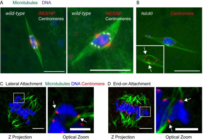
Figure 4. Lateral and end-on kinetochore attachments in Drosophila and mouse oocytes.

A) Wild-type Drosophila oocytes showing all kinetochores making end-on attachments, with DNA in blue, microtubules in green, central spindle protein INCENP in red and centromeres (CENP-C) in white. B) Oocyte lacking the kinetochore protein NDC80, which is required for end-on attachments, shows evidence of lateral attachments. DNA is in blue, microtubules in green, and centromeres (CENP-C) in red. Inset is a 10 μm region with the DNA removed showing most of the centromeres. C,D) Cold-treated mouse oocytes at metaphase I, showing examples of lateral (C) and end-on (D) kinetochore attachments. The boxed regions are shown at higher magnification. DNA is in blue, microtubules in green and centromeres (CREST) in red and the arrows point to lateral or end-on attachments. In all images, the scale bar is 10 μm.
