Abstract
The azygous lobe of the lung is an uncommon developmental anomaly. Its surgical importance is hardly being described in literature. Here, we are presenting a case of lung cancer with incidental azygous lobe, with its surgical relevance during lung cancer surgery.
Keywords: Azygous lobe, Developmental anomaly, Azygous vein, Azygous fissure, Squamous cell lung cancer
Introduction
The azygous lobe of the lung is a developmental anomaly of the lung, from the apical segment of bronchus of the right upper lobe. It is seen in 0.1–8% of population. It is most often an incidental finding on chest X-ray, CT chest, during surgery, and postmortem. It has not been associated with any disease process nor its clinical utility being described. We describe a case of azygous lobe associated with endobronchial squamous cell carcinoma of the right bronchus intermedius and its clinical utility.
Case Report
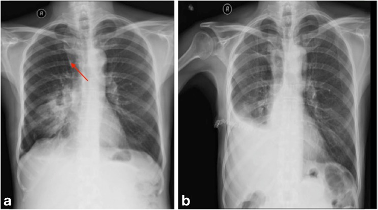
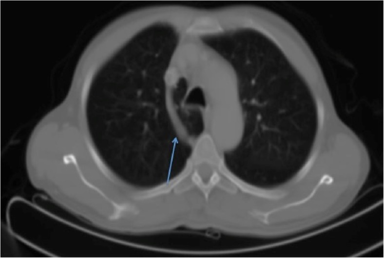
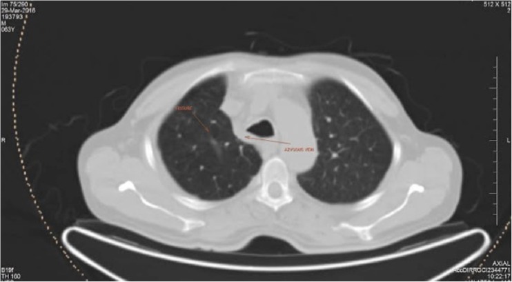
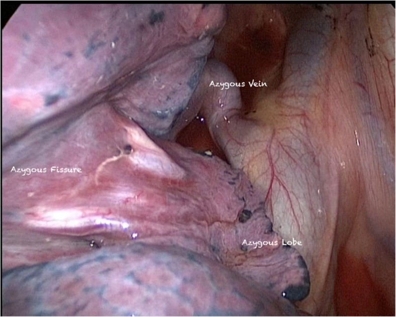
A 63-year-old smoker with no other comorbidities presented with complains of fever and cough of 4 months duration. On evaluation, he was having solid mass in the right middle lobe. Chest X-ray and CT chest showed azygous lobe, (Figs. 1a, 2, 3, and 4). FOB was done, which showed an endobronchial mass in the right bronchus intermedius, away from the right upper lobe bronchus. Endoscopic biopsy confirmed NSCLC, squamous cell carcinoma. Patient was staged using PET CT and histopathological confirmation of negative mediastinal nodes by EBUS staging from mediastinal nodes.
Fig. 1.
Chest X-ray showing faint azygous fissure (a) and postsurgery (b)
Fig. 2.
Chest CT scan (presurgery) showing azygous lobe with azygous vein (arrow)
Fig. 3.
CT chest (postsurgery) showing well-mediated azygous vein and faintly visible azygous fissure (as marked with arrows)
Fig. 4.
Intraoperative picture showing released azygous lobe, azygous fissure, and azygous vein
He underwent right posteriolateral thoracotomy with bilobectomy of the right middle and lower lobes with mediastinal lymph node dissection. During surgery, azygous lobe was observed, which was lying medial to the azygous vein. The azygous lobe was confined to the small azygous pleural cavity lined by azygous tissue along the superiormedial aspect of the azygous vein. The azygous fissure was opened above the azygous vein arch and the azygous pleural cavity was opened to the pleural cavity proper. The azygous lobe was released from the azygous pleural cavity by pulling it underneath the azygous vein and making it lying lateral to the azygous vein. Following this, mediastinal lymph node dissection of levels 2R, 4R, and 10R was done. This procedure further mobilizes the pleural reflection on the hilum and freeing more area of the lung for expansion. Following the completion of bilobectomy and lymph node dissection, medialization of the azygous vein was done. We could see the azygous lobe well expanded in the pleural cavity proper. The patient had uneventful recovery and was discharged on the seventh postoperative day.
A CT chest was done at 3-month post op and showed a well-expanded right upper lobe with the azygous lobe occupying its right full position at the apex of the pleural cavity.
Discussion
German anatomist, Heinrich Wrisberg, first described the azygous lobe in 1877. However, radiologically, Jaches identified it about 46 years later in 1923 [1].
It is a normal developmental anomaly found in 0.1 to 8% of population [2]. The proposed theory says that, it happens due to arrest in normal medial migration of the right posterior cardinal vein (future azygous vein) that leads to a persistant azygous vein arching over the right upper lobe of the lung. The part of the right upper lobe lying medial to the azygous vein is referred as the azygous lobe. Azygous vein itself lies in fissure called azygous fissure [3].
Azygous lobe, supplied by the apical segmental bronchus, as saying lobe is a misnomer [4]. It may occasionally be confused with certain disease like bulla, lung abscess, localized pneumothorax, or neoplasm [4].
Because of its rarity, clinical utility was not shown in literature. We found few series showing some importance of this entity. Chabot Naud et al. described it as curious lobe with description on diagnostic X-ray findings [5]. Betschart et al. reported two cases; an azygous lobe without azygous vein is highly suggestive of a previous pneumothorax. This observation may indicate an iatrogenic cause, because of the possible protective effect of an azygous lobe to the occurrence of spontaneous pneumothorax [6]. Mostly they are visible on plain chest X-ray and further confirmation can be made on cross-section imaging [1].
In a case report, Gill AJ et al. during thoracoscopic sympathectomy highlighted that it may compromise success of thoracoscopic sympathectomy mainly by hampering vision and increase risk of bleeding by injury to aberrantly placed azyguos vein [7]. In our case after the azygous pleura was opened, the mediastinal LN dissection along 2R, 4R, and 10R was carried out as a routine procedure without difficulty. Final staging was pT2N1M0, with no high-risk features. Patients received adjuvant chemotherapy and tolerated the same well. He is under routine follow-up.
Conclusion
The azygous lobe of the lung is a rare occurrence. It can be effectively used to increase lung volume following lobectomy by release of the lobe from azygous fissure and medialization of the azygous vein. This allows free expansion of this lobe in the pleural cavity proper.
Compliance with Ethical Standards
Conflict of Interest
The authors declare that they have no conflict of interest.
References
- 1.Felson B. The azygos lobe: its variation in health and disease. Sem Roentgen. 1989;24(1):56–66. doi: 10.1016/0037-198X(89)90054-0. [DOI] [PubMed] [Google Scholar]
- 2.Civetta JM, Daggett WM. The azygos lobe: clinical implications and an unusual variant. J Thor Cardiovasc Surg. 1968;56(3):430–434. [PubMed] [Google Scholar]
- 3.Streeter GL. Developmental horizons in human embryos. Contrib Embryo. 1948;32:27. [PubMed] [Google Scholar]
- 4.Patil SJ. Azygous lobe—a review. Int j clin surg adv. 2013;1(1):17–19. [Google Scholar]
- 5.Chabot Naud A et al (2011) A curious lobe: Can respir J 18(2):79–80 [DOI] [PMC free article] [PubMed]
- 6.Betschart T, Goerres GW. Azygos lobe without azygos vein as a sign of previous iatrogenic pneumothorax: two case reports. Surg Radiol Anat. 2009;31(7):559–562. doi: 10.1007/s00276-009-0479-x. [DOI] [PubMed] [Google Scholar]
- 7.Gill A. The azygos lobe: an anatomical variant encountered during thoracoscopic sympathectomy. Eur J Vasc Endovasc Surg. 2004;28:223–224. doi: 10.1016/j.ejvs.2004.02.011. [DOI] [PubMed] [Google Scholar]


