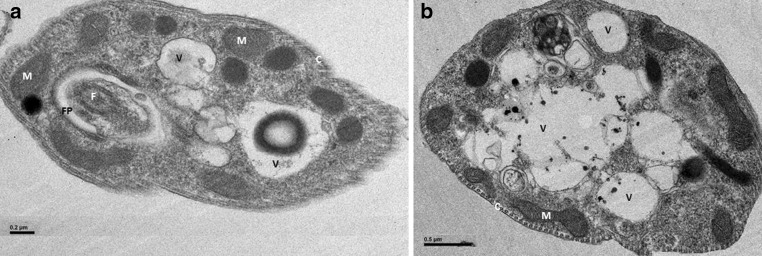
Fig. 1.
Electron microscopy of purified intracellular and axenic Leishmania amastigotes. a purified intracellular amastigotes of L. donovani, b purified intracellular amastigotes of L. infantum, the presence of vacuoles (V) in purified amastigotes could be explained by the observed cholesterol increase. F flagellum, K kinetoplast, N nucleus, M mitochondrion, FP flagellar pocket, C cytoskeleton

