Abstract
A 69-year old bed-bound woman presented with chest pain and diaphoresis. Diagnostic imaging led to the diagnosis of bilateral extensive pulmonary emboli extending into all segmental branches. Tissue plasminogen activator (tPA) was administered via 2 separate EKOS catheters. Repeat evaluation after 33 hours revealed improvement of right ventricular size and function. EKOS catheters are useful for administration of fibrinolytics in pulmonary embolism.
KEY WORDS: EKOS, thrombolysis, pulmonary embolus
INTRODUCTION
This is a case of a 69-year-old woman who presents to the ER with chest pain and diaphoresis. She had been mostly bedbound for the past 2 months due to a stroke 2 months ago with resulting residual hemiplegia. After diagnostic imaging was performed, she was diagnosed with extensive bilateral emboli involving the right and left main pulmonary arteries and extending into all segmental branches. The study decided to use catheter directed tPA via EKOS catheter. tPa was administered via 2 separate catheters over 12 hours for a total of 24mg. Then, the EKOS catheters were removed, and repeat echo done 33 hours after the initial echo showed improvement of right ventricular size and function. This case report aims to show benefits of using EKOS catheters for the administration of fibrinolytics in pulmonary embolism.
CASE REPORT
A 69-year-old woman presented to the emergency room with chest pain and diaphoresis. She had been mostly bedbound for the past 2 months due to stroke 2 months ago with residual hemiplegia.
Findings
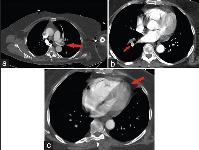
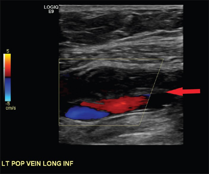
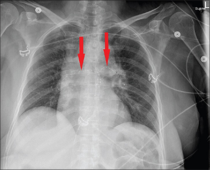
Chest X-ray on admission was normal. Computed tomography (CT) angiogram was performed and showed extensive bilateral emboli involving the right and left main pulmonary arteries and extending into all segmental branches [Figure 1a and b] and straightening of the interventricular septum [Figure 1c]. Bilateral lower extremity venous duplex ultrasound showed a deep venous thrombus in the left distal popliteal vein [Figure 2]. She was initially treated with heparin. Transthoracic echocardiography (echo) was performed to evaluate for right heart strain and showed severe dilation of the right ventricle and McConnell's sign (right ventricular dysfunction with akinesia of the mid-free wall and hypercontractility of the apex), which is a specific sign of acute pulmonary embolism (PE) [Video 1]. Risks and benefits were discussed with the patient about the use of pulmonary artery catheter-directed tissue plasminogen activator (tPA) in light of her recent stroke 2 months ago. It was decided to use catheter-directed tPA via EKOS catheter. EKOS catheters were placed in the bilateral pulmonary arteries as seen on the chest X-ray [Figure 3]. tPa was administered via two separate catheters over 12 h for a total of 24 mg. Then, the EKOS catheters were removed, and repeat echo done 33 h after the initial echo showed improvement of right ventricular size and function [Video 2].
Figure 1.

(a) Significant evidence of emboli in the left mainstem pulmonary artery can be seen in this contrast-enhanced axial computed tomography image (red arrow). Emboli in the subsegmental branches can also be noted just superior to the left mainstem. (b) Contrast-enhanced computed tomography image showing filling defect and emboli of the right mainstem pulmonary artery (red arrow). (c) Interventricular septum straightening (red arrow) is shown on this contrast-enhanced axial computed tomography image. This phenomenon is seen due to elevated right heart pressures from the resistance created by the massive emboli
Figure 2.

Lower extremity venous duplex ultrasound showing a deep venous thrombus in the left distal popliteal vein (red arrow)
Figure 3.

EKOS catheters can be visualized in bilateral pulmonary arteries (red arrows). Catheters were in place for 12 h to aid in the delivery of tissue plasminogen activator and thrombolysis
DISCUSSION
Although CT angiography remains the first-line imaging study for diagnosing acute PE, echo may be useful in cases of massive PE to evaluate right heart strain to determine the need for thrombolytics. On echo, McConnell's sign can be valuable to demonstrate the cardiac dysfunction caused by an acute PE.[1] In this case, it was particularly important to determine the effects of the clot burden given the patients increased risks associated with tPA administration because of a history of recent stroke. In this case, EKOS catheters were used to administer tPA. The EKOS catheters have ultrasound transducers within the wire of the catheter, which uses ultrasound waves to accelerate the fibrinolytic process, and therefore decrease treatment time and decrease the total dose of thrombolytic agent needed which should translate into decreased morbidity and mortality.[2] A prospective multicenter study on 150 patients with acute massive or submassive pulmonary emboli who underwent ultrasound-facilitated, catheter-directed, low-dose fibrinolysis showed decreased right ventricular dilatation, decreased pulmonary hypertension, reduced clot burden, and decreased intracranial hemorrhages in these patients. Efficacy outcomes were measured within 48 h of initiation of fibrinolytic therapy.[3] In the case presented, there was also improved cardiac function after administration of tPA via EKOS catheters within 48 h. Therefore, this case supports the benefits of using EKOS catheters for the administration of fibrinolytics in PE.
Financial support and sponsorship
Nil.
Conflicts of interest
There are no conflicts of interest.
Videos available on: www.lungindia.com
REFERENCES
- 1.Patra S, Math RS, Shankarappa RK, Agrawal N. McConnell's sign: An early and specific indicator of acute pulmonary embolism. BMJ Case Rep 2014. 2014:pii: Bcr2013200799. doi: 10.1136/bcr-2013-200799. [DOI] [PMC free article] [PubMed] [Google Scholar]
- 2.Owens CA. Ultrasound-enhanced thrombolysis: EKOS EndoWave infusion catheter system. Semin Intervent Radiol. 2008;25:37–41. doi: 10.1055/s-2008-1052304. [DOI] [PMC free article] [PubMed] [Google Scholar]
- 3.Piazza G, Hohlfelder B, Jaff MR, Ouriel K, Engelhardt TC, Sterling KM, et al. A prospective, single-arm, multicenter trial of ultrasound-facilitated, catheter-directed, low-dose fibrinolysis for acute massive and submassive pulmonary embolism: The SEATTLE II Study. JACC Cardiovasc Interv. 2015;8:1382–92. doi: 10.1016/j.jcin.2015.04.020. [DOI] [PubMed] [Google Scholar]
Associated Data
This section collects any data citations, data availability statements, or supplementary materials included in this article.


