Abstract
Patient: Female, 23
Final Diagnosis: Chronic tonsillitis with kincking of right internal carotid artery
Symptoms: Recurrant sore throat
Medication: —
Clinical Procedure: Tonsillotomy
Specialty: Otolaryngology
Objective:
Unusual clinical course
Background:
The internal carotid artery (ICA) is about 2.5 cm away from the tonsils. It has no branches in the cervical portion. ICA anomalies of the neck zone may result in a massive arterial bleeding during pharynx and neck surgery. Due to these anomalies, the surgeon must be aware of this risk during tonsillectomy, adenoidectomy, and pharyngeal operations.
Case Report:
A 23-year-old woman who was discovered to have an acute S curling-type anomaly of the ICA in contact with the lateral border of the right tonsil during a work-up for a tonsillectomy. This anomaly was incidentally discovered via computed tomography (CT) with contrast. In re-evaluating the course of treatment, we found a severe S-shape kink on the right side, bringing it close to the right tonsil by approximately 2 mm, and putting it at severe risk of injury during a simple tonsillectomy, possibly exposing the patient to serious bleeding. Partial tonsillectomy was performed for this patient with the aim to preserve and not expose the internal carotid artery. Pulsation of right tonsil was recorded. The patient made an uneventful postoperative recovery.
Conclusions:
Undetected ICA anomaly variation can lead to fatal bleeding during a simple procedure, like tonsillectomy. We recommend vigilance during tonsillectomy if one is using a hot dissection method versus a cold dissection method, which may allow for detection of a perioperative ICA anomaly. Tonsillectomy performed by a junior resident should be under direct supervision, particularly if the hot dissection method is used.
MeSH Keywords: Anatomy; Carotid Artery Diseases; Imaging, Three-Dimensional; Neck Dissection; Palatine Tonsil; Pharynx
Background
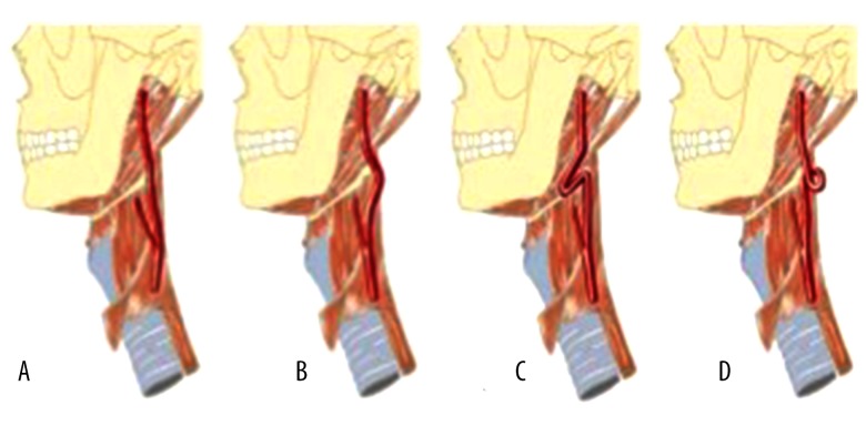
The internal carotid artery (ICA) is a branch of the common carotid artery. It has a straight course in the neck to the base of the skull, and does not have branches in this cervical portion. The human anatomy has variations between individuals, and ICA is one of the structures with anatomic variations, such as kinking, elbowing, and notches. In older individuals, such variations may be caused by arteriosclerosis and thrombosis, which can affect blood flow and cause encephalic ischemic processes [1]. The variations of the ICA include variability of pattern and degree (Figure 1). Some otolaryngology procedures carry a risk of ICA injury; these procedures include tonsillectomy, peritonsillar abscess drainage, soft palate impalement injuries, adenoidectomy, and velopharyngeoplasty [2]. The origin of these different variations has been controversial. Some are believed to represent congenital vascular anomalies, and others are related to arteriosclerotic pathology or fibromuscular dysplasia. Undetected ICA anomalies can result in fatal complications from biopsy, as well as surgical and anesthetic procedures [3]. Iatrogenic injury to the ICA is a rare complication of pharyngeal surgery. In addition, most anatomic variations of ICA are asymptomatic, and can be found incidentally during radiographic studies; these are done for unrelated reasons, or can remain undiscovered preoperatively [4]. In pediatrics, diagnosis of these variations must be considered, especially in patients indicated for tonsillectomy, in which iatrogenic injury during the surgical process may have catastrophic consequences [4,5]. Our rationale to report this case is to emphasize the fact that the surgeon must protect against vascular anomalies that may be confronted during head-neck surgery, and that preoperative examinations might be considered within this context.
Figure 1.
Schematic drawings of form and course variations of the cervical ICA: (A) straight course, (B) curved course, (C) kinking, and (D) coiling [1]. Curving and looping of the internal carotid artery in relation to the pharynx: Frequency, embryology and clinical implications. J Anat, 2000; 197: 373–81 by Paulsen F, Tillman B, Christofides C, et al. © 2000 Anatomical Society of Great Britain and Ireland. Reproduced with permission of John Wiley and Sons via Copyright Clearance Center.
Case Report
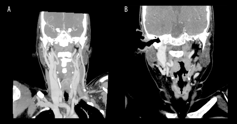
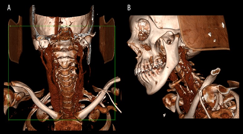
A 23-year-old Saudi female patient, with controlled sickle cell anemia, was seen in the otolaryngology clinic complaining of recurrent tonsillitis and nasal obstruction. No signs or symptoms suggested any jeopardy of cerebral blood flow. No history of stroke or other sickle cell anemia crisis, except vaso-occlusive crisis twice. On examination, the nose looked clear of polyps but the turbinate was hypertrophied, while the intra-nasal fiber optic examination was unremarkable apart from thick nasal secretion. On oropharyngeal examination, there was bilateral tonsillar hypertrophy, more prominent on the right side (grade 3), and the left side (grade 2). Our plan was to admit the patient for bilateral tonsillectomy. CT of the head and neck was requested to evaluate her paranasal sinuses; contrast was added to evaluate unilateral tonsillar enlargement and rule out other neck pathology. CT showed bilateral large tonsils, more enlarged on the right side, and overlaying a large vessel (ICA). In re-evaluating the course of treatment, we found a severe S-shape kink on the right side, bringing it close to the right tonsil by approximately 2 mm, and putting it at severe risk of injury during a simple tonsillectomy, possibly exposing the patient to serious bleeding (Figures 2, 3). The plan was thus changed to tonsillotomy as a tissue biopsy; following a right tonsillectomy, pulsation of that tonsil was noted and recorded (Video 1). The sample was sent to the histopathology laboratory, and results indicated a chronic inflammatory process. The patient made an uneventful postoperative recovery. No neurologic deficit or hemorrhage was detected during or after the operation. The patient and family were educated about this anatomic variation and told to be careful with future head and neck surgery.
Figure 2.
(A) CT scan of head and neck with contrast where the S-shape of the right ICA was detected (arrow). The external angle of the right ICA was 135° and the internal angle was 45° (type II based on Metz’s classification). The left ICA shows mild tortuosity with a posterior course to the jugular vein, more clarity with a 3D view. (B) Same CT of head and neck with contrast and a different view, where right ICA was detected and markedly touching the right tonsil, with no vessel calcification.
Figure 3.
3D view: CT scan of head and neck with contrast. (A) Right ICA with S-shaped kinking course (arrow). (B) Left ICA also has tortuous course, but milder than right ICA (arrow), with posterior course to left jugular vein.
Video 1.
Pulsation of right tonsil noted after tonsillotomy due to kincking of right internal carotid artery.
Discussion
Previous studies have reported the prevalence of ICA anomalies, ranging from 16% to 60% among different age groups (Table 1) [6–12]. Macchi C et al., is the only author who did the image study on one hundred of normal healthy individuals and found 26.5% has ICA anomaly [8]. These studies also report the ICA anomaly as seen more often in elderly women [6–12]. Types of ICA angle severity were first described by Metz et al. in 1961 (Table 2) [7]. Weibel and Fields first introduced the classification of tortuosity, kinking, and coiling in 1965. Tortuosity has been described as an S- or C-shaped elongation or undulation in the course of ICA. Coiling has been defined as elongation or redundancy of the ICA, resulting in a severe S-shaped curve or a circular configuration. Kinking has been the most frequently reported carotid abnormality (CA), and described as an angulation of the ICA, graded according to the severity of the angle between the two segments forming the kink [6].
Table 1.
| Author | Subjects | Study design | Image modality | Percentage of ICA anomaly |
|---|---|---|---|---|
| Metz H et al., 1961 | 1000 | Retrospective | Angiography | 16% |
| Macchi C et al., 1997 | 100 | Prospective | Duplex US | 26.5% |
| Del Corso L et al., 1998 | 469 | Prospective | Duplex US | 58% |
| TOGAY I et al., 2005 | 345 | Prospective | Duplex US | 24.6% |
| Sacco S et al., 2007 | 1217 | Prospective | Duplex US | 26.2% |
| Ekici F et al., 2012 | 607 | Retrospective | CTA | 60.3% |
| Yu C et al., 2013 | 923 | Prospective | Duplex US | 30% |
Table 2.
Classification of kinks according to angle severity [7].
| Mild kinking | Acute angulation of ICA with angle between two segments of the kink measured >60° |
| Moderate kinking | Acute angulation of ICA with angle between the two segments forming the kink, measured 30–60° |
| Severe kinking | Acute angulation of ICA with angle between the two segments forming the kink, measured <30° |
Aydogan et al. thought there were types of forces that might cause vessels elongation, buckling, and then tortuosity. The two forces identified were traction and pressure inside the lumen. Branches and perivascular tissues could thus stretch the main straight artery and cause traction, while the intraluminal pressure could push the vessels walls from inside and cause extension. The hyperdynamic circulation in sickle cell anemia patients could cause lengthening and tortuosity by high intraluminal pressure via the mechanism described above [13], as in our case.
Ekici et al. reported 607 cases of imaging evaluation of ICA. The carotid-pharyngeal distance (CPD) to the right and left ICA yielded an average of 11.13 mm. The shortest CPD was measured as 0.92 mm, while the longest CPD was 24.12 mm [11]. In our case report, the right ICA was kinking with a CPD of 2 mm. Cong et al. reported seven cases of ICA anomaly in China, of which four patients had pharyngeal bulge with pulsation, and normal pharynges in three other patients [14]. Bhandarkar et al. reported a case of a 45-year-old woman with right pulsating and swelling over the oropharynx, with normal mucosal lining. The distance between the ICA and oropharyngeal mucosa was 3 mm [15]. Another case reported by De Virgilio et al. involved a woman in her 70s with a right retropharyngeal pulsating mass, with 1 mm between the ICA and mucosal surface [16].
Some patients with ICA anomalies have episodes of cerebrovascular insufficiency related to the position of their heads [17,18], but this was not a concern in our patient.
ICAs have their embryonic origins in the third aortic arch and dorsal aorta. During normal embryonic development, the dorsal aortic root descends into the chest in the fifth to eighth week of fetal life, lengthening and straightening the course of the carotid artery. It is suggested that incomplete straightening and persistence of embryonic angulation can result in aberrant carotid arteries [19]. Postoperative bleeding is an adenotonsillectomy complication in normal anatomical circumstances. The risk of life-threatening bleeding increases with ICA anomalies. Therefore, the physicians must be careful in performing pharyngeal surgeries [11].
Conclusions
Undetected ICA anomaly variation can lead to fatal bleeding during a simple procedure, like tonsillectomy. We recommend vigilance during tonsillectomy if one is using a hot dissection method versus a cold dissection method, which may allow for detection of a perioperative ICA anomaly. Tonsillectomy performed by a junior resident should be under direct supervision, particularly if the hot dissection method is used.
Abbreviations:
- ICA
internal carotid artery
- CT
computed tomography
- CA
carotid anomaly
- CPD
carotid pharyngeal distance
References:
- 1.Paulsen F, Tillman B, Christofides C, et al. Curving and looping of the internal carotid artery in relation to the pharynx: Frequency, embryology and clinical implications. J Anat. 2000;197:373–81. doi: 10.1046/j.1469-7580.2000.19730373.x. [DOI] [PMC free article] [PubMed] [Google Scholar]
- 2.Cairney J. Tortuosity of the cervical segment of the internal carotid artery. J Anat. 1924;59:87–96. [PMC free article] [PubMed] [Google Scholar]
- 3.Agarwal R, Agarwal SK. Dangerous anatomic variation of internal carotid artery. Int J Anat Var. 2011;4:174–76. [Google Scholar]
- 4.Wasserman JM, Sclafani SJ, Goldstein NA. Intraoperative evaluation of a pulsatile oropharyngeal mass during adenotonsillectomy. Int J Pediatr Otorhinolaryngol. 2006;70:371–75. doi: 10.1016/j.ijporl.2005.07.002. [DOI] [PubMed] [Google Scholar]
- 5.Kay DJ, Mehta V, Goldsmith AJ. Perioperative adenotonsillectomy management in children: current practices. Laryngoscope. 2003;113:592–97. doi: 10.1097/00005537-200304000-00002. [DOI] [PubMed] [Google Scholar]
- 6.Togay I, Sikay C, Kim J, et al. Carotid artery tortuosity, kinking, coiling: stroke risk factor, marker, or curiosity. Acta Neurol Belg. 2005;105:68–72. [PubMed] [Google Scholar]
- 7.Metz H, Murray-Leslie RM, Bannister RG, et al. Kinking of the internal carotid artery in relation to cerebrovascular disease. Lancet. 1961;1:424–6. doi: 10.1016/s0140-6736(61)90004-6. [DOI] [PubMed] [Google Scholar]
- 8.Macchi C, Gulisano M, Giannelli F, et al. A statistical study in healthy subjects by echocolor Doppler. J Cardiovasc Surg. 1997;38:629–37. [PubMed] [Google Scholar]
- 9.Del Corso L, Moruzzo D, Conte B, et al. Tortuosity, kinking, and coiling of the carotid artery: Expression of atherosclerosis or aging? Angiology. 1998;49:361–71. doi: 10.1177/000331979804900505. [DOI] [PubMed] [Google Scholar]
- 10.Sacco S, Totaro R, Baldassarre M, et al. Morphological variations of the internal carotid artery: Prevalence, characteristics and association with cerebrovascular disease. Int J Angiol. 2007;16:59–61. doi: 10.1055/s-0031-1278249. [DOI] [PMC free article] [PubMed] [Google Scholar]
- 11.Ekici F, Tekbas G, Önder H, et al. Course anomalies of extracranial internal carotid artery and their relationship with pharyngeal wall: An evaluation with multislice CT. Surg Radiol Anat. 2012;34:625–31. doi: 10.1007/s00276-012-0958-3. [DOI] [PubMed] [Google Scholar]
- 12.Yu C, Xiong J-Q, Dai JP, et al. Independent risk factors for morphological abnormalities of the internal carotid artery. Acta Cardiol. 2013;68:481–87. doi: 10.1080/ac.68.5.2994471. [DOI] [PubMed] [Google Scholar]
- 13.Aydoğan B, Kiroğlu M, Talas D, et al. Kinking of the internal carotid artery. T Klin J ENT. 2001;1:42–44. [Google Scholar]
- 14.Cong TC, Duan X, Gao WH, et al. [Tortuosity and kinking of cervical segment of internal carotid artery: An analysis of 7 cases] Zhonghua Er Bi Yan Hou Tou Jing Wai Ke Za Zhi. 2012;47:913–17. [in Chinese] [PubMed] [Google Scholar]
- 15.Bhandarkar AM, Nayak R, Chidambaranathan N, et al. Beware of a pulsating oropharynx. BMJ Case Rep. 2014;2014 doi: 10.1136/bcr-2014-208184. pii: bcr2014208184. [DOI] [PMC free article] [PubMed] [Google Scholar]
- 16.De Virgilio A, Greco A, de Vincentiis M. A submucosal retropharyngeal pulsatile mass. JAMA Otolaryngol Head Neck Surg. 2015;141:1027–28. doi: 10.1001/jamaoto.2015.2398. [DOI] [PubMed] [Google Scholar]
- 17.Desat B, Toole JF. Kinks, coils, and carotids: a review. Stroke. 1975;6:649–53. doi: 10.1161/01.str.6.6.649. [DOI] [PubMed] [Google Scholar]
- 18.Leipzig TJ, Dohrmann GJ. The tortuous or kinked carotid artery: Pathogenesis and clinical considerations. A historical review. Surg Neurol. 1986;25:478–86. doi: 10.1016/0090-3019(86)90087-x. [DOI] [PubMed] [Google Scholar]
- 19.Kelly AB. Tortuosity of the internal carotid artery in relation to the pharynx. Journal of Laryngology and Otology. 1925;40:15–23. [Google Scholar]


