To the editors,
Olfactory Neuroblastomas (ONBs) are tumors of neuroectodermal origin which, according to World Health Organization, usually not express cytokeratin [1]. In their paper entitled “Neuroendocrine Neoplasm of the Sinonasal Tract: Neuroendocrine Carcinomas and Olfactory Neuroblastoma” Shah and Perez-Ordoñez [2] reported a cytokeratins positivity, observed in up to 30 % of ONBs, in accordance with other Authors who documented cytokeratins expression from 23 to 35 % of cases, both focal or diffuse with strongly intensity, but never showing a paranuclear or globoid stain (“dot-like” pattern). This latter being an important difference respect to Neuroendocrine Carcinoma (NEC) [1–5]. Furthermore another recent article by Holbrook et al. [6] demonstrated that cytokeratin 18 (CK18) was positive in ONB separating tumor cells into nests.
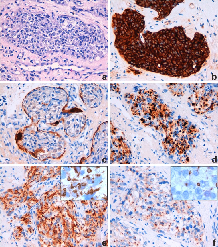
We recently observed a small biopsy performed on a stenotic right nasal cavity in a 44 years old man. Histological examination revealed a submucosal infiltration of epithelial-like neoplasia, with nested pattern of growth and high vascularity (Fig. 1a). The neoplastic cells had abundant cytoplasm and atypical nuclei with prominent nucleoli; the mitotic rate was low. Immunohistochemical staining showed diffuse and strong cytoplasmatic positivity for synaptophysin (Fig. 1b) and neuron specific enolase (NSE); the S-100 was positive in a sustentacular cell pattern (Fig. 1c), while EMA, GFAP, and TTF-1 were negative. Moreover a patchy and strong positivity, with a “dot-like” pattern for multicytokeratin (Novocastra™ Liquid Mouse Monoclonal Antibody Cytokeratin—5/6/8/18, clone 5D3 and LP34, dilution 1:100, full automated immunohistochemistry stainer BOND-III Leica Biosystems) was observed (Fig. 1d).
Fig. 1.
a H&E; b synaptophysin; c S100; d multicytokeratin (5/6/8/18); e CK18; f CK 8. Original magnification ×40 (a, b, c, d, e, f), ×100 (e inset, f inset)
In order to better understanding the specificity of this immunoreactivity we decided to perform the separate immunostaining for CK 5 (Leica Biosystems, clone XM26, ready to use, BOND-III Leica Biosystems), CK 6 (Invitrogen™, clone SP87, dilution 1:100, BOND-III Leica Biosystems), CK 8 (Leica Biosystems, clone TS1, ready to use, BOND-III Leica Biosystems), CK18 (Dako, clone DC10, dilution 1:15, BOND-III Leica Biosystems), multicytokeratin AE1/AE3 (Novocastra™, cocktail of two clones AE1 and AE3, ready to use, BOND-III Leica Biosystems) and multicytokeratin 34βE12 (Novocastra™, clone 34βE12, ready to use, BOND-III Leica Biosystems). CK5, CK6, multicytokeratin AE1/AE3 and 34βE12 were all negative. Instead the neoplasm showed CK18 positivity, focally dot-like (Fig. 1e) and, surprisingly, focally CK8 positivity too, never reported for ONB in Literature to our knowledge (Fig. 1f). The CK18 immunostaining result was in accordance with the article of Holbrook et al. [6]. The morphological and immunophenotypical features were consistent with the diagnosis of ONB, but with an aberrant expression of cytokeratins.
In conclusion our case represents the first demonstration of a “dot-like” cytokeratin expression in an ONB, a feature hitherto undescribed to our knowledge and supplements the Literature data [2–5]. Pathologists should be aware of this potential diagnostic pitfall in distinguishing a high grade ONB from a high grade NEC, particularly in scanty biopsies [2], in which is crucial an integration of immunohistochemical data with careful interpretation of morphological features.
Compliance with Ethical Standards
Conflict of interest
None.
References
- 1.Barnes L, Eveson JW, Reichart P, Sidransky D. World Health Organization classification of tumours, pathology and genetics, head and neck tumours. Lyon: IARC Press; 2005. [Google Scholar]
- 2.Shah K, Perez-Ordoñez B. Neuroendocrine neoplasm of the sinonasal tract: neuroendocrine carcinomas and olfactory neuroblastoma. Head Neck Pathol. 2016;10(1):85–94. doi: 10.1007/s12105-016-0696-7. [DOI] [PMC free article] [PubMed] [Google Scholar]
- 3.Hunt JL. Immunohistology of head and neck neoplasms. In: Dabbs DJ, editor. Diagnostic immunohistochemistry theranostic and genomic applications. 3. Philadelphia: Saunders Elsevier; 2010. pp. 256–290. [Google Scholar]
- 4.Argani P, Perez-Ordoñez B, Xiao H, et al. Olfactory neuroblastoma is not related to Ewing family of tumors. Absence of EWS/FLI1 gene fusion and MIC2 expression. Am J Surg Pathol. 1998;22:392–398. doi: 10.1097/00000478-199804000-00002. [DOI] [PubMed] [Google Scholar]
- 5.Hirose T, Scheithauer BW, Lopes MB, et al. Olfactory neuroblastoma. An immunohistochemical, ultrastructural, and flow cytometric study. Cancer. 1995;76(1):4–19. doi: 10.1002/1097-0142(19950701)76:1<4::AID-CNCR2820760103>3.0.CO;2-E. [DOI] [PubMed] [Google Scholar]
- 6.Holbrook EH, Wu E, Curry WT, et al. Immunohistochemical characterization of human olfactory tissue. Laryngoscope. 2011;121(8):1687–1701. doi: 10.1002/lary.21856. [DOI] [PMC free article] [PubMed] [Google Scholar]


