Abstract
Chronic Achilles tendinopathy (AT) is an overuse condition seen among runners. Eccentric exercise can decrease pain and improve function for those with chronic degenerative tendon changes; however, some individuals have continued pain requiring additional intervention. While joint mobilization and manipulation has not been studied in the management in Achilles tendinopathy, other chronic tendon dysfunction, such as lateral epicondylalgia, has responded well to manual therapy (MT). Three runners were seen in physical therapy (PT) for chronic AT. They were prescribed eccentric loading exercises and calf stretching. Joint mobilization and manipulation was implemented to improve foot and ankle mobility, decrease pain, and improve function. Immediate within-session changes in pain, heel raise repetitions, and pressure pain thresholds (PPT) were noted following joint-directed MT in each patient. Each patient improved in self-reported function on the Achilles tendon specific Victorian Institute for Sport Assessment questionnaire (VISA-A), pain levels, PPT, joint mobility, ankle motion, and single-leg heel raises at discharge and 9-month follow-up. The addition of MT directed at local and remote sites may enhance the rehabilitation of patients with AT. Further research is necessary to determine the efficacy of adding joint mobilization to standard care for AT.
Level of Evidence
Case series. Therapy, Level 4.
Keywords: Ankle, Manipulation, Mobilization, Running injury, Tendon
Background
Chronic Achilles tendinopathy (AT) can be a functionally limiting condition occurring in up to 11% of runners,1 with an incidence rate of injury reported to be 2.35 per 1000 adults.2 This condition is often characterized by limited function, stiffness, pain, and tenderness at the Achilles tendon, most commonly 2–7 cm proximal to the insertion site. Histopathological investigation of AT has revealed degeneration and disordered arrangement of collagen fibers, increased vascularity, an increase in mucoid ground substance and more cells with rounded nuclei.3 AT is typically related to overuse with a number of proposed risk factors4 including abnormal talocrural and subtalar joint motion.5 Subjective reports of pain and tenderness may be sufficient to arrive at a clinical diagnosis of AT.6 Physical therapy (PT) examination and treatment options are well detailed in clinical practice guidelines developed by the Orthopedic section of the American Physical Therapy Association, with strong evidence supporting eccentric exercise.5 However, despite strong evidence supporting conservative management for AT, up to 29% of patients go on to surgical intervention1 indicating the needs of some patients are not being met by previously described intervention approaches. In cases when 4–6 months of conservative treatment fails, surgery may be indicated to remove fibrotic adhesions and degenerative nodules to restore vascularity,4 with positive outcomes noted.5
While the mechanisms are still under investigation, eccentric exercise has been associated with decreased neovascularization, decreased tendon thickness, and improved tendon structure in individuals with chronic AT.7,8 This is interesting, as neurovascular ingrowth of the degenerated tendon has been linked to nociception in patients with AT.9 Different eccentric exercise protocols exist for AT, yet each has demonstrated positive outcomes.10–14 One case series reported that improvements were maintained at a five year follow-up, after using bilateral, unilateral, eccentric, and fast-rebounding heel raise exercises for patients with chronic AT.15 Although eccentric exercise alone has led to good outcomes, it remains unclear if a specific prescription is optimal.
While a single case report detailing the utilization of soft tissue mobilization in the management of AT has been published,16 joint mobilization and manipulation have not been studied in the management of AT. Numerous mechanisms for the efficacy of manual therapy (MT) have been described, involving biomechanical, neurophysiological, and psychosocial components.17 Joint mobilization specifically has been associated with decreased nociceptive reflex excitability,18 enhanced conditioned pain modulation,19 and reduction of bilateral hyperalgesia following unilateral joint mobilization.20 Spinal thrust manipulation has been associated with immediate changes in pain sensitivity21,22 indicating both thrust and non-thrust techniques can alter pain processing mechanisms, potentially enhancing rehabilitation programs. Considering that limited ankle dorsiflexion motion may contribute to the development of AT,23 and that dorsiflexion mobility improved after joint mobilization in other patient populations,24,25 it seems reasonable that using joint mobilization to improve ankle mobility may decrease overuse of the Achilles tendon when walking or running. While not well described in the AT population, MT has been associated with positive outcomes in other tendinopathies,26–33 and may be beneficial for AT as well.
The purpose of this report is to describe the addition of joint mobilization and manipulation to an eccentric exercise program in three patients with AT.
Examination
Three patients were seen in an outpatient PT setting for chronic AT, confirmed by Magnetic Resonance Imaging (MRI). This work was deemed exempt from IRB approval, as fewer than four patients are described. Informed consent was received and the rights of the patients were protected.
A standardized examination was performed by the same therapist (DJ) on each patient. The examination included a detailed subjective history and objective tests and measures. Each patient denied a history of ankle sprains or other ankle injuries. Each patient took ibuprofen as needed for pain relief. None of the patients had prior treatment for their condition. Their goals were to return to running without symptoms.
Objective testing included examination of symptom provoking functional activities and functional movements, regional screening, balance, range of motion (ROM), force generation capacity, joint physiologic and accessory mobility, self-report outcome measures, and quantitative sensory testing. Pertinent examination findings are presented in Table 1.
Table 1.
Initial examination findings
| Patient 1 | Patient 2 | Patient 3 | |
|---|---|---|---|
| Location of primary symptoms | Left Achilles tendon | Right Achilles tendon | Right Achilles tendon |
| BMI | 24.2 | 21.4 | 23.8 |
| Mechanism of injury | Overuse | Overuse | Overuse |
| Tenderness | (+) 4 cm proximal to insertion | (+) 2 cm proximal to insertion | (+) 4 cm proximal to insertion |
| Running gait | Increased pronation through left mid/terminal stance | Early heel rise on right | Increased pronation through right mid/terminal stance |
| VISA-A | 32/100 | 51/100 | 47/100 |
| Pain rating (NPRS) | Best: 0/10 | Best: 0/10 | Best: 0/10 |
| Worst: 6/10 | Worst: 8/10 | Worst: 5/10 | |
| Heel raise repetitions | < 1, able to get 20o plantarflexion (6/10 pain) | 5 (6/10 pain) | 6 (4/10 pain) |
| PPT (kPa) | Left: 152 | Left: 420 | Left: 415 |
| Right: 308 | Right: 314 | Right: 360 | |
| Ankle DF AROM | 0–6o | 0–8o | 0–7o |
| Joint mobility | Hypomobile AP glide of talus | Hypomobile AP glide of talus | Hypomobile AP glide of talus |
| Hypomobile lateral glide at subtalar joint | Hypomobile lateral glide at subtalar joint |
Abbreviations: BMI: body mass index; VISA-A: victorian institute of sports, Australia – Achilles tendon; NPRS: numerical pain rating scale; PPT: pressure pain thresholds, in kilopascals; DF: dorsiflexion; AROM: active range of motion; MRI: magnetic resonance imaging; AP: anterior to posterior.
Active Motion Testing
Posture, single limb stance balance, bilateral and unilateral squats were examined for static and dynamic lower extremity alignment, and were unimpaired. Walking and running gait were evaluated on a treadmill. Lumbar, hip, and knee screens, including active and passive ROM including overpressures,34 were negative for Achilles pain reproduction in all three cases. Neuroprovocation assessment using the straight leg raise and slump test,35 was negative for symptom reproduction. Hip abduction (gluteus medius bias) and extension (gluteus maximus bias) strength were tested in sidelying and prone, respectively, and each patient tested strong. Ankle plantarflexion endurance was tested using a repeated single-limb heel raise.5 If a patient was unable to complete a full repetition due to pain, the angle that the patient’s heel raised off the ground was measured, using the first metatarsalphalangeal (MTP) joint as the fulcrum.
Passive Mobility Assessment
Ankle dorsiflexion ROM was tested in supine using a goniometer. Calf length was tested in supine by passively dorsiflexing the ankle with the knee bent and straight. Restriction of the gastrocnemius was noted on the involved side in each patient, as demonstrated by decreased ankle DF noted with the knee straight, and more motion present with the knee bent. Tenderness and thickening was noted with palpation of the mid-portion of the Achilles tendon in each case, yet the insertion site and retrocalcaneal bursa were not tender, and no active trigger points were noted in the calf. The Thompson test was negative.36 Ligamentous stress testing at the talocrural and subtalar joints was negative. Joint physiologic and accessory motion testing was performed at the proximal and distal tibiofibular joints, talocrural and subtalar joints, midfoot and first MTP, with restrictions present at the rearfoot in each case.37
Outcome Measures
Several outcomes were used to measure the effectiveness of the interventions. The Victorian Institute for Sport Assessment developed a valid and reliable self-reported functional scale for disability related to Achilles tendon dysfunction (VISA-A). The VISA-A is comprised of eight questions, scored 0 to 100, with lower scores indicating greater disability. It was shown to have good test-retest (r = 0.93), intrarater (r = 0.90), and interrater reliability (r = 0.90).38 Pain was measured using the 11-point 0–10 NPRS, where 0 indicates no pain, and 10 represents the worst pain imaginable. The minimal clinically important difference (MCID) for the NPRS differs based on the patient population examined, but has been reported to be 1 point for patients with chronic musculoskeletal pain.39 Pressure pain threshold (PPT) measurements were taken at the primary location of pain on the Achilles tendon using an algometer (Wagner FPXTM). PPT is quantified using an algometer and applying gradually increasing pressure against the target tissue until the patient first reports pain. It is commonly used to determine the presence of mechanical hyperalgesia and demonstrates good intrarater reliability (ICC = 0.94–0.97) and an intrarater minimal detectable change (MDC) ranging from 42.7 to 137.0 kPa depending on the region tested.40 While PPT is often measured at boney or muscular sites, the Achilles tendon was used in these cases. Normative PPT values at tendon sites were not found during a literature review so the contralateral Achilles was used as a reference point.41 At discharge, the Global Rating of Change (GROC) scale was administered. The GROC scale is a self-reported 15-point Likert scale with −7 being a ‘a very great deal worse,’ 0 being ‘no change,’ and +7 being ‘a very great deal better.’ A change in 3 or more points was determined to be a clinically important difference.42
Individual Case Presentations
Patient 1 was a 52-year-old female referred from her primary care physician with a diagnosis of left Achilles pain. Central, deep, aching low back pain (LBP) was also reported; however, this was long-standing and she did not believe it to be contributory to her Achilles pain. Otherwise, her past medical history (PMH) was unremarkable. She stated that her symptoms started insidiously 6 months prior when she transitioned to outdoor running from a treadmill. On the numeric pain rating scale (NPRS), her pain was 0/10 at rest and 6/10 at worst. The pain was described as a superficial dull ache, sharp with certain movements, and was worsened by running, walking more than 30 mins, and getting up from sitting. Rest, ice, medication and calf stretching decreased her pain. She would usually run three times a week on a treadmill for 2–3 miles each run. Her LBP was reproduced with overpressure into extension, although it did not recreate her Achilles pain.
Patient 2 was a 38-year-old male presenting to physical therapy with a diagnosis of right AT from his orthopedic physician. Aside from an anterior cruciate ligament reconstruction 15 years prior on the contralateral limb, his PMH was unremarkable. He reported ongoing Achilles problems for the past three years with 1–2 flare-ups each year. He reported pain to be 0/10 at best and 8/10 at worst. It was aggravated by running, especially when doing speed workouts, and the calf raise machine at the gym. Alleviating activities included icing, rest, taking medication, and cross training. He stated the pain began when training for his first marathon likely due to increasing mileage too quickly. Typical running habits included both trail and treadmill running, 5–6 days each week, between 3 and 12 miles/run depending on training purpose.
Patient 3 was a 45-year-old male referred by his orthopedic doctor with a diagnosis of right Achilles tendinitis. The patient had hypertension controlled with medication. Otherwise his PMH was unremarkable. He reported an insidious onset nearly one year prior, when training for his first half marathon. At rest, pain was reported at 0/10 and, at worst, it would escalate to 5/10. While the pain diminished following the race, discomfort remained when getting up from sitting or when jogging for fitness. Rest and gentle stretching helped but did not eliminate symptoms. Prior to symptom onset, he was running on treadmills 3–4 times each week, between 3 and 5 miles each time.
Clinical Impression
Based on subjective and objective data, each patient’s signs and symptoms appeared consistent with non-insertional AT. The primary PT diagnosis was dysfunction of the plantarflexor mechanism including restricted gastrocnemius length and hypomobility of the talocrural and subtalar joints. Symptoms were activity dependent, suggesting a musculoskeletal condition. Each patient had isolated tenderness at the mid-portion of the Achilles tendon, with no tenderness at the tendon’s insertion or retrocalcaneal bursa. Patients had no reproduction of posterior calf or ankle pain with lumbar, hip, or knee screens, making a proximal joint pain referral less likely. Neuroprovocation testing was negative making a neural condition less likely. Systemic inflammatory disorders did not seem to be present based on subjective examination. The clinical diagnosis was consistent with MRI findings which were available in each case showing chronic tendinopathic changes at the involved Achilles tendon.
Because patients presented with signs and symptoms appearing consistent with mid-portion AT, they were prescribed an eccentric loading program, which has been shown to be generally effective for this population.5 In addition to eccentric exercise, each patient demonstrated joint restrictions that the therapist believed to be contributory to the patients’ symptoms. Subsequently, impairment based joint thrust and non-thrust mobilizations were added, with therapist discretion as to the location and type of application.
Intervention
Specific interventions utilized at each visit and the response to treatment is detailed for each patient in Table 2. All interventions for each session were provided by the same therapist (DJ) who performed the initial examination.
Table 2.
Interventions
| Patient | Visit | Exercise | Pre-treatment values | Manual therapy | Post-treatment values |
|---|---|---|---|---|---|
| 1 | 1 (evaluation) | Eccentric heel drop, 3 × 15 | NA | None | None |
| Calf stretch, 3 × 30” | |||||
| 2 | Same as above | Heel Raises: < 1 rep, 20o off ground | Supine talocrural distraction thrust manipulation | Heel Raises: < 1, 47o off ground | |
| PPT: 154 | PPT: 175 | ||||
| 3 | Same as above | Heel Raises: 1 | Right sidelying, Left L5-S1 rotational thrust manipulation | Heel Raises: 3 | |
| PPT: 178 | PPT: 188 | ||||
| 4 | Same as above | NA | None | None | |
| 5 (discharge) | Same as above | NA | None | None | |
| 2 | 1 (evaluation) | Eccentric heel drop, 3 × 15 | NA | None | None |
| Calf stretch, 3 × 30” | |||||
| 2 | Same as above | Heel Raises: 5 | AP glide right talus, grade III+, ×4 min | Heel Raises: 7 | |
| PPT: 317 | Lateral glide right subtalar joint, grade III+, ×4 min | PPT: 323 | |||
| 3 | Same as above | Heel Raises: 7 | Standing mobilization with movement with belt for ankle dorsiflexion, ×4 min | Heel Raises: 9 | |
| PPT: 331 | Lateral glide right subtalar joint, grade IV, ×4 min | PPT: 338 | |||
| 4 | Same as above | NA | None | None | |
| 5 (discharge) | Same as above | NA | None | None | |
| 3 | 1 (evaluation) | Eccentric heel drop, 3 ×15 | NA | None | None |
| Calf stretch, 3 × 30” | |||||
| 2 | Same as above | Heel Raises: 6 | AP glide right talus, grade III+, ×4 min | Heel Raises: 7 | |
| PPT: 355 | Lateral glide right subtalar joint, grade III+, ×4 min | PPT: 374 | |||
| 3 | Same as above | Heel Raises: 7 | Supine talocrural distraction thrust manipulation | Heel Raises: 8 | |
| PPT: 372 | PPT: 380 | ||||
| 4 | Same as above | NA | None | None | |
| 5 (discharge) | Same as above | NA | None | None |
Abbreviations: AP: anterior to posterior; NA: not applicable; PPT: pressure pain threshold readings taken at involved tendon, in kilopascals.
Visit 1 – Initial session
At the initial examination, patients were educated on Achilles tendon anatomy, clinical pathology, pain science, proper training, footwear while running, and what patients could expect with physical therapy. Each patient was trained in eccentric loading for the Achilles tendon, modified from Alfredson et al.10 Alfredson et al’s protocol calls for 3 sets of 15 repetitions with the knee both flexed and extended, twice each day for 12 weeks. Given the positive outcomes with varied exercise prescriptions,10–12 patients were instructed to perform eccentric loading as described by Alfredson et al.10 only with knees straight, as the therapist believed this position to be more comfortable for the patient. Additionally, patients were prescribed stretching for the gastrocnemius standing flat on the floor in subtalar neutral, to be performed 3 times each day, 3 sets of 30 s each. Static stretching and eccentric exercise were found to be more effective than eccentric exercise alone in a small study of patients with patellar tendinopathy.43
Visit 2 – Week 2
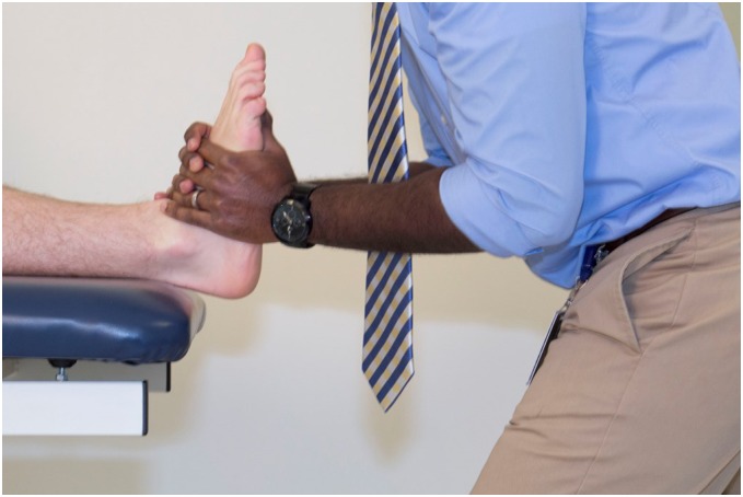
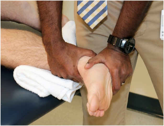
Patients reported compliance with eccentric training and stretching exercises. As each patient had restrictions in joint motion, joint thrust and non-thrust mobilizations were performed to improve ankle mobility and decrease pain. Single-limb heel raises and PPT values were assessed immediately before and after joint mobilization. Joint accessory glides were re-assessed at the onset of each session. Given patient 1’s mild mobility restriction, but low PPT values, she received a high velocity low amplitude distraction thrust manipulation (Fig. 1) to the left talocrural joint. Patients 2 and 3 had greater joint restriction with less symptom irritability, so high-grade mobilization was performed, as described by Maitland et al.37 Grade III + anterior–posterior (AP) mobilization to the talocrural joint and a grade III + subtalar joint lateral glide (Fig. 2) were performed until less restriction was noted, which turned out to be approximately 4 mins each. While subjective in nature, ‘less restriction’ was operationally defined as decreased tissue resistance to the same force application, and increased movement noted prior to end of available motion. Each patient demonstrated improvements in post-treatment PPT readings and heel raises. They were instructed to continue with their HEP as previously described.
Figure 1.

Talocrural joint long-axis thrust manipulation. With the patient in supine, the therapist grasps the plantar aspect of the foot with his thumbs, while grasping the talus with the ring fingers. Talocrural distraction is added, while simultaneously dorsiflexing the ankle. Ankle inversion and eversion is added, as needed, to increase tissue resistance. A long-axis thrust is performed. Source: Author
Figure 2.

Subtalar joint lateral glide mobilization. With the patient sidelying on the involved side, the therapist stabilizes the distal tibia and fibula with one hand. With the other hand, the therapist grasps the calcaneus, distal to the talus, and provides a mobilization force perpendicular to the ground. Source: Author
Visit 3 – Week 3
Patients reported continued compliance with their HEP, and noted slight improvement in symptoms. Patient 1 stated that, when walking, she nearly slipped on ice and aggravated her back pain. Increased lumbar extension was noted during gait observation with a shortened stride length bilaterally. Following examination, the therapist believed she may benefit from lumbar manipulation, as she had three positive predictor variables (local pain that did not radiate past the knee, hypomobility of multiple lumbar spine segments, and Fear Avoidance Belief Questionnaire work subscale score less than 19) on the validated clinical prediction rule described by Childs et al.44 for successful response. Beyond strictly manipulating a location of dysfunction, it was believed that improving lumbar mobility would improve gait mechanics by decreasing proximal compensation. A right side-lying, rotational lumbar thrust manipulation was directed at the left L5-S1 segment, as this was the symptomatic side. The patient was able to perform more heel raises with less pain reported, and with improved PPT values noted at the Achilles tendon. This is consistent with the work of Coronado et al.,22 which reported improved remote site PPT after spinal manipulation.
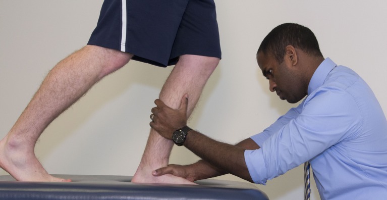
Patient 2 demonstrated improved but continued hypomobility at the talocrural and subtalar joints. Symptoms were starting later during her runs and the therapist believed that weight bearing joint mobility should be addressed, hopefully enhancing functional gait. Standing mobilization with movement with a posterior talar glide was added (Fig. 3), in addition to grade IV subtalar joint lateral glides. Each technique was performed for approximately 4 mins, which is when the joint was felt to be more mobile. Patient 3’s joint restriction was improved, but hypomobility remained when compared bilaterally. A supine talocrural distraction thrust manipulation was performed to normalize mobility. Heel raise repetitions and PPT readings increased after joint mobilizations, while pain decreased. Patients were instructed to continue with their HEP.
Figure 3.
Talocrural joint weight bearing mobilization with movement (anterior to posterior glide). The therapist places the web space of the application hand over the anterior aspect of the talus, while the other hand is placed proximally on the posterior aspect of the tibia. The patient lunges forward to the involved leg, while the therapist applies a posteriorly directed force to stabilize the talus. The opposite hand can guide the tibia into anterior translation as needed. A mobilization belt can also be applied to help guide motion. Source: Author
Visit 4 – Week 4
Each patient demonstrated normal joint accessory motion throughout the rearfoot as compared to their uninvolved side. Pain, VISA-A, PPT values and heel raise repetitions were also re-assessed. Patient 1 reported jogging a mile with minimal pain, a task she was previously unable to do, however at a mile and a half she would stop due to pain (4/10). Patient 2 remained active in running at a decreased mileage throughout the course of care. He could run 5 miles without increased pain and continue for 5 more miles with 2/10 pain, before pain increased to 4/10 which made him stop. Patient 3 could jog 2 miles prior to discomfort, which he opted not to push through.
Given the lack of joint restriction, symptomatic and functional improvements, and 12 week duration eccentric exercise protocol,10 the treating therapist believed the patients would benefit from consistent performance of their HEP, without the need for continued manual treatment. They were instructed to continue with eccentric exercise and stretching and were scheduled for a follow-up re-evaluation at 12 weeks. Patients were instructed to contact the therapist with any questions, and to return prior to 12 weeks if symptoms increased.
Visit 5 – Week 12
Patients were re-evaluated again at week 12 for progress. Each patient reported compliance with their HEP. Outcome measures were reassessed, with improvements noted throughout. Walking and running gait were evaluated, with no remarkable impairments noted. Patients were symptom free, had normal talocrural and subtalar joint mobility, accomplished all goals, and were, therefore, discharged from physical therapy. Patients were advised to continue to perform stretching and eccentric exercises as part of a regular HEP.
Outcomes
Outcome measures for all patients are presented in Table 3. Immediate pre-post measurements improved in heel raises, PPT, and pain levels following each of the joint thrust and non-thrust mobilizations. At discharge, each patient markedly improved their scores on the VISA-A questionnaire and PPT values, and exceeded the MCID for both NPRS and GROC scores. Symptomatic and functional improvements were maintained at a nine month email follow up.
Table 3.
Patient Outcomes
| Patient 1 |
Patient 2 |
Patient 3 |
|||||||
|---|---|---|---|---|---|---|---|---|---|
| Baseline | 4 weeks | Discharge | Baseline | 4 weeks | Discharge | Baseline | 4 weeks | Discharge | |
| VISA-A | 32/100 | 58/100 | 88/100 | 51/100 | 72/100 | 92/100 | 47/100 | 80/100 | 96/100 |
| Pain rating: | |||||||||
| Best | 0/10 | 0/10 | 0/10 | 0/10 | 0/10 | 0/10 | 0/10 | 0/10 | 0/10 |
| Worst | 6/10 | 3/10 | 1/10 | 8/10 | 2/10 | 0/10 | 5/10 | 2/10 | 0/10 |
| Heel raises | < 1, 6/10 pain | 5, 1/10 pain | 10, 0/10 pain | 5, 6/10 pain | 10, 0/10 pain | 14, 0/10 pain | 6, 4/10 pain | 8, 1/10 pain | 12, 0/10 pain |
| PPT (kPa): | |||||||||
| Left | 152 | 191 | 287 | 420 | 419 | 461 | 415 | 417 | 432 |
| Right | 308 | 310 | 320 | 314 | 368 | 415 | 360 | 381 | 411 |
| Ankle DF (AROM) | 0–6° | 0–10° | 0–13° | 0–8° | 0–11° | 0–12° | 0–7° | 0–10° | 0–11° |
| Joint mobility | Hypomobile talocrural joint | Normal mobility | Normal talocrural joint | Hypomobile talocrural and subtalar joints | Normal mobility | Normal talocrural and subtalar joints | Hypomobile talocrural and subtalar joints | Normal mobiity | Normal talocrural and subtalar joints |
| GROC | NA | NA | +7 | NA | NA | +6 | NA | NA | +7 |
Abbreviations: VISA-A: victorian institute of sports assessment - achilles tendon; PPT: pressure pain thresholds, in kilopascals; DF: dorsiflexion; AROM: active range of motion; NA: not available; GROC: global rating of change scale.
Discussion
This case series describes the inclusion of joint mobilization and manipulation with positive immediate effects in pain and function in three patients with chronic AT. To date, no studies have examined the effects of joint thrust and non-thrust mobilization in the treatment of individuals with Achilles tendinopathy.
In runners, limited ankle dorsiflexion motion, subtalar joint hypomobility, decreased ankle plantarflexion strength, training errors, and improper footwear may contribute to the development of Achilles tendinopathy.4 Interestingly, a 3.5-fold increase in AT has been seen in patients with less than 11.5 degrees of ankle dorsiflexion.23 Each patient in this case series had restrictions in rearfoot joint accessory mobility that responded positively to joint thrust and non-thrust mobilization, with immediate improvements in pain, PPT values, and plantarflexor endurance. A variety of models may explain this response.17 This may be associated with a reduction in pain-associated muscle inhibition,45 via improved motor activation46,47 and activation of local, spinal, and supraspinal pain modulatory systems.47–49 Joint treatment has been shown to be effective in reducing local concentrations of inflammatory and neuromodulatory cytokines,50,51 which can also affect local tissue hyperalgesia. Improved joint mobility may have allowed for improved joint mechanics52,53 during functional tasks thereby reducing aberrant loading though the painful tendon. The increased PPT readings could be associated with decreased peripheral sensitization of nociceptors.54
While eccentric exercise has been useful in the management of patients with AT,5,10–15 addressing mobility deficits may be the link required to address persistence of symptoms in individuals who may otherwise be unresponsive to standard care or may accelerate the rehabilitation process with regard to pain. If foot and ankle joint mechanics are altered, individuals may be overloading the ankle plantarflexors and the Achilles tendon, which are essential to the gait cycle. By utilizing joint mobilization and manipulation for Achilles tendinopathy with associated talocrural and subtalar joint hypomobility, as was done in these cases, it is possible that patients may recover more completely than those using exercise alone. Tendinopathy has been addressed in other body regions using various MT techniques, with positive responses. In patients with lateral epicondylagia, mobilization with movement has been associated with pain reduction,55 improved shoulder mobility,26 and improved grip strength.27 Cervical spine thrust manipulation has also reduced pain for patients with lateral epicondylalgia.30,56 MT has additionally been shown to be beneficial in the management of rotator cuff tendinopathy.28,31 As motion restriction is a possible risk factor for AT5 and joint mobilization can be beneficial in managing other tendinopathic conditions,29,31,32 it seems sensible to include joint mobilizations and manipulation in conservative care of Achilles tendinopathy as well.
A number of limitations exist within this report. With three patients, the ability to generalize this work is limited. Given the non-experimental study design, and the lack of a control group, causal relationships cannot be inferred, and factors other than joint thrust and non-thrust mobilization may have been responsible for patient improvement. However, each patient immediately improved pain and function following joint treatment indicating at least immediate benefit for these patients, and patients reported good outcomes at discharge (12 weeks) and follow-up (nine months following initiation of treatment). Eight minutes of high-grade mobilization may have a different treatment effect than a single thrust manipulation (with or without cavitation), making comparison between manual treatments less valid. However, MT remains an intervention requiring sound patient-specific clinical judgment, and therefore, variability between similar patient cases is likely to exist in non-controlled trials.
In the cases presented, joint mobilization/manipulation was utilized in addition to eccentric exercise in the rehabilitation of three runners with Achilles tendinopathy. Immediate improvements in pain levels, PPT readings, and single-leg heel raise performance were noted with joint mobilization and manipulation performed both locally and at remote sites. Subjective, objective, and functional improvements were consistently seen throughout the course of care, in addition to a nine month follow-up. While the long-term efficacy of this treatment approach cannot be determined, the findings suggest that a more rigorous investigation of the role of joint-directed MT in the management of AT is warranted. It would also be interesting to see if MT, to normalize joint mobility, may have a preventative effect on the development of tendon dysfunction.
Conclusion
Abnormal ankle motion and joint mobility may contribute to the development or persistence of Achilles tendinopathy.5,23 Three runners were seen in physical therapy for Achilles tendinopathy, with rearfoot hypomobility noted. Joint mobilization and manipulation were utilized in addition to eccentric exercise, with immediate improvements in symptoms and function noted, which were maintained at discharge (12 weeks) and follow-up (nine months). MT appears to be a safe and effective intervention in the rehabilitation of chronic tendinopathic dysfunction.
Disclosure statement
Contributors
DJ: conceived and designed the study, attained ethical approval, analyzed the data, wrote and revised the article; MK, DA & JS: analyzed the data, wrote and revised the article.
Conflicts of interest
No potential conflict of interest was reported by the authors.
Funding
The authors received no grant funding for this work, and have no conflicts of interest to report. The George Washington University Hospital IRB was contacted, and this work was deemed exempt from review, as fewer than four subjects are described.
References
- 1.Zafar MS, Mahmood A, Maffulli N. Basic science and clinical aspects of achilles tendinopathy. Sports Med Arthrosc. 2009 Sep;17(3):190–197. 10.1097/JSA.0b013e3181b37eb7 [DOI] [PubMed] [Google Scholar]
- 2.de Jonge S, van den Berg C, de Vos RJ, van der Heide HJ, Weir A, Verhaar JA, et al. Incidence of midportion Achilles tendinopathy in the general population. Br J Sports Med. 2011 Oct;45(13):1026–1028. 10.1136/bjsports-2011-090342 [DOI] [PubMed] [Google Scholar]
- 3.Khan KM, Cook JL, Bonar F, Harcourt P, Astrom M. Histopathology of common tendinopathies. Update and implications for clinical management. Sports Med. 1999 Jun;27(6):393–408. 10.2165/00007256-199927060-00004 [DOI] [PubMed] [Google Scholar]
- 4.Rompe JD, Furia JP, Maffulli N. Mid-portion Achilles tendinopathy–current options for treatment. Disabil Rehabil. 2008;30(20–22):1666–1676. 10.1080/09638280701785825 [DOI] [PubMed] [Google Scholar]
- 5.Carcia CR, Martin RL, Houck J, Wukich DK. Orthopaedic section of the American Physical Therapy Association. Achilles pain, stiffness, and muscle power deficits: achilles tendinitis. J Orthop Sports Phys Ther. 2010 Sep;40(9):A1–26. 10.2519/jospt.2010.0305 [DOI] [PubMed] [Google Scholar]
- 6.Hutchison AM, Evans R, Bodger O, Pallister I, Topliss C, Williams P, et al. What is the best clinical test for Achilles tendinopathy? Foot Ankle Surg. 2013 Jun;19(2):112–117. 10.1016/j.fas.2012.12.006 [DOI] [PubMed] [Google Scholar]
- 7.Ohberg L, Lorentzon R, Alfredson H. Eccentric training in patients with chronic Achilles tendinosis: normalised tendon structure and decreased thickness at follow up. Br J Sports Med [cited 2004 Feb];38(1):8–11; discussion 11. [DOI] [PMC free article] [PubMed] [Google Scholar]
- 8.Ohberg L, Alfredson H. Effects on neovascularisation behind the good results with eccentric training in chronic mid-portion Achilles tendinosis? Knee Surg Sports Traumatol Arthrosc. 2004 Sep;12(5):465–470. [DOI] [PubMed] [Google Scholar]
- 9.van Sterkenburg MN, van Dijk CN. Mid-portion Achilles tendinopathy: why painful? An evidence-based philosophy. Knee Surg Sports Traumatol Arthrosc. 2011 Aug;19(8):1367–1375. 10.1007/s00167-011-1535-8 [DOI] [PMC free article] [PubMed] [Google Scholar]
- 10.Alfredson H, Pietila T, Jonsson P, Lorentzon R. Heavy-load eccentric calf muscle training for the treatment of chronic Achilles tendinosis. Am J Sports Med. [cited 1998 May–Jun];26(3):360–366. [DOI] [PubMed] [Google Scholar]
- 11.Stanish WD, Rubinovich RM, Curwin S. Eccentric exercise in chronic tendinitis. Clin Orthop Relat Res. [cited 1986 Jul];208:65–68. [PubMed] [Google Scholar]
- 12.Stevens M, Tan CW. Effectiveness of the alfredson protocol compared with a lower repetition-volume protocol for midportion Achilles tendinopathy: a randomized controlled trial. J Orthop Sports Phys Ther. 2014 Feb;44(2):59–67. 10.2519/jospt.2014.4720 [DOI] [PubMed] [Google Scholar]
- 13.Habets B, van Cingel RE. Eccentric exercise training in chronic mid-portion Achilles tendinopathy: a systematic review on different protocols. Scand J Med Sci Sports 2015 Feb;25(1):3–15. [DOI] [PubMed] [Google Scholar]
- 14.Meyer A, Tumilty S, Baxter GD. Eccentric exercise protocols for chronic non-insertional Achilles tendinopathy: how much is enough? Scand J Med Sci Sports. 2009 Oct;19(5):609–615. 10.1111/sms.2009.19.issue-5 [DOI] [PubMed] [Google Scholar]
- 15.Silbernagel KG, Brorsson A, Lundberg M. The majority of patients with Achilles tendinopathy recover fully when treated with exercise alone: a 5-year follow-up. Am J Sports Med. 2011 Mar;39(3):607–613. 10.1177/0363546510384789 [DOI] [PubMed] [Google Scholar]
- 16.Christenson RE. Effectiveness of specific soft tissue mobilizations for the management of Achilles tendinosis: single case study – experimental design. Man Ther. 2007 Feb;12(1):63–71. 10.1016/j.math.2006.02.012 [DOI] [PubMed] [Google Scholar]
- 17.Bialosky JE, Bishop MD, Price DD, Robinson ME, George SZ. The mechanisms of manual therapy in the treatment of musculoskeletal pain: a comprehensive model. Man Ther. 2009 Oct;14(5):531–538. 10.1016/j.math.2008.09.001 [DOI] [PMC free article] [PubMed] [Google Scholar]
- 18.Courtney CA, Witte PO, Chmell SJ, Hornby TG. Heightened flexor withdrawal response in individuals with knee osteoarthritis is modulated by joint compression and joint mobilization. J Pain. 2010 Feb;11(2):179–185. 10.1016/j.jpain.2009.07.005 [DOI] [PubMed] [Google Scholar]
- 19.Courtney CA, Steffen AD, Fernandez-de-Las-Penas C, Kim J, Chmell SJ. Joint mobilization enhances mechanisms of conditioned pain modulation in individuals with osteoarthritis of the knee. J Orthop Sports Phys Ther. 2016 Jan;1:1–30. [DOI] [PubMed] [Google Scholar]
- 20.Sluka KA, Skyba DA, Radhakrishnan R, Leeper BJ, Wright A. Joint mobilization reduces hyperalgesia associated with chronic muscle and joint inflammation in rats. J Pain. 2006 Aug;7(8):602–607. 10.1016/j.jpain.2006.02.009 [DOI] [PubMed] [Google Scholar]
- 21.Bialosky JE, Bishop MD, Robinson ME, Zeppieri G Jr, George SZ. Spinal manipulative therapy has an immediate effect on thermal pain sensitivity in people with low back pain: a randomized controlled trial. Phys Ther. 2009 Dec;89(12):1292–1303. 10.2522/ptj.20090058 [DOI] [PMC free article] [PubMed] [Google Scholar]
- 22.Coronado RA, Gay CW, Bialosky JE, Carnaby GD, Bishop MD, George SZ. Changes in pain sensitivity following spinal manipulation: a systematic review and meta-analysis. J Electromyogr Kinesiol. 2012 Oct;22(5):752–767. 10.1016/j.jelekin.2011.12.013 [DOI] [PMC free article] [PubMed] [Google Scholar]
- 23.Kaufman KR, Brodine SK, Shaffer RA, Johnson CW, Cullison TR. The effect of foot structure and range of motion on musculoskeletal overuse injuries. Am J Sports Med. [cited 1999 Sep–Oct];27(5):585–93. [DOI] [PubMed] [Google Scholar]
- 24.Cruz-Diaz D, Lomas Vega R, Osuna-Perez MC, Hita-Contreras F, Martinez-Amat A. Effects of joint mobilization on chronic ankle instability: a randomized controlled trial. Disabil Rehabil. 2015;37(7):601–610. [DOI] [PubMed] [Google Scholar]
- 25.Harkey M, McLeod M, Van Scoit A, Terada M, Tevald M, Gribble P, et al. The immediate effects of an anterior-to-posterior talar mobilization on neural excitability, dorsiflexion range of motion, and dynamic balance in patients with chronic ankle instability. J Sport Rehabil. 2014 Nov;23(4):351–359. 10.1123/JSR.2013-0085 [DOI] [PubMed] [Google Scholar]
- 26.Abbott JH. Mobilization with movement applied to the elbow affects shoulder range of movement in subjects with lateral epicondylalgia. Man Ther. 2001 Aug;6(3):170–177. 10.1054/math.2001.0407 [DOI] [PubMed] [Google Scholar]
- 27.Abbott JH, Patla CE, Jensen RH. The initial effects of an elbow mobilization with movement technique on grip strength in subjects with lateral epicondylalgia. Man Ther. 2001 Aug;6(3):163–169. 10.1054/math.2001.0408 [DOI] [PubMed] [Google Scholar]
- 28.Bang MD, Deyle GD. Comparison of supervised exercise with and without manual physical therapy for patients with shoulder impingement syndrome. J Orthop Sports Phys Ther. 2000 Mar;30(3):126–137. 10.2519/jospt.2000.30.3.126 [DOI] [PubMed] [Google Scholar]
- 29.Herd CR, Meserve BB. A systematic review of the effectiveness of manipulative therapy in treating lateral epicondylalgia. J Man Manip Ther. 2008;16(4):225–237. 10.1179/106698108790818288 [DOI] [PMC free article] [PubMed] [Google Scholar]
- 30.Isabel de-la-Llave-Rincon A, Puentedura EJ, Fernandez-de-Las-Penas C. Clinical presentation and manual therapy for upper quadrant musculoskeletal conditions. J Man Manip Ther. 2011 Nov;19(4):201–211. [DOI] [PMC free article] [PubMed] [Google Scholar]
- 31.Senbursa G, Baltaci G, Atay OA. The effectiveness of manual therapy in supraspinatus tendinopathy. Acta Orthop Traumatol Turc. 2011;45(3):162–167. 10.3944/AOTT.2011.2385 [DOI] [PubMed] [Google Scholar]
- 32.Vicenzino B, Cleland JA, Bisset L. Joint manipulation in the management of lateral epicondylalgia: a clinical commentary. J Man Manip Ther. 2007;15(1):50–56. 10.1179/106698107791090132 [DOI] [PMC free article] [PubMed] [Google Scholar]
- 33.Hensley CP, Kavchak AJ. Novel use of a manual therapy technique and management of a patient with peroneal tendinopathy: a case report. Man Ther. 2012 Feb;17(1):84–88. 10.1016/j.math.2011.04.004 [DOI] [PubMed] [Google Scholar]
- 34.Maitland G, Hengeveld E, Banks K, English K, editors. Maitland’s vertebral manipulation. 7th ed Edinburgh: Elsevier; 2005. [Google Scholar]
- 35.Walsh J, Hall T. Agreement and correlation between the straight leg raise and slump tests in subjects with leg pain. J Manipulative Physiol Ther. 2009. Mar–Apr;32(3):184–192. [DOI] [PubMed] [Google Scholar]
- 36.Maffulli N. The clinical diagnosis of subcutaneous tear of the Achilles tendon. A prospective study in 174 patients. Am J Sports Med 1998. Mar–Apr;26(2):266–270. [DOI] [PubMed] [Google Scholar]
- 37.Hengeveld E, Banks K, editors. Maitland’s peripheral manipulation. 4th ed Edinburgh: Elsevier; 2005. [Google Scholar]
- 38.Robinson JM, Cook JL, Purdam C, Visentini PJ, Ross J, Maffulli N, et al. The VISA-A questionnaire: a valid and reliable index of the clinical severity of Achilles tendinopathy. Br J Sports Med. 2001 Oct;35(5):335–341. 10.1136/bjsm.35.5.335 [DOI] [PMC free article] [PubMed] [Google Scholar]
- 39.Salaffi F, Stancati A, Silvestri CA, Ciapetti A, Grassi W. Minimal clinically important changes in chronic musculoskeletal pain intensity measured on a numerical rating scale. Eur J Pain. 2004 Aug;8(4):283–291. 10.1016/j.ejpain.2003.09.004 [DOI] [PubMed] [Google Scholar]
- 40.Walton DM, MacDermid JC, Nielson W, Teasell RW, Chiasson M, Brown L. Reliability, standard error, and minimum detectable change of clinical pressure pain threshold testing in people with and without acute neck pain. J Orthop Sports Phys Ther. 2011 Sep;41(9):644–650. 10.2519/jospt.2011.3666 [DOI] [PubMed] [Google Scholar]
- 41.Rolke R, Magerl W, Campbell KA, Schalber C, Caspari S, Birklein F, et al. Quantitative sensory testing: a comprehensive protocol for clinical trials. Eur J Pain. 2006 Jan;10(1):77–88. 10.1016/j.ejpain.2005.02.003 [DOI] [PubMed] [Google Scholar]
- 42.Wang YC, Hart DL, Stratford PW, Mioduski JE. Baseline dependency of minimal clinically important improvement. Phys Ther. 2011 May;91(5):675–688. 10.2522/ptj.20100229 [DOI] [PubMed] [Google Scholar]
- 43.Dimitrios S, Pantelis M, Kalliopi S. Comparing the effects of eccentric training with eccentric training and static stretching exercises in the treatment of patellar tendinopathy. A controlled clinical trial. Clin Rehabil. 2012 May;26(5):423–430. [DOI] [PubMed] [Google Scholar]
- 44.Childs JD, Fritz JM, Flynn TW, Irrgang JJ, Johnson KK, Majkowski GR, et al. A clinical prediction rule to identify patients with low back pain most likely to benefit from spinal manipulation: a validation study. Ann Intern Med. 2004 Dec 21;141(12):920–928. 10.7326/0003-4819-141-12-200412210-00008 [DOI] [PubMed] [Google Scholar]
- 45.Stackhouse SK, Eisennagel A, Eisennagel J, Lenker H, Sweitzer BA, McClure PW. Experimental pain inhibits infraspinatus activation during isometric external rotation. J Shoulder Elbow Surg. 2013 Apr;22(4):478–484. 10.1016/j.jse.2012.05.037 [DOI] [PubMed] [Google Scholar]
- 46.Dunning J, Rushton A. The effects of cervical high-velocity low-amplitude thrust manipulation on resting electromyographic activity of the biceps brachii muscle. Man Ther. 2009 Oct;14(5):508–513. 10.1016/j.math.2008.09.003 [DOI] [PubMed] [Google Scholar]
- 47.Sterling M, Pedler A, Chan C, Puglisi M, Vuvan V, Vicenzino B. Cervical lateral glide increases nociceptive flexion reflex threshold but not pressure or thermal pain thresholds in chronic whiplash associated disorders: a pilot randomised controlled trial. Man Ther. 2010 Apr;15(2):149–153. 10.1016/j.math.2009.09.004 [DOI] [PubMed] [Google Scholar]
- 48.Martins DF, Bobinski F, Mazzardo-Martins L, Cidral-Filho FJ, Nascimento FP, Gadotti VM, et al. Ankle joint mobilization decreases hypersensitivity by activation of peripheral opioid receptors in a mouse model of postoperative pain. Pain Med. 2012 Aug;13(8):1049–1058. 10.1111/j.1526-4637.2012.01438.x [DOI] [PubMed] [Google Scholar]
- 49.Martins DF, Mazzardo-Martins L, Cidral-Filho FJ, Gadotti VM, Santos AR. Peripheral and spinal activation of cannabinoid receptors by joint mobilization alleviates postoperative pain in mice. Neuroscience. 2013;255:110–121. 10.1016/j.neuroscience.2013.09.055 [DOI] [PubMed] [Google Scholar]
- 50.Ferretti M, Gassner R, Wang Z, Perera P, Deschner J, Sowa G, et al. Biomechanical signals suppress proinflammatory responses in cartilage: early events in experimental antigen-induced arthritis. J Immunol. 2006 Dec 15;177(12):8757–8766. 10.4049/jimmunol.177.12.8757 [DOI] [PMC free article] [PubMed] [Google Scholar]
- 51.Madhavan S, Anghelina M, Sjostrom D, Dossumbekova A, Guttridge DC, Agarwal S. Biomechanical signals suppress TAK1 activation to inhibit NF-kappaB transcriptional activation in fibrochondrocytes. J Immunol. 2007 Nov 1;179(9):6246–6254. 10.4049/jimmunol.179.9.6246 [DOI] [PMC free article] [PubMed] [Google Scholar]
- 52.Kang MH, Oh JS, Kwon OY, Weon JH, An DH, Yoo WG. Immediate combined effect of gastrocnemius stretching and sustained talocrural joint mobilization in individuals with limited ankle dorsiflexion: A randomized controlled trial. Man Ther 2015 Dec;20(6):827–834. [DOI] [PubMed] [Google Scholar]
- 53.Manske RC, Meschke M, Porter A, Smith B, Reiman M. A randomized controlled single-blinded comparison of stretching versus stretching and joint mobilization for posterior shoulder tightness measured by internal rotation motion loss. Sports Health. 2010 Mar;2(2):94–100. 10.1177/1941738109347775 [DOI] [PMC free article] [PubMed] [Google Scholar]
- 54.Courtney CA, Kavchak AE, Lowry CD, O’Hearn MA. Interpreting joint pain: quantitative sensory testing in musculoskeletal management. J Orthop Sports Phys Ther. 2010 Dec;40(12):818–825. 10.2519/jospt.2010.3314 [DOI] [PubMed] [Google Scholar]
- 55.Paungmali A, O’Leary S, Souvlis T, Vicenzino B. Hypoalgesic and sympathoexcitatory effects of mobilization with movement for lateral epicondylalgia. Phys Ther. 2003 Apr;83(4):374–383. [PubMed] [Google Scholar]
- 56.Gonzalez-Iglesias J, Cleland JA, del Rosario Gutierrez-Vega M, Fernandez-de-las-Penas C. Multimodal management of lateral epicondylalgia in rock climbers: a prospective case series. J Manipulative Physiol Ther. 2011 Nov;34(9):635–642. 10.1016/j.jmpt.2011.09.003 [DOI] [PubMed] [Google Scholar]


