Abstract
We explored the applicability of Facescan three-dimensional (3D) facial reconstruction technology for adjunctive diagnosis and therapeutic evaluation of cheilitis granulomatosa (CG) in 33 patients with CG and 29 healthy controls at the Dept. of Oral Medicine, Peking University, School and Hospital of Stomatology (PKUSS), from January 2015 to May 2016. The Facescan structured-light 3D facial reconstruction scanner was used to scan the scope of lips in both groups, in order to acquire 3D morphological data of the lips. The lengths of six characteristic line segments were measured from the 3D lip model of the two groups, and the acquired data were compared. The results showed that the distance between the labiale superius and labiale inferius, and the lengths of the upper and lower vermilion borders showed significant differences between the CG and control groups, by using the 3D lip model. Thus, Facescan 3D facial reconstruction technology showed good reproducibility in the evaluation of lip swelling in CG patients, and it can be used to analyse the degree of lip swelling and evaluate the therapeutic efficacy of different treatments for CG.
Introduction
Cheilitis granulomatosa (CG), also known as Miescher cheilitis, is a chronic granulomatous disease characterized by repeated swelling of the lips and refractory lesions1, 2. Its main clinical presentation includes swelling of the lips, face, and/or gums3, 4. Current understanding on the aetiology and pathogenesis of CG are still incomplete5, 6. Some studies have shown that CG may be related to several factors such as infection, genetics, and allergic reactions7, 8. To date, standardized and curative treatment methods for CG are yet to be identified. Moreover, the evaluation of lip swelling and therapeutic efficacy of CG does not follow a standard criteria or achieve consensus. The evaluation of therapeutic effects across most studies has been mainly based on the patients’ self-perception and doctors’ subjective evaluation9–11. Objective criteria for evaluation of treatment are still lacking.
The morphological features of various facial diseases have always drawn attention in orthodontics, orthognathic surgery, prosthodontics, and plastic surgery12, 13. Dental practitioners have long focused their research on the measurement of human facial anatomical features14–16. In recent years, optical 3D measurement techniques have been extensively applied in dental areas17. The objective evaluation of facial morphology is the fast and accurate three-dimensional (3D) measurement of the real morphology of facial soft tissues. The 3D measurement technique is regarded as optical measurement for facial soft tissues, because of the high image resolution of data acquisition and the ability to add real-skin–texture information.
Optical 3D facial measurement techniques include line laser, stereoscopic photography, and structured-light technology18. The mainstream 3D scanners based on these three principles include Faro line laser scanners, 3D-MD stereoscopic photography, and Facescan structured-light scanners. Zhao et al. conducted a horizontal comparison of the imaging accuracy of three facial scanners based on different scanning principles18–20. Their research results indicated that there were no statistically significant differences between the measurement values and standard value for these three mainstream scanners. Furthermore, the measurement data of all three scanners had good reliability21.
This study applied 3D reconstruction technology to acquire and compare the 3D facial information of patients with CG and healthy controls with the aim of identifying characteristic landmarks that can reflect the 3D lip morphology of patients with CG. We hypothesized that our results may provide a preliminary confirmation for the characteristic 3D lip morphology of patients with CG and likely be considered as an objective index for the clinical diagnosis and evaluation of therapeutic efficacy in CG.
Results
Characteristics of the two study groups
The characteristics of both study groups is shown in Table 1. In the CG group, 33 patients were included. Among them, 14 had swelling of the upper lip, 14 had swelling of the lower lip, and 5 had swelling of both lips. The control group included 29 age- and gender-matched healthy subjects (16 male and 13 female; age range, 18–59 years).
Table 1.
Baseline information of the CG and control groups.
| Number of subjects | Mean age (X ± S) | Gender | Swelling of lips | ||||
|---|---|---|---|---|---|---|---|
| F | M | Lower lip | Upper lip | Both lips | |||
| CG group | 33 | 42.28 ± 11.70 | 15 | 18 | 14 | 14 | 5 |
| Control group | 29 | 41.00 ± 12.04 | 13 | 16 | 0 | 0 | 0 |
Repeatability test
Line data (Sn-Ls) were collected from the healthy control group for repeatability analysis. The measured mean values and multiple comparisons are shown in Tables 2 and 3. The statistical results of multiple comparisons showed no significant differences among the pairwise comparisons for the three sets of data (P > 0.05). Therefore, we concluded that the experimental repeatability was reliable.
Table 2.
Measurements of Sn-Ls in the healthy control group.
| No. | Gender | Age | Sn-Ls1 | Sn-Ls2 | Sn-Ls3 | Mean |
|---|---|---|---|---|---|---|
| 1 | F | 30 | 11.65 | 11.73 | 11.60 | 11.66 |
| 2 | M | 26 | 17.01 | 15.93 | 16.25 | 16.40 |
| 3 | F | 28 | 12.96 | 13.01 | 12.85 | 12.94 |
| 4 | M | 28 | 16.11 | 15.97 | 16.38 | 16.15 |
| 5 | F | 47 | 15.84 | 15.09 | 15.04 | 15.32 |
| 6 | F | 50 | 13.19 | 13.19 | 13.39 | 13.26 |
| 7 | F | 46 | 15.30 | 15.28 | 14.39 | 14.99 |
| 8 | F | 44 | 18.90 | 19.26 | 19.00 | 19.05 |
| 9 | F | 44 | 15.94 | 15.74 | 15.81 | 15.83 |
| 10 | M | 27 | 22.43 | 23.17 | 22.81 | 22.80 |
| 11 | M | 49 | 19.06 | 18.75 | 19.02 | 18.94 |
| 12 | M | 50 | 21.34 | 22.77 | 22.12 | 22.08 |
| 13 | M | 59 | 28.13 | 27.95 | 28.45 | 28.18 |
| 14 | M | 55 | 18.82 | 18.96 | 18.48 | 18.75 |
| 15 | M | 50 | 15.85 | 15.03 | 16.41 | 15.76 |
| 16 | M | 30 | 16.73 | 16.81 | 15.66 | 16.40 |
| 17 | M | 36 | 18.23 | 17.83 | 17.71 | 17.92 |
| 18 | M | 52 | 18.85 | 18.96 | 17.63 | 18.48 |
| 19 | M | 48 | 22.90 | 22.91 | 23.07 | 22.96 |
| 20 | F | 48 | 13.62 | 14.35 | 13.46 | 13.81 |
| 21 | M | 46 | 15.48 | 15.35 | 16.48 | 15.77 |
| 22 | M | 55 | 18.76 | 18.27 | 18.77 | 18.60 |
| 23 | M | 50 | 15.01 | 16.53 | 15.94 | 15.83 |
| 24 | F | 27 | 14.77 | 14.50 | 15.17 | 14.81 |
| 25 | F | 20 | 14.44 | 14.41 | 14.82 | 14.56 |
| 26 | F | 49 | 17.91 | 17.92 | 17.73 | 17.85 |
| 27 | F | 18 | 11.58 | 12.15 | 11.97 | 11.90 |
| 28 | F | 52 | 16.61 | 15.58 | 16.11 | 16.10 |
| 29 | M | 25 | 16.47 | 17.07 | 16.27 | 16.60 |
Table 3.
Multiple comparisons among the mean values of the three measurements.
| Group | Group | Difference in means | Standard error | Significance | 95% confidence interval | ||
|---|---|---|---|---|---|---|---|
| Lower limit | Upper limit | ||||||
| Sn-Ls | 1 | 2 | −0.02 | 0.93 | 0.98 | −1.90 | 1.86 |
| 3 | 0.04 | 0.94 | 0.97 | −1.84 | 1.91 | ||
| 2 | 1 | 0.02 | 0.94 | 0.98 | −1.86 | 1.90 | |
| 3 | 0.06 | 0.94 | 0.95 | −1.82 | 1.93 | ||
| 3 | 1 | −0.04 | 0.94 | 0.97 | −1.91 | 1.84 | |
| 2 | −0.06 | 0.94 | 0.95 | −1.93 | 1.82 | ||
Measurement results of Sn-Ls, Ls-Li, CphL-CphR, ChL-ChR, and length of vermilion border of the two groups
In the CG group, there were 19 cases of swelling of the upper lip (14 with swelling of the upper lip only +5 with swelling of both lips) and 19 cases of swelling of the lower lip (14 with swelling of the lower lip only +5 with swelling of both lips). Descriptive statistics of the line data from both groups were obtained. The data generally showed a normal distribution. Among the 6 indices compared between both groups, statistically significant differences were found with respect to the measurements of Ls-Li, length of upper vermilion border, and length of lower vermilion border (Table 4).
Table 4.
Statistical values for the lengths of characteristic lines of the two groups.
| CG group (X ± s) | Control group (X ± s) | T value | P value | |
|---|---|---|---|---|
| Sn-Ls | 17.02 ± 3.58 | 17.16 ± 2.98 | 0.165 | 0.870 |
| Ls-Li | 15.87 ± 2.98 | 22.85 ± 5.59 | 6.238 | 0.000 |
| CphL-CphR | 12.94 ± 1.78 | 13.56 ± 2.66 | 1.075 | 0.287 |
| ChL-ChR | 51.05 ± 3.82 | 52.41 ± 4.76 | 1.227 | 0.225 |
| Length of upper vermilion border | 69.12 ± 5.50 | 75.24 ± 8.49 | 4.350 | 0.000 |
| Length of lower vermilion border | 63.78 ± 5.39 | 68.08 ± 9.50 | 4.503 | 0.000 |
Three-dimensional lip measurements of patients with CG before and after treatment
Independent samples t-tests results showed that the indicator reflective of lip thickness—Ls-Li—showed a significant difference before and after treatment (P = 0.000). The test also showed a significant difference in vermilion border length before and after treatment (P = 0.000). These results indicated that the vermilion border length showed downward trends after treatment. Patients’ lip changes before and after treatment are shown in Figs 4 and 5.
Figure 4.
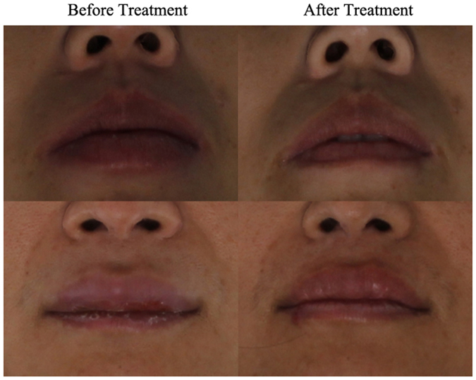
Lip changes before and after treatment.
Figure 5.
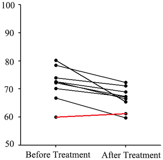
Trend of vermilion border lengths before and after treatment.
Discussion
Cheilitis granulomatosa (CG) is a chronic granulomatous disease characterized by repeated swelling of the lips. Its clinical presentations include local swelling that feels soft and rubber-like, no pain or itching, and non-pitting oedema under pressure22. This disease reportedly shows no significant biases with regard to gender or age, and has a low incidence in the general population. Studies have indicated that the annual incidence of CG is 0.3–8/100,00023, and past reports on CG have been relatively rare. At present, the aetiology and pathogenesis of CG are still unclear24. Swelling of the lips is a typical symptom of CG, but there is still a lack of objective indicators to measure its degree, assess grading, and determine the severity. So far, there is no quantitative method to evaluate lip swelling in patients with CG. Moreover, the evaluation of therapeutic efficacy of CG does not have any standard criteria or achieve consensus. We believe that this study provides an objective method for this purpose. We used Facescan 3D facial reconstruction technology to evaluate the degree of lip swelling in CG patients, and assess whether the 3D Scan could be used as an objective indicator to evaluate therapeutic efficacy.
The lip landmarks selected in this study are consistent with those used in other similar studies on maxillofacial symmetry25, 26. However, thus far, no studies have referred to the 3D Scanner being applied in mucosal disease or CG. Hence, we consider this study to be highly representative. Furthermore, the results of consistency tests performed prior to the formal experiment indicated that the measurement methods used in this study had good reliability. The six sets of line data measured and compared between the healthy control and CG groups showed significant differences with respect to Ls-Li and the lengths of the upper and lower vermilion borders. The three remaining sets of data (sn-Ls, CphL-CphR, and ChL-ChR) did not show such significant differences. Therefore, our results indicate that Ls-Li and the lengths of the upper and lower vermilion borders can be used as objective indicators for the degree of lip swelling in CG. This can serve as an objective index for the clinical assessment of degree of lip swelling. The acquisition process of the facial morphological data via Facescan 3D facial reconstruction technology was simple, quick, and had a high level of accuracy27. Hence, this method could meet the needs of clinical diagnosis as a simple and objective diagnostic method for assessing the degree of lip swelling.
To date, effective treatment methods for CG are still lacking6, 28–31. The treatment methods that are commonly employed in clinics include local injection of glucocorticoids; subcutaneous injection of adalimumab; and oral doses of metronidazole, roxithromycin, clofazimine, or dapsone. Triamcinolone acetonide (TA), a long-acting glucocorticoid, is the most commonly used drug to treat CG. Its specific mechanisms in the treatment of CG might be related to the reduction of local inflammatory reactions and interstitial oedema31. We used the 3D Facescan to evaluate the therapeutic efficacies for CG, and significant differences were found in Ls-Li and the vermilion border length before and after treatment with TA; whereas, the other characteristic line data were not found to be significantly different after treatment. The results showed that TA local injection was an effective treatment for CG. The thickness of both lips and the vermilion border lengths of the affected lip showed differences before and after local injections of TA and can be used as an objective parameter for evaluating the effectiveness of therapy. In addition, we found that the vermilion border lengths of the patients showed a decreasing trend. Furthermore, clinical examination revealed that the degree of lip swelling reduced and the texture became soft after treatment.
This study had some limitations. Owing to the low incidence of CG in the general population, our sample size was relatively small. Future studies in larger cohorts will likely enable a more accurate understanding and analysis on the characteristics of lip swelling in patients with CG. In addition, the observation period should be extended in order to more accurately evaluate the therapeutic efficacy of different drugs via the 3D scan.
To our knowledge, this is the first study to use Facescan 3D facial reconstruction technology to objectively evaluate the degree of lip swelling in CG. The preliminary results showed that 3D scan technology had good repeatability in the evaluation of lip swelling in patients with CG. Ls-Li and the lengths of the upper and lower vermilion borders can be used to evaluate the degree of lip swelling in CG. The data of lip morphology via 3D Facescan can be used as objective indicators for the adjunctive diagnosis of the degree of lip swelling and the evaluation of therapeutic efficacy of CG.
Materials and Methods
Research Design
This case-control study was approved by the institutional review board of Peking University School of Stomatology (PKUSSIRB-201520016). Written informed consent were obtained from all subjects before the study.
Case group
33 patients confirmed as CG by clinical and pathological examination, who were admitted to the Dept. of Oral Medicine, Peking University School and Hospital of Stomatology (PKUSS) from January 2015 to May 2016, were included in this study. Among them, 8 patients were treated with local injection of triamcinolone acetonide (TA), 2–4 times after establishing the diagnosis.
Control group
29 age- and gender-matched healthy volunteers were enrolled in the study during the same study period.
Inclusion criteria for CG patients
Patients in the age range of 10–70 years who fulfilled the clinical and pathological diagnostic criteria for CG and without mental disorders, sarcoidosis, Crohn’s disease, tuberculosis, and possessing both autonomous behaviour and decision-making capacity were included in this study. We had no restrictions on gender or ethnicity.
Exclusion criteria for CG patients
Patients who did not agree to participate in this study or were under long-term, systemic glucocorticoid treatment due to other diseases or received local or systemic glucocorticoid therapy within 1 month before admission were excluded. In addition, patients having severe cardiovascular diseases or acute, systemic infectious diseases were also excuded.
Experimental equipment and software
Facescan structured-light 3D scanner (Fig. 1) was purchased from 3D Systems GmbH, Germany; its theoretical scanning accuracy was 0.1 mm. The principle of this scanner involves projecting structured-light from a light source onto the target surface, which generates deformations in the structured-light. The patterns of the deformed structured-light are then captured by a camera for analysis. Then, the corresponding 3D reconstruction algorithm is implemented, in order to obtain the 3D measurement of the target surface morphology.
Figure 1.
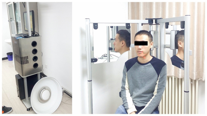
Facescan structured-light 3D scanner.
The software used in this study included: 3D reverse engineering software, Geomagic Studio 12; electronic spreadsheet, Microsoft Excel 2013; and statistical analysis software package, SPSS 22.0.
Selection of characteristic landmarks
Subnasale: sn; Labiale superius: Ls; Labiale inferius: Li; Crista philtri Left/Right: Cph L/R; Cheilion Left/Right: Ch L/R (Fig. 2).
Figure 2.
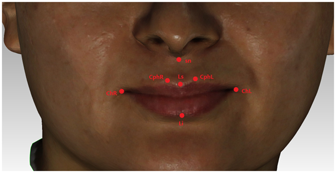
Selection of facial landmarks.
Measurement of lip data
Line data: Sub nasale–labiale superius: sn-Ls; labiale superius–labiale inferius: Ls-Li; distance between the left and right crista philtra: CphL-CphR; width of labial fissure: ChL-ChR; length of upper vermilion border; length of lower vermilion border.
Data acquisition were conducted by Dr.Mu and/or Wang. Before the study, Dr.Mu and Wang were trained by professional worker in Lab for Digital and Material technology of PKUSS. The length of each characteristic line was measured in triplicate by two independent researchers, using Geomagic Studio with reliable consistency (Kappa value: 0.87).
The characteristic facial landmarks of the subjects in the CG and control groups were labelled according to the above methods, and the relevant data measurements were performed.
Measurement of characteristic facial morphology in CG patients before and after TA treatment via the Facescan structured-light 3D scanner
Eight patients with CG, after diagnosis, received local injections of TA hormone (40 mg: 1 mL) + 2% lidocaine (100 mg: 5 mL). A 1-mL mixture was then injected into the lesion site of the lip, at a dose of 0.5 mL per site (Fig. 3)32, 33. Three local injections were administered at 0 wk, 1 wk, and 2 wk after the diagnosis was confirmed. Treatment Evaluation for CG patients via the Facescan structured-light 3D scanner.
Figure 3.
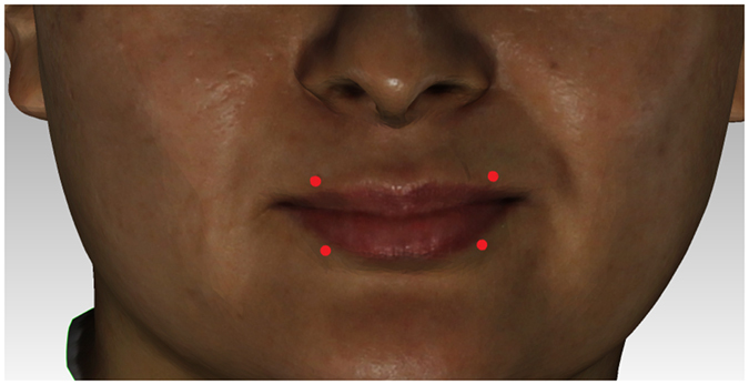
Schematic diagram of injection sites.
Then, the Facescan structured-light 3D scanner was used to acquire characteristic 3D facial data in order to compare the effectiveness before and after treatment.
Statistical analysis and methods
The values were recorded in an Excel spreadsheet, and the mean value was calculated. The lengths of the upper and lower vermilion borders were measured after segmentation and projection using Geomagic Studio, and the respective sum totals were obtained. The measurement was repeated three times, and the mean value was recorded.
SPSS was used to calculate the mean values of the data from the two groups. The basic information and each set of line data were compared between the experimental and control groups using t-tests. The differences before and after treatment among CG patients were compared using paired-samples t-test. A significance level of P < 0.05 indicated that the difference was statistically significant.
Ethical approval and consent to participate
This case-control study was approved by the institutional review board of Peking University School of Stomatology (PKUSSIRB-201520016). The methods used in this study were carried out in accordance with ethical principles of research. And informed consent was obtained from all subjects.
Acknowledgements
We thank Prof. Yong Wang, National Engineering Laboratory for Digital and Material Technology of Stomatology, for a careful guidelines for the use of 3D facescan. This work was supported by the National Natural Science Foundation of China (grant number 81300921).
Author Contributions
C.M. contributed to design, data acquisition, and drafted the manuscript; S.W. contributed to data acquisition, analysis, interpretation and drafted the manuscript; H.H. contributed to concept, design, and critically revised the manuscript; X.H. selected the item and contributed to data acquisition; C.L. selected the item and contributed to data acquisition; and Y.L. contributed to design, analysis and interpretation of the data. All authors reviewed and approved the final manuscript.
Competing Interests
The authors declare that they have no competing interests.
Footnotes
Chang-Qing Mu and Shi-Qin Wang contributed equally to this work.
Publisher's note: Springer Nature remains neutral with regard to jurisdictional claims in published maps and institutional affiliations.
References
- 1.van der Waal RI. Cheilitis Granulomatosa. Ned Tijdschr Tandheelkd. 2012;119:355. [PubMed] [Google Scholar]
- 2.White IR, Souteyrand P, MacDonald DM. Granulomatous Cheilitis (Miescher) Clin Exp Dermatol. 1981;6:391–397. doi: 10.1111/j.1365-2230.1981.tb02323.x. [DOI] [PubMed] [Google Scholar]
- 3.Raskovic S, et al. Clinical Characteristics and Treatment of Melkersson-Rosenthal Syndrome‒Overview of Six Patients. Med Pregl. 2015;68:401–404. doi: 10.2298/MPNS1512401R. [DOI] [PubMed] [Google Scholar]
- 4.Nagel F, Foelster-Holst R. Cheilitis Granulomatosa Melkersson-Rosenthal Syndrome. Hautarzt. 2006;57:121–126. doi: 10.1007/s00105-005-0981-y. [DOI] [PubMed] [Google Scholar]
- 5.Friedrich W, Timmermann J. Miescher’s Granulomatous Cheilitis. Diagnostic and Therapeutic Aspects. Laryngorhinootologie. 1990;69:564–568. doi: 10.1055/s-2007-998254. [DOI] [PubMed] [Google Scholar]
- 6.Samimi M. Cheilitis: Diagnosis and Treatment. Presse Med. 2016;45:240–250. doi: 10.1016/j.lpm.2015.09.024. [DOI] [PubMed] [Google Scholar]
- 7.Al-Hamad A, Porter S, Fedele S. Orofacial Granulomatosis. Dermatol Clin. 2015;33:433–446. doi: 10.1016/j.det.2015.03.008. [DOI] [PubMed] [Google Scholar]
- 8.Franz R, Andres C. Cheilitis Granulomatosa and Melkersson-Rosenthal Syndrome. Intralymphatic Histiocytosis as Valuable Diagnostic Indication. Pathologe. 2014;35:177–181. doi: 10.1007/s00292-013-1850-6. [DOI] [PubMed] [Google Scholar]
- 9.Critchlow WA, Chang D. Cheilitis Granulomatosa: A Review. Head Neck Pathol. 2014;8:209–213. doi: 10.1007/s12105-013-0488-2. [DOI] [PMC free article] [PubMed] [Google Scholar]
- 10.Miest R, Bruce A, Rogers RR. Orofacial Granulomatosis. Clin Dermatol. 2016;34:505–513. doi: 10.1016/j.clindermatol.2016.02.024. [DOI] [PubMed] [Google Scholar]
- 11.Raskovic S, et al. Clinicl Characteristics and Treatment of Melkersson-Rosenthal Syndrom–Overview of Six Patients. Med Pregl. 2015;68:401–404. doi: 10.2298/MPNS1512401R. [DOI] [PubMed] [Google Scholar]
- 12.Philipp Meyer-Marcotty Georg W, Alpers ABMG. Impact of Facial Asymmetry in Visual Perception: A 3-Dimensional Data Analysis. Am J Orthod Dentofacial Orthop. 2010;137:161–168. doi: 10.1016/j.ajodo.2008.11.023. [DOI] [PubMed] [Google Scholar]
- 13.Rogers BCTRJ. Computerized Manufacturing of Transparent Face Masks for the Treatment of Facial Scarring. J Burn Care Rehabil. 2003;24:91–96. doi: 10.1097/01.BCR.0000054171.82007.7A. [DOI] [PubMed] [Google Scholar]
- 14.Wilde F, Schramm A. Computer-Aided Reconstruction of the Facial Skeleton: Planning and Implementation in Clinical Routine. HNO. 2016;64:641–649. doi: 10.1007/s00106-016-0220-0. [DOI] [PubMed] [Google Scholar]
- 15.Pascali MA, et al. Face Morphology: Can It Tell Us Something About Body Weight and Fat? Comput Biol Med. 2016;76:238–249. doi: 10.1016/j.compbiomed.2016.06.006. [DOI] [PubMed] [Google Scholar]
- 16.Li P, et al. Accuracy of Three-Dimensional Facial Soft Tissue Simulation in Post-Traumatic Zygoma Reconstruction. Int J Oral Maxillofac Surg. 2016;45:1665–1670. doi: 10.1016/j.ijom.2016.06.021. [DOI] [PubMed] [Google Scholar]
- 17.Huang J, Xue Q, Wang Z, Gao J. Analysis and Compensation for Lateral Chromatic Aberration in a Color Coding Structured Light 3D Measurement System. Sensors (Basel). 2016;16:E1426. doi: 10.3390/s16091426. [DOI] [PMC free article] [PubMed] [Google Scholar]
- 18.Xiong Y, Zhao Y, Yang H, Sun Y, Wang Y. Comparison Between Interactive Closest Point and Procrustes Analysis for Determining the Median Sagittal Plane of Three-Dimensional Facial Data. J Craniofac Surg. 2016;27:441–444. doi: 10.1097/SCS.0000000000002376. [DOI] [PMC free article] [PubMed] [Google Scholar]
- 19.Sun Y, et al. Evaluation of the Accuracy of a Common Regional Registration Method for Three-Dimensional Reconstruction of Edentulous Jaw Relation by a 7-Axis Three-Dimensional Measuring System. Biomed Mater Eng. 2014;24:1275–1287. doi: 10.3233/BME-130930. [DOI] [PubMed] [Google Scholar]
- 20.Zhao Y, Wang Y, Huang M, Zhang J, Zhang X. Computer Aided Design Method of Digital Individual Guide for Brachytherapy. Zhonghua Kou Qiang Yi Xue Za Zhi. 2014;49:115–118. [PubMed] [Google Scholar]
- 21.Ye H, Lv L, Liu Y, Liu Y, Zhou Y. Evaluation of the Accuracy, Reliability, and Reproducibility of Two Different 3D Face-Scanning Systems. Int J Prosthodont. 2016;29:213–218. doi: 10.11607/ijp.4397. [DOI] [PubMed] [Google Scholar]
- 22.Savas J, Sokoloff D, Sanchez KM, Lichtstein DM. Granulomatous Cheilitis: A Stiff Upper Lip. Cutis. 2015;96:E18–E20. [PubMed] [Google Scholar]
- 23.Fessatou S, et al. Chronic Lip Swelling as the Sole Presentation of Crohn’s Disease: Three Case Reports. Turk J Pediatr. 2015;57:98–101. [PubMed] [Google Scholar]
- 24.Delir G, Strohal R, Haring N. Unclear Swelling of the Lips and Left Cheek. J Dtsch Dermatol Ges. 2015;13:1176–1177. doi: 10.1111/ddg.12833. [DOI] [PubMed] [Google Scholar]
- 25.Wang SW, et al. Evaluation of Three Methods for Constructing Craniofacial Mid-Sagittal Plane Based On the Cone Beam Computed Tomography. Beijing Da Xue Xue Bao. 2016;48:330–335. [PubMed] [Google Scholar]
- 26.Xiong YX, Yang HF, Zhao YJ, Wang Y. Comparison of Two Kinds of Methods Evaluating the Degree of Facial Asymmetry by Three-Dimensional Data. Beijing Da Xue Xue Bao. 2015;47:340–343. [PubMed] [Google Scholar]
- 27.Djordjevic J, et al. Three-Dimensional Longitudinal Assessment of Facial Symmetry in Adolescents. Eur J Orthod. 2013;35:143–151. doi: 10.1093/ejo/cjr006. [DOI] [PubMed] [Google Scholar]
- 28.Serarslan G, Aksakal M. Cutaneous Leishmaniasis Mimicking Granulomatous Cheilitis and Treated Successfully with Oral Fluconazole in a Boy. Ann Parasitol. 2015;61:197–199. doi: 10.17420/ap6103.08. [DOI] [PubMed] [Google Scholar]
- 29.Ridder GJ, Fradis M, Lohle E. Cheilitis Granulomatosa Miescher: Treatment with Clofazimine and Review of the Literature. Ann Otol Rhinol Laryngol. 2001;110:964–967. doi: 10.1177/000348940111001013. [DOI] [PubMed] [Google Scholar]
- 30.Kesler A, Vainstein G, Gadoth N. Melkersson-Rosenthal Syndrome Treated by Methylprednisolone. Neurology. 1998;51:1440–1441. doi: 10.1212/WNL.51.5.1440. [DOI] [PubMed] [Google Scholar]
- 31.Gu L, et al. Intralesional Injection of Pingyangmycin Plus Corticosteroids May be an Effective Treatment for Cheilitis Granulomatosa. Med Hypotheses. 2013;81:729–730. doi: 10.1016/j.mehy.2013.07.037. [DOI] [PubMed] [Google Scholar]
- 32.Oudrhiri L, Chiheb S, Marnissi F, Zamiati S, Benchikhi H. Successful Treatment of Miescher’s Cheilitis in Melkersson-Rosenthal Syndrome with Betamethasone Injections and Doxycycline. Pan Afr Med J. 2012;13:75. [PMC free article] [PubMed] [Google Scholar]
- 33.Okudo J, Oluyide Y. Melkersson-Rosenthal Syndrome with Orofacial Swelling and Recurrent Lower Motor Neuron Facial Nerve Palsy: A Case Report and Review of the Literature. Case Rep Otolaryngol. 2015;2015:214946. doi: 10.1155/2015/214946. [DOI] [PMC free article] [PubMed] [Google Scholar]


