Abstract
A series of 5-aryl-1-alkylimidazole derivatives was synthesized using the van Leusen multicomponent reaction. The chemotype is the first example of low-basicity scaffolds exhibiting high affinity for 5-HT7 receptor together with agonist function. The chosen lead compounds 3-(1-ethyl-1H-imidazol-5-yl)-5-iodo-1H-indole (AGH-107, 1o, K i 5-HT7 = 6 nM, EC50 = 19 nM, 176-fold selectivity over 5-HT1AR) and 1e (5-methoxy analogue, K i 5-HT7 = 30 nM, EC50 = 60 nM) exhibited high selectivity over related CNS targets, high metabolic stability and low toxicity in HEK-293 and HepG2 cell cultures. A rapid absorption to the blood, high blood-brain barrier permeation and a very high peak concentration in the brain (Cmax = 2723 ng/g) were found for 1o after i.p. (5 mg/kg) administration in mice. The compound was found active in novel object recognition test in mice, at 0.5, 1 and 5 mg/kg. Docking to 5-HT7R homology models indicated a plausible binding mode which explain the unusually high selectivity over the related CNS targets. Halogen bond formation between the most potent derivatives and the receptor is consistent with both the docking results and SAR. 5-Chlorine, bromine and iodine substitution resulted in a 13, 27 and 89-fold increase in binding affinities, respectively, and in enhanced 5-HT1AR selectivity.
Introduction
Within the serotonergic system, 5-HT7, the last identified serotonin receptor, is one of the most valuable drug targets1. Discovered independently by three laboratories in 1993 2–4, 5-HT7R was thought to function by being coupled to Gs to stimulate intracellular production of cAMP, but it was later found to also be coupled to G12 which is an alternative signal transduction pathway5. 5-HT7R is expressed in the central nervous system (thalamus, hypothalamus, hippocampus and cortex) as well as peripherally (pancreas, spleen, coronary artery, ileum and intestine)3–6. Experiments using animal models have shown that the receptor is involved in many physiological processes, i.e. the regulation of body temperature7, smooth muscle relaxation of cerebral arteries8, circadian rhythm, learning and memory9–13, as well as pathophysiological processes such as mood disorders, anxiety14, inflammatory processes in the CNS15, schizophrenia16 and pain17, 18. 5-HT7R antagonists have been proposed as potential drugs targeting depression19, 20. There have been less conclusive reports on potential anxiolytic21, 22, analgesic17 and antipsychotic properties of the receptor ligands23, 24. It was established that 5-HT7R blockade can induce promnesic effects indicating the possibility of developing atypical procognitive antidepressants25.
There are several 5-HT7R agonists available to serve as molecular probes. These include small, low-weight molecules, e.g. AS-1926, RA-77, 5-carboxyamidotryptamine (5-CT), 5-methoxytryptamine, 8-OH-DPAT27, and lysergic acid derivatives28 and long-chain diphenylpiperazines (LP-12, LP-44, LP-211) developed by Leopoldo et al.29–31. None of these compounds qualify as perfect radioligand candidates, mainly due to their poor selectivity. 5-HT7R agonists often bind to 5-HT1AR and 5-HT5AR32. Some promising 5-HT7R molecular probes suffer from poor metabolic stability33. In the case of larger molecules, membrane penetration may be an issue as it is considerably harder to find an optimal mass-lipophilicity ratio. Animal models suggest several possible indications of 5-HT7R agonists as therapeutics. The stimulation of 5-HT7R in adolescent rats by LP-211 causes plastic rearrangements within the forebrain networks, accounting for long-lasting behavioural changes in adulthood34. The activation of 5-HT7R was found to be beneficial in mouse models of Rett syndrome35 and fragile X syndrome36. Compound AS-19, which was the first selective agonist of the 5-HT7R, as well as LP-12, LP-44, LP-211 and E-55888 have been used in numerous in vivo experiments which led to the characterization of the receptor. It was found, that 5-HT7 agonists can enhance long-term memory formation. In animal models of pain, AS-19, LP-44 and LP-211 were found antinociceptive. The antinociceptive properties of morphine can be dramatically enhanced by the co-administration of the E-55888, while the compound does not produce any analgesic effect on its own37.
Non-basic aminergic GPCR ligands have recently emerged as pharmacological rule breakers. Despite lacking the capability of forming a salt bridge (strong, charge assisted hydrogen bond) with Asp3.32 such chemical entities have been shown to bind to 5-HT2AR38 and later to 5-HT6 and 5-HT1B receptors with nanomolar and subnanomolar potencies39, 40. Interestingly, there have been no published examples of low-basicity ligands of the 5-HT7 receptor41. A hypothesis that an aromatic basic moiety can successfully serve as the aminergic fragment of a serotonin receptor ligand was not yet tested. Till date there were numerous basic scaffolds (piperazine, piperidine, pyrrolidine, polycyclic amines, alkylamines) incorporated into serotonin receptor ligands. The imidazole fragment, which is a privileged structure commonly used in medicinal chemistry, has not been tested as a basic fragment in the aminergic GPCR field and was hardly used as a part of serotonin receptor ligands42.
Our initial hypothesis was that multicomponent reaction (MCR) protocols can be successfully applied to the field of GPCR ligand synthesis. We believed that the close serotonin analogues 1a (AGH-38) and 1b (AGH-39) (Figs 1 and 2), which can be concisely prepared via van Leusen tosylmethylisocyanide (TosMIC) imidazole synthesis43, 44, would exhibit activity at some serotonin receptors.
Figure 1.
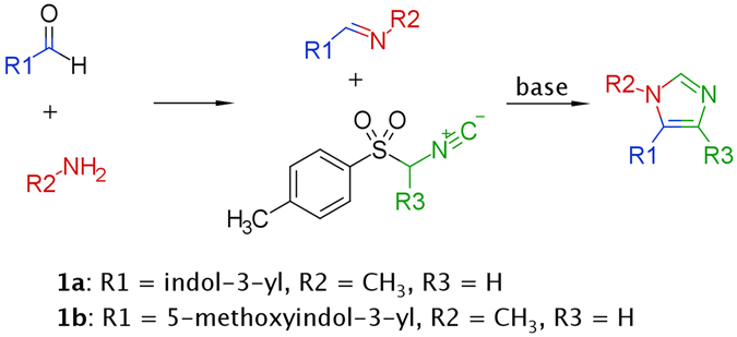
Van Leusen imidazole synthesis.
Figure 2.
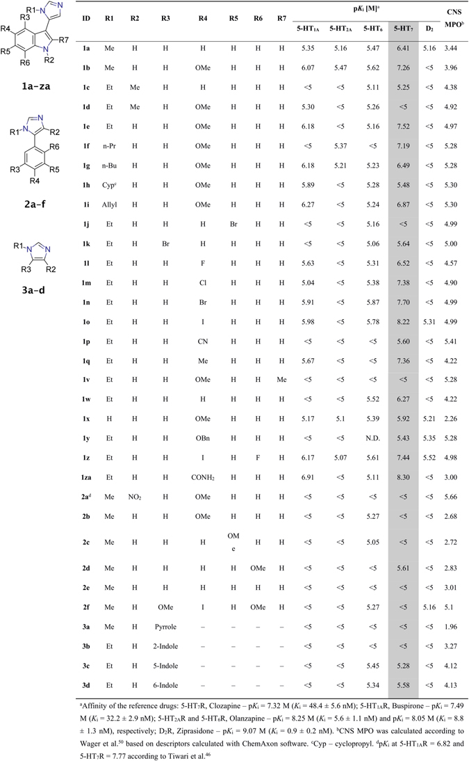
Structure, binding data and Central Nervous System Multiparameter Optimization score (CNS MPO) of compounds 1a–1za, 2a–2f and 3a–3d.
Owing to the click-chemistry properties of van Leusen imidazole synthesis, different 1-substituted, 5-arylated imidazoles were conveniently prepared and evaluated. The MCR protocol enabled the systematic SAR exploration by first optimizing the core containing carbonyl component, then optimizing the amine component for the most active entities. Over the course of the project it became clear that potent and selective, non-basic 5-HT7R ligands can arise via the SAR exploration of the close chemical space of arylimidazoles.
Results and Discussion
Chemistry
Compounds 1a–1q, 1s–1w, 1y–1za, 2b–2f and 3a–3d (Fig. 2) were synthesized using a concise multicomponent protocol (Fig. 1). The van Leusen reaction proceeds via the stepwise cycloaddition of TosMIC to a polarized double bond of the preformed imine. The elimination of p-toluenesulfonic acid from the cyclic intermediate affords imidazole. Aromatic aldehydes were combined with appropriate amines in methanol. When the imine formation equilibrium was achieved, TosMIC and potassium carbonate were added to yield the desired products. Most of the indole-3-carboxaldehydes used were synthesized by the Vilsmeier-Haack formylation of the appropriate indoles. Derivatives 1r and 1x were synthesized by reductive debenzylation of 1y and 1 s, respectively. Compound 1za was synthesized from 1p by the controlled hydrolysis of the cyano group in basic conditions.
Pharmacology
All the synthesized compounds were tested in a radioligand binding assays to determine their affinity for five receptors: 5-HT1A, 5-HT2A, 5-HT6, 5-HT7 and D2. The assays were performed via the displacement of the respective radioligands from cloned human receptors, all stably expressed in HEK293 cells (except for 5-HT2A which was expressed in CHO cells): [3H]-8-OH-DPAT for 5-HT1AR, [3H]-ketanserin for 5-HT2AR, [3H]-LSD for 5-HT6R, [3H]-5-CT for 5-HT7R and [3H]-raclopride for D2R. Ligands with a high affinity for the 5-HT7 receptor were selected for a functional assay (as hydrochloride salts). The experiment was performed in HEK293 cells overexpressing 5-HT7R, and the ability of tested compounds to increase cAMP production was measured. The cAMP concentration was measured using TR-FRET and a cAMP specific antibody. Lead compounds 1e (AGH-44) and 1o (AGH-107) were screened toward several CNS targets (Eurofins Cerep, Le Bois l’Eveque, 86600 Celle L’Ev-escault, France, www.cerep.fr): α1, α2C, CB1, H1, D3, 5-HT1B, 5-HT2B and 5-HT5A. The intrinsic clearance of 1e and 1o were determined using human liver microsomes according to Obach et al.45 (see Supporting Information for detailed results). The HEK-293 and HepG2 cell lines viabilities were tested after 72 h of incubation with tested compounds. Doxorubicin (DX), and carbonyl cyanide 3-chlorophenylhydrazone (CCCP) were used as internal standards (see Supporting Information). The pharmacokinetics of compound 1o was tested on CD-1 mice after intraperitoneal injection at 5 mg/kg dose (see Supporting Information). The compound 1o was administered at 0.5, 1 and 5 mg/kg to investigate the ability of the compound to reverse MK-801 induced impairment in novel object recognition test. The experiment was conducted on Albino Swiss mice (see Supporting Information).
Structure-Activity Relationship
After acquiring highly encouraging initial results for compounds 1a and 1b (K i 5-HT7 = 385 nM and 50 nM, respectively, selectivity over 5-HT1AR > 11-fold, Fig. 2) a literature search made us realize that an arylimidazole 5-HT7R ligand was recently described, although no SAR studies were reported46. The compound, 5-(4-([11C]methoxyphenyl)-1-methyl-4-nitro-1H-imidazole, 2a, has been developed as a PET radioligand. The high and rapid uptake of 2a to the rat brain after injection was reported which would suggest at least decent ADME properties. The finding further encouraged us to explore the series, so 2a was resynthesized to serve as a reference compound. However, we were not able to reproduce the results presented by Tiwari et al.46. In our radioligand displacement binding assays compound 2a did not bind to 5-HT7R and 5-HT1AR (binding curves of compound 2a can be found in Supporting Information). Moreover, a very close analogue of 2a (the only structural difference being the lack of the nitro group) – 2b – was also found to be completely inactive. A compound based on the 2,5-dimethoxy-4-iodophenyl core, which is a scaffold common to many 5-HT2R ligands47, 48, 2f was expected to bind to 5-HT2AR, but this assumption was not validated. Compounds synthesized from unsubstituted and differently methoxy-substituted benzaldehydes (entries 2c–2e) were also not active. Compounds 3a–3d were synthesized to answer the question of whether methylimidazoles coupled to different aromatic cores (3-pyrrole, 2-indole, 5-indole, or 6-indole) could show any 5-HT7R activity. This group of compounds did not bind to 5-HT7R at all, even 3a which is a close, simplified analogue of 1a did not possess any activity. It was thus evident that indole-3-imidazole (the core structure of series 1) is the molecular scaffold of choice.
The role of the indole nitrogen atom in the binding of the active compounds to 5-HT7R was determined by the synthesis of two derivatives. Entry 1c (K i 5-HT7 = 5623 nM) which is a methylated analogue of 1a, and 1d (K i 5-HT7 > 10000 nM) which is a methylated counterpart of 1b clearly showed that the unsubstituted pyrrole fragment plays an important role in receptor binding. Homologues 1b, and 1e–1 g and compounds 1 h, and 1i were synthesized to optimize the length and shape of the amine component. Apparently, the ethyl substituted derivative exhibited optimal binding affinity (1e, K i 5-HT7 = 30 nM). Interestingly, 1 h (R1 = cyclopropyl) and 1 s (R1 = benzyl) were found to be non-active which would suggest a very narrow space in the binding pocket reserved for an alkyl fragment. The modification of the indole containing component resulted in compounds 1j–1 v. Forbidden positions 2-, 6-, and 7- (R7, R5, and R6) can be clearly observed in entries 1k, 1 u, and 1 v, respectively. Compound 1t exhibited moderate activity toward 5-HT7R (K i = 122 nM) indicating that appropriate substituents are acceptable at the fourth position of the indole nucleus. Although 1j and 1k (4-bromo- and 6-bromoindole cores, respectively) were found to possess low affinity for 5-HT7R, 1n (5-bromo) was a hit compound with K i = 20 nM. Successively, a series of 5-substituted indole-containing derivatives were synthesized. The activity of compounds 1l–1q and 1w showed a very probable involvement of halogen bonding in the binding of the most active derivatives 1m–1o. 5-HT7R affinities of methyl and fluoro derivatives 1q and 1 l (44 nM and 302 nM, respectively) were considerably lower than for the halogen series (42 nM, 20 nM, and 6 nM for Cl, Br and I, respectively), indicating the hydrophobicity to be of secondary importance. Compound 1o (5-iodo) became the lead in the series exhibiting K i = 6 nM at the human 5-HT7 receptor, and 176-fold selectivity over 5-HT1AR. A very close serotonin analogue 1r bound to 5-HT7R with K i = 39 nM, whereas an analogue of 5-CT, 1za displayed K i = 5 nM.
In vitro pharmacology
Functional assay results for the chosen compounds are shown in Table 1. Compounds 1b, 1e, 1m, 1n, 1o, 1q and 1r acted as full agonists at the human 5-HT7 receptor. The strongest agonist function was observed for 1o (5-iodo) and 1r (5-hydroxy).
Table 1.
Results of functional activity for 5-HT7R, CYP3A4 inhibition assay and metabolic stability for the selected compounds.
| Compound ID | R1 | R4 | K i | 5-HT7REC50 [nM] | CYP3A4 IC50 [μM] | Clint a [ml/min/kg] |
|---|---|---|---|---|---|---|
| 1b | Me | OMe | 55 | 150 | — | — |
| 1e | Et | OMe | 30 | 60 | 0.24 | 3.69 |
| 1m | Et | Cl | 42 | 149 | — | — |
| 1n | Et | Br | 20 | 154 | — | — |
| 1o | Et | I | 6 | 19 | 0.17 | 6.3 |
| 1q | Et | Me | 44 | 45 | — | — |
| 1r | Et | OH | 39 | 22 | — | — |
aMetabolic clearance (human liver microsomes).
The receptor profile of the tested compounds shows high selectivity over the tested CNS targets. All compounds from chemotype 1 showed K i > 1000 nM at the 5-HT2A, 5-HT6 and D2 receptors. 1e and 1o were found to be selective toward a set of CNS targets (Table 2). Within the extended receptor panel tested, 1e and 1o showed affinity only for 5-HT2B (% of inhibition at 1 µM = 93 and 87, respectively), but at a 10 nM concentration, the level of receptor binding was very low (6 and 7%, respectively). Additionally, compounds 1e and 1o showed very promising metabolic stability with metabolic clearance 3.69 and 6.3 ml/min/kg, respectively.
Table 2.
Anti-target profiles of selected compounds 1e and 1o.
| Compound | Concentra-tion [M] | % Inhibition of control bindinga | |||||||
|---|---|---|---|---|---|---|---|---|---|
| α1 | α2C | CB1 | D3 | H1 | 5-HT1B | 5-HT2B | 5-HT5A | ||
| 1e | 10−6 | 5 | 9 | 8 | 8 | 6 | 24 | 93 | 9 |
| 10−8 | — | — | — | — | — | — | 6 | — | |
| 1o | 10−6 | — | — | — | — | — | 40 | 87 | 7 |
| 10−8 | — | — | — | — | — | — | 7 | — | |
aExperiments performed at Eurofins Cerep.
The cytotoxicity of the compounds 1e and 1o was tested by checking the viability of HEK-293 and HepG2 cells after incubation with different concentrations of the compounds in the medium. 1o was found toxic for the cells only in the highest concentration (100 μM), while with 1e the cells remained viable in all tested concentrations (Fig. 3).
Figure 3.
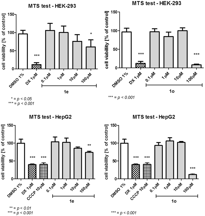
The HEK-293 and HepG2 cell lines viability in the presence of 1e and 1o after 72 h of incubation.
Preliminary pharmacokinetics and behavioural study
The pharmacokinetic analysis performed by a non-compartmental approach has shown a rapid absorption of 1o after i.p. administration (tmax = 15 min) with maximum plasma concentration of 749 ng/mL (~2.2 μM) (Fig. 4). Half of the compound was eliminated from the body within 90 min but brain concentrations of compound 1o decreased slowly with t0.5 = 130 min. Large volume of distribution (Vd/F = 46.6 L/kg) indicated the penetration of 1o into the peripheral compartments. The compound 1o was measurable to the last sampling time (6 h) both in plasma and brain. The compound penetrated the blood-brain barrier after i.p. administration and peaked in the brain and plasma at the same time but the maximum concentration in the brain was higher (Cmax = 2723 ng/g, approx. 8 μM). The mean residence time (MRT) for compound 1o in brain was longer than in plasma and equalled 60 min and 27 min, respectively. The brain to plasma ratio was high and equal 6.34. The detailed description of the pharmacokinetic experiment can be found in the Supporting Information. In a recent paper by Leopoldo et al.31 a disposition study for 5-HT7R agonists LP-44 and LP-211 after i.p. administration at a dose of 10 mg/kg in mice was reported. Despite 1o reached the Cmax in approx. 15 min, which is roughly the same for both LP compounds, the maximum concentrations of LP-44 and LP-211 in the brain were found several-fold lower than for 1o, despite the dose used was twice as high as that of 1o. The LP compounds were measurable in plasma for a very short time compared to over 6 hours for 1o which can be attributed to their very low metabolic stability. The blood/plasma ratio for 1o was three-fold higher compared to LP-44 and 9 times higher than for LP-211.
Figure 4.
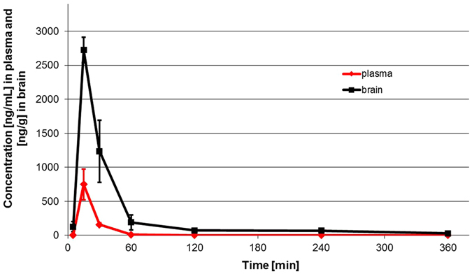
Concentration – time profiles for 1o in plasma and brain after intraperitoneal administration to mice at a dose of 5 mg/kg (linear plots). Concentrations of 1o in plasma and brain are expressed as ng/mL and ng/g, respectively.
Considering the PK study and suitable brain penetration we concluded that 1o has desirable pharmacokinetic profile to be characterized as a compound of choice to study the in vivo pharmacology. The compound dose-dependently reversed MK-801-induced disruption in novel object recognition (NOR) in mice (Fig. 5). This finding is in line with the results obtained earlier for 5-CT49. MK-801-induced disruption of novel object recognition in rodents reflects impairment of working memory, regarded as one of the cognitive symptoms of schizophrenia.
Figure 5.
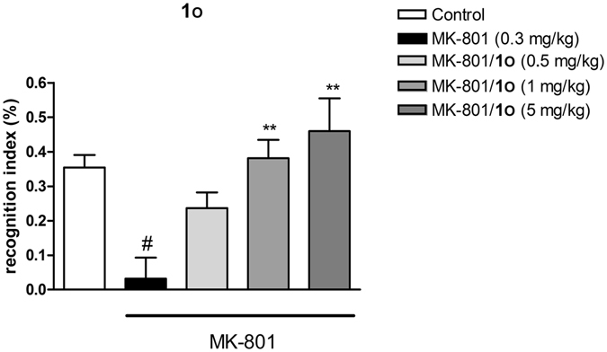
Reversal of MK-801 induced disruption in novel object recognition. Statistically significant effect was found at 1 mg/kg and 5 mg/kg doses.
Molecular modelling
For all compounds in the series, Multiparameter Optimization (MPO) based on six factors (molecular weight, number of hydrogen bond donors, calculated: basic pK a, topological polar surface area, logP, logD) was performed. MPO parameter which was calculated according to Wager et al.50 was found to lie between 4–6 for most of the compounds in the series (Fig. 2).
A very promising group of 5-HT7 receptor agonists has been developed. To make the search for highly active molecules more rational and explain the unusually high selectivity, a binding mode model had to be explored. The proposed binding mode was based on the analysis of the mutual spatial arrangement of particular ligands at the 5-HT7 receptor homology models. The final analysis and visualizations were performed using a model built on a 5-HT1BR (4IAR, complex with agonist, ergotamine) template, because it showed the most coherent binding mode, combined with low binding free energies (see Experimental Section). The observed structure-activity relationships were in agreement with the docking results. Quantum chemical calculations of pK a for 1w (performed in Jaguar using DFT functional B3PW91 and the cc-pVTZ basis set) showed an imidazole fragment pK a = 6.9, indicating that the molecules at physiological pH = 7.4 occur in protonated (33%) as well as deprotonated (67%) forms. Two protonation states for each ligand were considered in docking study. The virtual ligand-receptor complexes of the protonated states did not follow the observed SAR, in addition, a more coherent binding mode was found for the unprotonated forms of the compounds. Thus in discussion and visualizations only deprotonated ligand-receptor complexes were used. The indole nucleus in all the docked ligands formed a hydrogen bond with Asp3.32 and aromatic interactions (CH-π or π-π stacking) with the Phe6.51 or Phe6.52 residues (Fig. 6A), whereas the imidazole ring was frequently hydrogen bonded to Arg6.58. It should be emphasized, that among all targets tested in vitro, the arginine in position 6.58 is a unique feature of 5-HT7R (Table 3)51, which could explain the high selectivity of the discussed compounds.
Figure 6.
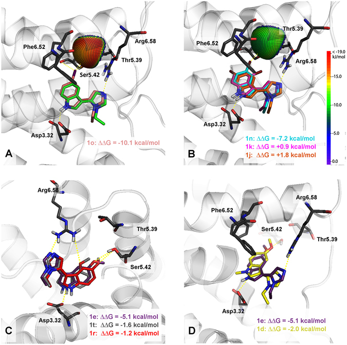
Representative complexes (top scores based on ΔG) of selected ligands with the 5-HT7 receptor model. Amino acids that were selected as crucial for the binding of the presented compounds are shown as sticks. The ∆∆G [kcal/mol] value shows the difference between the ∆G of complexes of a particular compound (1d, 1 f, 1j, 1k, 1n, 1r, 1t) and an unsubstituted analogue 1w (green in Fig. A). In all cases except 1d (R2 = Me, yellow in Fig. D), the ∆∆G values correspond to the binding affinity. (A) – halogen bond formation between the iodine atom of 1o and the receptor. (B) – 1j (R5 = Br) and 1k (R3 = Br) which do not pass geometrical parameters needed for halogen bond formation and 1n halogen bonded to the receptor. (C) – differences in binding modes of compounds 1e (R4 = OMe), 1r (R4 = OH) and 1t (R3 = OMe), the lack of interaction of 1t with Ser5.42 is compensated by the formation of additional hydrogen bond with Arg6.58. (D) – the inability of 1d (R2 = Me) to interact with Asp3.32.
Table 3.
Amino acid residue at 6.57, 6.58 and 6.59 positions in relevant targets.
| Target | 5-HT1AR | 5-HT2AR | 5-HT2BR | 5-HT6R | 5-HT7R | D2R |
|---|---|---|---|---|---|---|
| 6.57 residue | Val | Met | Thr | Val | Ala | Leu |
| 6.58 residue | Leu | Ala | Leu | Gln | Arg | Asn |
| 6.59 residue | — | Val | Val | Ala | — | Ile |
The substitution of hydrogen to a halogen atom in position 5 of the indole ring, proportionally increased the 5-HT7R affinity to the size of the halogen atom. The analysis of binding modes, showed that a significant contribution to the binding can be attributed to the halogen bond created between the ligand and carbonyl oxygen of one or several amino acids of transmembrane helix 5 (TMH5). The geometric parameters for the 5-iodine derivative showed the significant contribution of Ser5.42 (d(I···O) = 3.1 Å, sigma hole angle (C–I···O) = 155°) and Thr5.39 (d(I···O) = 4.4 Å, sigma hole angle (C-I···O) = 171°) (Fig. 6A). Additionally, the order of the binding free energy changes (after chlorine, bromine and iodine substitution ∆∆G was −6.6, −7.2 and −10.1 kcal/mol, respectively) and the interaction sphere plotted onto the relevant backbone carbonyl oxygen (all X···O=C interactions were within the energetically favourable areas of the sphere, Fig. 6A and B) confirmed the role of halogen bonding in the formation of the L-R complexes. The interaction spheres plotted onto the backbone carbonyl oxygen of Ser5.42 were also used to explain the decreased activity upon shifting the bromine atom from position 5 to 4 or 6 in the indole ring (Fig. 6B). The obtained interaction sphere showed that 5-Br derivative (d(Br···O) = 3.2 Å, sigma hole angle (C-Br···O) = 159°) lies within the energetically favourable areas of the sphere, while the 4- and 6-Br derivatives bromine atoms were pointed outside the sphere, ruling out the formation of halogen bond. This observation is consistent with calculated binding free energy values: only 5-bromo derivative 1n showed a gain in the binding energy (ΔΔG = −7.6 kcal/mol), compared to the 4-bromo and 6-bromo analogues (ΔΔG = +0.9 and +1.8 kcal/mol, respectively).
The significance of the hydrogen bond formed between the indole NH group and the Asp3.32 side chain can be observed by comparing compounds 1e and 1d (Figs 2 and 6D). The substitution of hydrogen with a −CH3 group resulted in an approximately 580-fold decrease in binding affinity because no hydrogen bond can be formed with Asp3.32. The calculated ΔΔG for the methylated derivative (1d) showed the stabilization of the L-R complex which is in sharp contrast with the experimental K i value (>10000 nM).
Because the discussed compounds are structurally very similar to the class of tryptamines that are known to be promiscuous serotonergic agents, representative tryptamine derivatives were purchased and their binding affinities for 5-HT7, 5-HT1A, 5-HT2A, 5-HT6 and D2 receptors were determined (Fig. 7).
Figure 7.
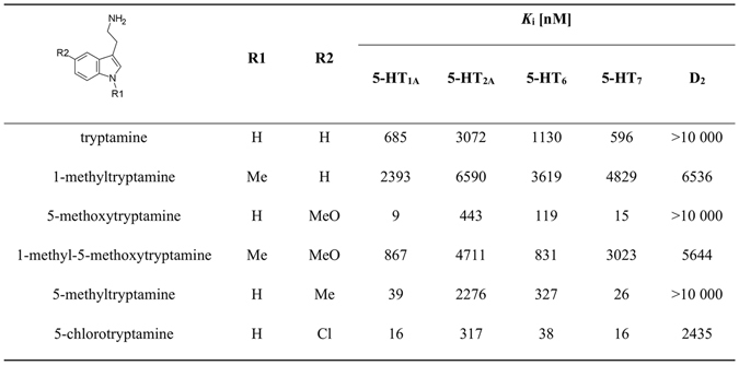
Structure and pharmacological profile of tryptamine analogues.
It is clear that tryptamines (tryptamine as an analogue for 1a; 5-methoxytryptamine for 1b, 1e; 5-chlorotryptamine for 1m, and 5-methyltryptamine for 1q) exhibit multi-receptor profiles, i.e. a high affinity for 5-HT7, 5-HT1A, 5-HT2A, and 5-HT6 receptors. The introduction of halogen at the 5th position of indole in tryptamines resulted in an enhancement of the affinity for 5-HT7R, but not nearly as much as the introduction of a methoxy group (introduction of the chlorine and methoxy groups had very similar impacts on the binding affinity of the title imidazoles). The methylation of the indole NH group in tryptamines resulted in a dramatic drop in potency for all four tested serotonin receptors. It was rather unusual that the tryptamines exhibited very similar 5-HT7R SARs while exhibiting distinctive multi-receptor profiles. The proposed binding mode for the imidazole compounds was compared with several tryptamines, which were docked to the best 5-HT7R homology models using the same procedure (Fig. 8). The binding mode of the tryptamine derivatives was similar to the findings of Vermeulen et al.52 and was different from the one found for the title compounds. The tryptamines formed a salt bridge (charge assisted hydrogen bond) between the protonated amine moiety and Asp3.32, but did not interact with Arg6.58. Vermeulen suggested Thr5.43 to be the anchoring point of 5th position substituent of tryptamines. The sharp drop in potencies of 1-methylated tryptamines at 5-HT7R contributes to the significance of indole NH – Thr3.37 interaction (Fig. 8A–D) Despite different binding modes (Fig. 8D), the 5-HT7R SARs of tryptamines and imidazoles are very similar due to involvement of indole 5-substitutent interaction with Ser5.42 (or Thr5.39) in both series (compare Figs 6C and 8B). Ser5.42 can act as a donor/acceptor of hydrogen bonding through the side chain hydroxyl group. The docking results of the imidazole derivatives substituted in position 5 of the indole by –OCH3 and –OH (1 f (AGH-87) and 1r, Fig. 6C) indicated the formation of a hydrogen bond between the hydroxyl group of Ser5.42 and the oxygen atom of substituents. Interestingly, shifting the –OCH3 group from position 5 to 4, resulted in only a slight decrease in activity, which can be linked to creation of a hydrogen bond with Arg6.58 (Fig. 6C).
Figure 8.
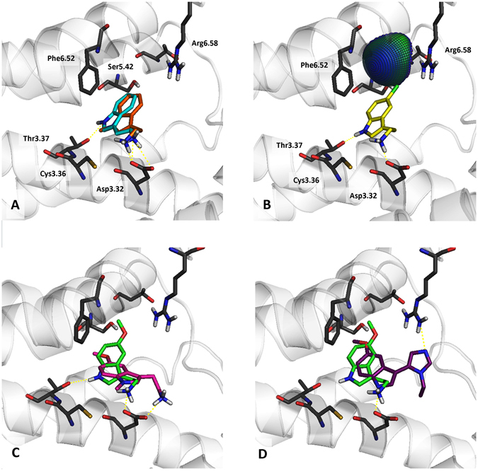
Representative complexes of selected tryptamines with 5-HT7 receptor. Amino acids that were selected as crucial for the binding of the presented compounds are shown as sticks. (A) – the superposition of tryptamine (cyan) and 1-methyltryptamine (orange). (B) – the halogen bond formed between Ser5.42 and 5-chlorotryptamine (yellow). (C) – the superposition of 5-methoxytryptamine (green) and 1-methyl-5-methoxytryptamine (magenta). (D) – a comparison of the binding modes of 1f (violet) and the analogous 5-methoxytryptamine.
Apparently, it is possible to synthesize heterocyclic analogues of tryptamines that maintain high affinity and agonist function at 5-HT7R. Modelling studies revealed that the incorporation of an amine moiety into an imidazole ring resulted in a distinctive binding mode. The difference may be attributed partially to the lower basicity of imidazoles versus tryptamines. An interaction with Arg6.58, which is a residue unique for 5-HT7R may underlay the observed dramatic effect on the selectivity.
Possible application as a PET radioligand
5-HT7 receptor is yet to be PET imaged in humans53. No suitable 5-HT7R agonist radioligand candidates were reported till date54, 55. Serotonin receptors exist in high and low agonist affinity states. The antagonist ligands bind to the high affinity (HA) and low affinity (LA) conformations with similar affinity, whereas agonist ligands bind preferentially to the HA state of the receptor, which is coupled to G-protein and therefore agonist binding provides a more meaningful functional measure of the serotonin receptors activity. Although antagonist PET tracers can measure the total receptor binding, they cannot detect changes in the high affinity receptor binding in disease states. Antagonist binding is insensitive to the changes in the intra-synaptic 5-HT concentration and antagonist PET tracers are less sensitive for measuring agonist receptor occupancy in clinical studies to guide new drug development in dose-finding56.
Because of rapid penetration to the brain and quick elimination from the CNS, 1o showed desirable properties as perspective PET radiotracer. Therefore, a potential synthetic pathway leading to the 11C-labeled version of one of the lead compounds (1e) starting with commercially available substrates with only two concise steps involving “hot” chemicals has been developed (Fig. 9). The developed synthetic scheme included the sequential introduction of protecting groups to achieve the desired regioselectivity (Fig. 9). 5-Benzyloxyindole-3-carboxaldehyde was subjected to the MCR protocol to yield 1y. This compound was N-Boc substituted to protect the indole NH group from alkylation during one of the last steps. The derivative was reductively debenzylated yielding tert-butyl 3-(1-ethyl-1H-imidazol-5-yl)-5-hydroxy-1H-indole-1-carboxylate, ready for alkylation with 11CH3I. After alkylation with a non-radioactive CH3I, Boc group was easily cleaved using either hydrochloric or trifluoroacetic acid. A similar strategy, involving synthesis of a N-Boc protected indole derivative, ready for the introduction of an isotopically labelled alkyl fragment to the 1-position of imidazole, may be employed for the synthesis of 1o, or other 5-HT7R agonists.
Figure 9.
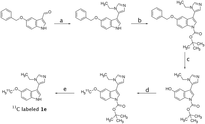
Proposed synthetic scheme for the synthesis of radiolabeled 1e. Conditions: (a) – EtNH2, TosMIC, K2CO3, MeOH (85% - yield); (b) – Boc2O, DMAP, THF (85% yield); (c) – Pd/C, H2, MeOH (36% yield); (d) – 11CH3I, NaOH, toluene, H2O, TBAB (25% yield); (e) – HCl, MeOH (97% yield).
Conclusions
To date there have been no examples of low-basicity agonists of serotonin receptors. We report a series of highly drug-like, low-basicity 5-HT7R agonists synthesized using MCR imidazole synthesis. The procedure described by van Leusen proceeded under various conditions: a range of solvents and bases are tolerable. Notably, the scope of the carbonyl and amine reagents is very broad. Hydroxy-, amino-, amido-, ester and other reactive groups do not interfere with the reaction.
There are several highly active, selective 5-HT7R ligands within the presented series. Hit compound 1o exhibits high affinity towards the receptor (K i = 6 nM), intrinsic activity (EC50 = 19 nM), low intrinsic clearance (Clint = 6.3 ml/min/kg, human liver microsomes) and high predicted drug-likeness. These facts combined with an exceptional ease of synthesis may contribute to the use of radiolabeled 1o and 1e as potential molecular probes. 1e has 5-HT1A/5-HT7 receptor selectivity comparable to known 5-HT7R agonists, whereas 1o is 176-fold selective. Both compounds are very selective towards 5-HT5A which is not a common feature of 5-HT7R agonists. The compounds are metabolically stable in human liver microsomes, exhibit very low toxicity in HEK-293 and HepG2 cells and are water soluble. The blood-brain barrier permeation of 1o was found very high, which is consistent with its high activity in vivo, where all three tested i.p. doses (0.5, 1 and 5 mg/kg) reversed MK-801 induced novel object recognition impairment in mice.
The proposed binding mode was developed by SAR analysis and homology modelling and involves the formation of a hydrogen bond between the indole NH group and Asp3.32. The imidazole fragment is proposed to interact with Arg6.58 which could explain the unusually high selectivity that is not observed in tryptamines. The similar 5-HT7R SAR patterns of tryptamines and the described compounds can be attributed to the interaction of the substituents of indole at the 5th position with Ser5.42 leading to comparable binding affinity gains for the same substituents in the two series.
Structures of NCE’s in the area of medicinal chemistry have become increasingly complex due to the widespread application of the hit-to-lead strategy and the rising palette of structural modifications that enable ADME optimization. We suppose that a better strategy for radioligand searches may be to find simple scaffolds exhibiting moderate to high activity that upon decoration with substituents would retain or improve their affinity for a given target, rather then finding sophisticated structures via virtual or high-throughput screening that upon simplification may not yield any active scaffolds. Because the available chemical space of compounds with low molecular mass (MW < 250 Da) that pass drug likeness filters can be considered inexhaustible taking current synthetic methods into account, we suppose that there are still numerous privileged structures to be discovered. Quoting Richard Feynman: “There’s plenty of room at the bottom”.
Methods
Chemicals
All organic reagents were purchased from Sigma-Aldrich, Apollo Scientific Apollo Scientific Ltd, or Combi-Blocks and were used without purification. Solvents and inorganic reagents were acquired from Chempur. Reaction progress was monitored by TLC on Merck Silica Gel 60 F 254 on aluminium plates. Column chromatography was performed on Merck Silica Gel 60 (0.063–0.200 mm; 70–230 mesh ASTM).
Analytical Methods
UPLC/MS analysis was performed on Waters TQD spectrometer combined with UPLC Acquity H-Class with PDA eLambda detector. Waters Acquity UPLC BEH C18 1.7 μm 2.1 × 100 mm chromatographic column was used, at 40 °C, 0.300 ml/min flow rate and 1.0 μL injection volume (the samples were dissolved in LC-MS grade acetonitrile, typically at a concentration of 0.1–1 mg/ml prior to injection). All mass spectra were recorded under electrospray ionization in positive mode (ESI+) and chromatograms were recorded with UV detection in the range of 190–300 nm. The gradient conditions used were: 80% phase A (water+ 0.1% formic acid) and 20% phase B (acetonitrile + 0.1% formic acid) to 100% phase B (acetonitrile + 0.1% formic acid) at 3.0 minutes, kept till 3.5 minutes, then to initial conditions until 4.0 minutes and kept for additional 2.0 minutes. Total time of analysis – 6.0 minutes.
1H and 13C NMR spectra were recorded on a Bruker Avance III HD 400 NMR spectrometer. All samples were dissolved in DMSO-d6 with TMS as the internal standard. The spectral data of the compounds refer to their free bases.
All presented compounds were of at least 95% purity as determined by LC-MS. Syntheses and characterization details for intermediate products and final compounds as well as the spectral data for all compounds is included in the Supporting Information.
In vivo experiments
The pharmacokinetic experimental procedures were carried out in accordance with EU Directive 2010/63/EU and approved by the I Local Ethics Committee for Experiments on Animals of the Jagiellonian University in Krakow, Poland (approval number: 123/2015). The behavioural experiments were carried out in accordance with EU Directive 2010/63/EU and approved by the Local Ethics Committee for Experiments on Animals of the Institute of Pharmacology, Polish Academy of Sciences (approval number: 181/2016).
General Procedure 1 for the Synthesis of Compounds 1a–1q, 1s–1 v, 1y–1za, 2b–2f, and 3a–3d
Aromatic aldehyde (3 mmol) was mixed with amine (15 mmol) in 20 ml dry methanol. Reaction mixture was left overnight to complete the imine formation although it can be TLC monitored (SiO2/CHCl3). Anhydrous K2CO3 (3 mmol) and TosMIC (tosylmethylisocyanide, 3 mmol) were subsequently added. The mixture was stirred for an additional 8 hours, diluted with 50 ml H2O, and extracted three times with 20 ml ethyl acetate. The combined extracts were washed twice with 20 ml H2O, and once with 20 ml brine, treated with anhydrous magnesium sulfate and evaporated. The final products were purified either by trituration under a 2:1 hexane:isopropanol mixture, or chromatography on a short silicagel bed. The unreacted aldehydes were eluted with ethyl acetate or chloroform, and then a mixture of AcOEt:MeOH or CHCl3:MeOH was applied to elute the product.
General Procedure 2 for the Synthesis of Substituted Indole-3-carboxaldehydes
The Vilsmeier-Haack reagent was generated by the addition of 2 ml of POCl3 over the course of 15 minutes to 8 ml of dry DMF cooled in an ice-salt bath. After the addition was complete, the ice bath was removed and the contents of the flask were left to warm to room temperature over approx. 30 minutes. The substituted indole (21.9 mmol) was dissolved in 10 ml of DMF and added over a period of 15 minutes to the formylating mixture. The stirring was continued for an hour during which the flask contents were heated to 40 °C in a hot water bath. A total of 20 ml of 5 M NaOH was added, and the mixture was diluted with H2O, quickly brought to a boil and left to cool slowly. The crystals were removed by filtration, washed with cold water and vacuum dried. The products thus obtained were in most cases sufficiently pure for the subsequent reactions. And the impure aldehydes were recrystallized from ethanol-water mixtures. The yields varied from 48 to 90%.
General Procedure 3 for the Synthesis of Substituted 1-Alkylindole-3-carboxaldehydes
A total of 10 mmol of an appropriate indole-carboxaldehyde, 50 ml of toluene, 36 ml of 30% NaOH solution, 1 mmol (322 mg) tetrabutylammonium bromide (TBAB) and 10.5 mmol alkyl iodide (0.66 ml of methyl iodide, for instance) were placed in a stoppered flask. The mixture was stirred until the substrate disappeared on TLC (on average 12 h). After completion of the reaction, the phases were separated, next the organic extract was washed with 30 ml of H2O followed by 30 ml of brine, and was dried over MgSO4, and the toluene was stripped off in a rotary evaporator. The crude product was triturated twice with 5 ml of hexane:isopropanol 2:1 mixture, and was vacuum filtered and dried. The yields varied from 85 to 97%.
Unsubsituted indole-3-carboxaldehyde was synthesized at full scale according to the OrgSyn procedure56.
General Procedure 4 for the Synthesis of 1r and 1x (Reductive Debenzylation)
The benzyl protected compound was dissolved in methanol in a pressure reactor, and 10 mol% Pd/C was added. The reactor was sealed and pressurized to 7 bar with H2, and the completion of the reaction was monitored with LC-MS. The mixture was then filtered through Celite which was then washed with MeOH. The solvent was stripped off on a rotary evaporator and the resulting solid was triturated under a hexane:isoporopanol:acetone 3:1:1 mixture. The yields were not lower than 90%.
3-(1-Ethyl-1H-imidazol-5-yl)-1H-indole-5-carboxamide (1za)
The compound was synthesized from 1p, the nitrile group hydrolysis was accomplished according to a modified procedure outlined by Agarwal et al.57.
A suspension of 1p (3.4 mmol, 0.8 g) in 1 ml MeOH and 5 ml THF was stirred in an ice-salt bath for 15 minutes until 0 °C was reached. Hydrogen peroxide (30% solution, 5.4 ml) was added dropwise keeping the temperature below 10 °C. Stirring was continued for 15 minutes and sodium hydroxide (20% solution, 5.4 ml) was added dropwise keeping temperature below 10 °C. The mixture was allowed to warm to room temperature and stirred for 24 hours. The product was extracted from the reaction mixture with chloroform and purified by trituration with acetone. Yield: 81 mg (9%).
Radiosynthesis of 1e
Tert-butyl 5-(benzyloxy)-3-(1-ethyl-1H-imidazol-5-yl)-1H-indole-1-carboxylate (4)
To a round bottom flask there was added: THF (15 ml), 1y (410 mg, 1.29 mmol), di-tert-butyl dicarbonate (590 mg, 2.7 mmol, 2.1 eq) and a catalytic amount of DMAP. The mixture was stirred until disappearance of the substrate on TLC. H2O (20 ml) and ethyl acetate (20 ml) were added, the phases separated, the aquous phase extracted twice with 10 ml ethyl acetate. The organic phase was washed with brine, dried over MgSO4 and evaporated. Crude product was of >95% purity and was used in the next step without purification. Yield: 485 mg (85%). An analytical sample was obtained via column chromatography (SiO2, ethyl acetate:hexane 2:1, then ethyl acetate).
Tert-butyl 3-(1-ethyl-1H-imidazol-5-yl)-5-hydroxy-1H-indole-1-carboxylate (5)
4 (200 mg, 0.48 mmol) was dissolved in 50 ml MeOH in a glass reactor. 10% Pd/C (50 mg) was added and the reactor was pressurized to 7 bar with hydrogen. The mixture was stirred overnight, filtered through celite and the celite bed was washed thrice with 20 ml MeOH. The solvent was evaporated and the residue was subjected to column chromatography (SiO2, ethyl acetate). Yield: 57 mg (36%).
Tert-butyl 3-(1-ethyl-1H-imidazol-5-yl)-5-methoxy-1H-indole-1-carboxylate (6)
To a round bottom flask there were added: 3 ml toluene, 5 (100 mg, 0.305 mmol), CH3I (20 μL, 0.32 mmol, 1.05 eq), TBAB (10 mg, 0.0305 mmol, 0.1 eq.) and 1 ml 30% NaOH solution. The mixture was stirred and the progress of the reaction was TLC monitored. The mixture was diluted with 10 ml H2O, extracted three times with 10 ml ethyl acetate, the combined organic phase was washed with 10 ml H2O, 10 ml brine, dried over MgSO4 and evaporated. The crude product was purified via column chromatography (SiO2, ethyl acetate). Yield: 26 mg (25%).
3-(1-Ethyl-1H-imidazol-5-yl)-5-methoxy-1H-indole hydrochloride (1e)
6 (10 mg, 0.0293 mmol) was suspended in 2 ml MeOH and one drop of concentrated hydrochloric acid was added. The mixture was refluxed for 3 h, and the solvent was evaporated. Yield: 7.9 mg (97%).
In Vitro Pharmacology. Cell Culture
HEK293 cells with stable expression of human serotonin 5-HT1AR, 5-HT6 and 5-HT7bR or dopamine D2LR (obtained using of Lipofectamine 2000, Invitrogen) or CHO-K1 cells with plasmid containing the sequence coding for the human serotonin 5-HT2A receptor (Perkin Elmer) were maintained at 37 °C in a humidified atmosphere with 5% CO2 and were grown in Dulbecco’s Modified Eagle’s Medium containing 10% dialyzed fetal bovine serum and 500 μg/ml G418 sulfate. For membranes preparations, cells were subcultured into 150 cm2 cell culture flasks, grown to 90% confluence, washed twice with phosphate buffered saline (PBS) prewarmed to 37 °C, pelleted by centrifugation (200 g) in PBS containing 0.1 mM EDTA and 1 mM dithiothreitol, and stored at −80 °C57–59.
5-HT1A/5-HT2A/5-HT6/5-HT7/D2 Radioligand Binding Assays
The membrane preparation and general assay procedures for the cloned receptors were adjusted to 96-microwell format as described in our former papers58–61. The cell pellets were thawed and homogenized in 10 volumes of assay buffer using an Ultra Turrax tissue homogenizer and were centrifuged twice at 35,000 g for 15 min at 4 °C, with incubation for 15 min at 37 °C in between the rounds of centrifugation. The composition of the assay buffers was as follows: for 5-HT1AR: 50 mM Tris–HCl, 0.1 mM EDTA, 4 mM MgCl2, 10 μM pargyline and 0.1% ascorbate; for 5-HT2AR: 50 mM Tris–HCl, 0.1 mM EDTA, 4 mM MgCl2 and 0.1% ascorbate; for 5-HT6R: 50 mM Tris–HCl, 0.5 mM EDTA and 4 mM MgCl2, and for 5-HT7bR: 50 mM Tris–HCl, 4 mM MgCl2, 10 μM pargyline and 0.1% ascorbate; for dopamine D2LR: 50 mM Tris–HCl, 1 mM EDTA, 4 mM MgCl2, 120 mM NaCl, 5 mM KCl, 1.5 mM CaCl2 and 0.1% ascorbate.
All assays were incubated in a total volume of 200 μL in 96-well microtiter plates for 1 h at 37 °C, except for 5-HT1AR and 5-HT2AR, which were incubated at room temperature and 27 °C, respectively. The equilibration process was terminated by rapid filtration through Unifilter plates with a 96-well cell harvester and the radioactivity retained on the filters was quantified using a Microbeta plate reader (PerkinElmer, USA).
For the displacement studies, the assay samples contained the following as radioligands (PerkinElmer, USA): 1.5 nM [3H]-8-OH-DPAT (135.2 Ci/mmol) for 5-HT1AR; 2 nM [3H]-ketanserin (53.4 Ci/mmol) for 5-HT2AR; 2 nM [3H]-LSD (83.6 Ci/mmol) for 5-HT6R, 0.6 nM [3H]-5-CT (39.2 Ci/mmol) for 5-HT7R or [3H]-Raclopride (74.4 Ci/mmol). Non-specific binding was defined using 10 μM of 5-HT in 5-HT1AR and 5-HT7R binding experiments, whereas 20 µM of mianserin, 10 μM of methiothepine or 1 μM of (+) butaclamol was used in the 5-HT2AR, 5-HT6R and D2LR assays, respectively. Each compound was tested in triplicate at 7–8 concentrations (10–11–10–4 M). The inhibition constants (K i) were calculated using the Cheng-Prusoff equation62 and the results were expressed as the means of at least two independent experiments.
Functional Assay
The functional properties of compounds in HEK293 cells overexpressing 5-HT7R were evaluated for their ability to increase cAMP production for the agonists or to inhibit 10 nM 5-CT at a concentration producing 90% (EC90) of the maximum agonist activation for the antagonists.
Each compound was tested at 8 concentrations in the range of 10−11–10−4 M. Cells (prepared with the use of Lipofectamine 2000) were maintained at 37 °C in a humidified atmosphere with 5% CO2 and were grown in DMEM containing 10% dialyzed (FBA) and 500 µg/ml G418 sulfate. For the functional experiments, the cells were subcultured in 25 cm2 diameter dishes, grown to 90% confluence, washed twice with PBS prewarmed to 37 °C and centrifuged for 5 min (160 × g). The supernatant was aspirated, and the cell pellet was resuspended in stimulation buffer (1 × HBSS, 5 mM HEPES, 0.5 mM IBMX, and 0.1% BSA). The total cAMP was measured using the LANCE cAMP detection kit (PerkinElmer), according to the manufacture’s directions. For the cAMP level quantification, cells (5 µl) were incubated with compounds (5 µl) for 30 min at room temperature in a 384-well white opaque microtiter plate. After incubation, 10 µl working solution (5 µl Eu-cAMP and 5 µl ULight-anti-cAMP) was added to stop the reaction and induced cells lysis. The assay plate was incubated for 1 h at room temperature and time-resolved fluorescence resonance energy transfer (TR-FRET) was detected by an Infinite M1000 Pro (Tecan, Männedorf, Switzerland) using instrument settings from the LANCE cAMP detection kit manual.
Molecular Modeling. Homology Modeling
Homology models of the 5-HT7 receptor were built on a GPCR crystal structures co-crystalized with agonist molecules, i.e. 5-HT1B (PDB ID: 4IAR, 4IAQ), 5-HT2B (PDB ID: 4IB4), A2A (PDB ID: 3QAK, 2YDV, 4UHR), β2 (PDB ID: 3P0G, 4LDE, 3PDS) and M2 (PDB ID: 4MQS) receptors, retrieved from the Protein Data Bank63. Sequences of the modeled receptor and selected templates were aligned manually using Accelrys Discovery Studio64, making sure that the most conserved amino acid in each helix, and the motif characteristic for class A GPCRs were in equivalent positions. The ranges of helices were determined on the basis of the crystal structures, and the loop regions were modeled, but not refined. For each template, 20 models were generated using Modeller software65, and were tested by the docking of active and inactive compounds, and enrichment calculation. For every model, ROC curves were calculated based on the Glide Score values of the docked compounds (the undocked actives and inactives were considered as false negatives and true negatives, respectively). The quality of the models was determined by the Area Under the ROC curve (AUROC). Three models per template with the highest AUROC value were selected for further studies. The final validation of the selected receptor models was performed by using the QM/MM molecular docking (QPLD Protocol) of the whole set of studied molecules. The models for which binding modes coherent for the whole set of compounds and explaining the main structure-activity relationships were observed were kept.
CNS MPO Calculation
The Central Nervous System Multiparameter Optimization (CNS MPO) scores were calculated based on the six basic physicochemical molecular descriptors: logP, logD, molecular weight (MW), topological polar surface area (TPSA), the count of hydrogen bond donor (HBD), and the strongest basic pKa. All descriptors were calculated using Chemaxon’s Calculator Plugins66. A monotonically decreasing function was used to transform the values of the descriptors into dimensionless scale logP, logD, MW, HBD, and pKa whereas a hump function was used to transform TPSA. The most desirable and least desirable ranges for each physicochemical descriptor were adopted from the original paper50. Transformed values (desirability score) of the six descriptors were determined and summed up for each compound, to obtain the final CNS MPO, which can range from 0 (the worst CNS drug) to 6 (best CNS drug).
QM/MM Docking Protocol
The Quantum Mechanics/Molecular Mechanics (QM/MM) docking of all synthesized compounds to 5-HT7R homology models were performed by means of the QM-Polarized Ligand Docking Protocol (QPLD)67 from Schrödinger Suite. At the first stage of the QPLD procedure the ligands were initially docked into a rigid protein using Glide. The resulting binding modes of the ligands were then used for the calculation of the partial charges of the ligand by a single-point calculation in QSite treating the ligand with the ab initio (B3PW91/cc-pVTZ) and the receptor with MM (OPLS-2005) level of theory. The partial atomic charges of the ligand molecules were recalculated using the electrostatic potential fitting method under the influence of external fields exerted by the surrounding protein atoms within and near the binding site. At the final stage, Glide re-docked the ligands using new charges, and the 10 most energetically favorable poses were returned for each ligand.
Binding Free Energy Calculation
MM-GBSA (Generalized-Born/Surface Area) was used to calculate the binding free energy for all ligand-receptor complexes to select the correct poses between possible binding modes. The binding free energy was calculated using ligand charges obtained via the QM/MM calculations using Prime software from Schrodinger. To assess the influence of a given substituent on the binding, the ΔΔG was calculated as a difference between binding free energy (ΔG) of unsubstituted (1w) and substituted analogues.
Plotting Interaction Spheres for Halogen Bonding
To visualize (plotting interaction spheres) the possible contribution of halogen bonding for the resulting ligand-receptor complexes, the halogen bonding webserver was used (access 1.4.2016, http://www.halogenbonding.com/).
Associated Content
Syntheses and characterization details for intermediate products and final compounds, cell culture and transfection methods, pharmacological data for compound 2a, detailed description of: metabolic stability assays for compound 1e and 1o, cytotoxicity assays, metabolic stability assays, pharmacokinetic study, novel object recognition experiment, 1H NMR, 13C NMR and LC-MS spectra (PDF).
Molecular formula strings (CSV).
Electronic supplementary material
Acknowledgements
We would like to thank Prof. Alexander Dömling for the MCR introductory course that was held in the Faculty of Chemistry, Jagiellonian University, Kraków, Poland, 2014. J.H., K.K.-K. and A.J.B. participate in the European Cooperation in Science and Technology (COST) Action CM1207 ‘‘GLISTEN: GPCR-Ligand Interactions, Structures, and Transmembrane Signalling: a European Research Network”. The study was supported by the Polish-Norwegian Research Programme operated by the National Centre for Research and Development under the Norwegian Financial Mechanism 2009–2014 in the frame of the Project PLATFORMex (Pol-Nor/198887/73/2013), by the Polish National Science Centre grants 2014/15/D/NZ7/01782 and 2014/13/B/NZ7/02210, and by statutory funds K/ZDS/005593.
Author Contributions
Andrzej J. Bojarski supervised the study, performed data analysis and wrote the manuscript text, Adam Hogendorf wrote the manuscript draft, designed and synthesized a part of the library, was responsible for SAR study, Agata Hogendorf synthesized a part of the library, was responsible for purity assessment, Rafał Kurczab prepared the molecular modelling part, Grzegorz Satała performed all in vitro radioligand displacement assays and functional assays, Tomasz Lenda prepared the cell lines, Maria Walczak performed the pharmacokinetic analysis, Gniewomir Latacz performed the metabolic stability and cytotoxicity assays, Jadwiga Handzlik partly contributed in the metabolic stability and cytotoxicity assays. Katarzyna Kieć-Kononowicz supervised the ADMET study, Monika Woźniak and Paulina Cieślik performed the Novel Object Recognition experiments, Joanna Wierońska supervised the behavioural study, Ryszard Bugno synthesized a part of the library, Jakub Staroń synthesized a part of the library, all authors reviewed the manuscript.
Competing Interests
The authors declare that they have no competing interests.
Footnotes
Electronic supplementary material
Supplementary information accompanies this paper at doi:10.1038/s41598-017-00822-4
Publisher's note: Springer Nature remains neutral with regard to jurisdictional claims in published maps and institutional affiliations.
References
- 1.Hedlund PB. The 5-HT7 receptor and disorders of the nervous system: an overview. Psychopharmacology. 2009;206:345–354. doi: 10.1007/s00213-009-1626-0. [DOI] [PMC free article] [PubMed] [Google Scholar]
- 2.Hoyer D, Hannon JP, Martin GR. Molecular, pharmacological and functional diversity of 5-HT receptors. Pharmacol. Biochem. Behav. 2002;71:533–554. doi: 10.1016/S0091-3057(01)00746-8. [DOI] [PubMed] [Google Scholar]
- 3.Ruat M, et al. Molecular cloning, characterization, and localization of a high-affinity serotonin receptor (5-HT7) activating cAMP formation. Proc. Natl. Acad. Sci. USA. 1993;90:8547–8551. doi: 10.1073/pnas.90.18.8547. [DOI] [PMC free article] [PubMed] [Google Scholar]
- 4.Lovenberg TW, et al. A novel adenylyl cyclase-activating serotonin receptor (5-HT7) implicated in the regulation of mammalian circadian rhythms. Neuron. 1993;11:449–458. doi: 10.1016/0896-6273(93)90149-L. [DOI] [PubMed] [Google Scholar]
- 5.Kvachnina E, et al. 5-HT7 receptor is coupled to Gα subunits of heterotrimeric G12-protein to regulate gene transcription and neuronal morphology. J. Neurosci. 2005;25:7821–7830. doi: 10.1523/JNEUROSCI.1790-05.2005. [DOI] [PMC free article] [PubMed] [Google Scholar]
- 6.Bard JA, et al. Cloning of a novel human serotonin receptor (5-HT7) positively linked to adenylate cyclase. J. Biol. Chem. 1993;268:23422–23426. [PubMed] [Google Scholar]
- 7.Hedlund PB, et al. LP-211 is a brain penetrant selective agonist for the serotonin 5-HT7 receptor. Neurosci. Lett. 2010;481:12–16. doi: 10.1016/j.neulet.2010.06.036. [DOI] [PMC free article] [PubMed] [Google Scholar]
- 8.Terron JA, Falcon-Neri A. Br. Pharmacological evidence for the 5-HT7 receptor mediating smooth muscle relaxation in canine cerebral arteries. J. Pharmacol. 1999;127:609–616. doi: 10.1038/sj.bjp.0702580. [DOI] [PMC free article] [PubMed] [Google Scholar]
- 9.Roberts AJ, et al. Mice lacking 5-HT7 receptors show specific impairments in contextual learning. Eur. J. Neurosci. 2004;19:1913–1922. doi: 10.1111/j.1460-9568.2004.03288.x. [DOI] [PubMed] [Google Scholar]
- 10.Gasbarri A, Cifariello A, Pompili A, Meneses A. Effect of 5-HT7 antagonist SB-269970 in the modulation of working and reference memory in the rat. Behav. Brain Res. 2008;195:164–170. doi: 10.1016/j.bbr.2007.12.020. [DOI] [PubMed] [Google Scholar]
- 11.Eriksson TM, Golkar A, Ekström JC, Svenningsson P, Ögren SO. 5-HT7 receptor stimulation by 8-OH-DPAT counteracts the impairing effects of 5-HT1A receptor stimulation on contextual learning in mice. Eur. J. Pharmacol. 2008;596:107–110. doi: 10.1016/j.ejphar.2008.08.026. [DOI] [PubMed] [Google Scholar]
- 12.Sarkisyan G, Hedlund PB. The 5-HT7 receptor is involved in allocentric spatial memory information processing. Behav. Brain Res. 2009;202:26–31. doi: 10.1016/j.bbr.2009.03.011. [DOI] [PMC free article] [PubMed] [Google Scholar]
- 13.Meneses A, et al. The effects of the 5-HT6 receptor agonist EMD and the 5-HT7 receptor agonist AS19 on memory formation. Behav. Brain Res. 2008;195:112–119. doi: 10.1016/j.bbr.2007.11.023. [DOI] [PubMed] [Google Scholar]
- 14.Hedlund PB, Sutcliffe JG. Functional, molecular and pharmacological advances in 5-HT7 receptor research. Trends Pharmacol. Sci. 2004;25:481–486. doi: 10.1016/j.tips.2004.07.002. [DOI] [PubMed] [Google Scholar]
- 15.Mahe C, et al. Serotonin 5-HT7 receptors coupled to induction of interleukin-6 in human microglial MC-3 cells. Neuropharmacology. 2005;49:40–47. doi: 10.1016/j.neuropharm.2005.01.025. [DOI] [PubMed] [Google Scholar]
- 16.Dean B, Pavey G, Thomas D, Scarr E. Cortical serotonin 7, 1D and 1F receptors: effects of schizophrenia, suicide and antipsychotic drug treatment. Schizophr. Res. 2006;88:265–267. doi: 10.1016/j.schres.2006.07.003. [DOI] [PubMed] [Google Scholar]
- 17.Rocha-Gonzalez HI, Meneses A, Carlton SM, Granados-Soto V. Pronociceptive role of peripheral and spinal 5-HT7 receptors in the formalin test. Pain. 2005;117:182–192. doi: 10.1016/j.pain.2005.06.011. [DOI] [PubMed] [Google Scholar]
- 18.Matthys A, Haegeman G, van Craenenbroeck K, Vanhoenacker P. Role of the 5-HT7 receptor in the central nervous system: from current status to future perspectives. Mol. Neurobiol. 2011;43:228–253. doi: 10.1007/s12035-011-8175-3. [DOI] [PubMed] [Google Scholar]
- 19.Monsma FJ, Shen Y, Ward RP, Hamblin MW, Sibley D. R Cloning and expression of a novel serotonin receptor of high affinity for tricyclic psychotropic drugs. Mol. Pharmacol. 1993;43:320–327. [PubMed] [Google Scholar]
- 20.Mullins UL, Gianutsos G, Eison AS. Effects of antidepressants on 5-HT7 receptor regulation in the rat hypothalamus. Neuropsychopharmacol. 1999;21:352–367. doi: 10.1016/S0893-133X(99)00041-X. [DOI] [PubMed] [Google Scholar]
- 21.Wesolowska A, Nikiforuk A, Stachowicz K, Tatarczynska E. Effect of the selective 5-HT7 receptor antagonist SB 269970 in animal models of anxiety and depression. Neuropharmacology. 2006;51:578–586. doi: 10.1016/j.neuropharm.2006.04.017. [DOI] [PubMed] [Google Scholar]
- 22.Wesolowska A, Tatarczynska E, Nikiforuk A, Chojnacka-Wojcik E. Enhancement of the anti-immobility action of antidepressants by a selective 5-HT7 receptor antagonist in the forced swimming test in mice. Eur. J. Pharmacol. 2007;555:43–47. doi: 10.1016/j.ejphar.2006.10.001. [DOI] [PubMed] [Google Scholar]
- 23.Pouzet B, Didriksen M, Arnt J. Effects of the 5-HT7 receptor antagonist SB-258741 in animal model for schizophrenia. Pharmacol. Biochem. Behav. 2002;71:655–665. doi: 10.1016/S0091-3057(01)00744-4. [DOI] [PubMed] [Google Scholar]
- 24.Semenova S, Geyer MA, Sutcliffe JG, Markou A, Hedlund PB. Inactivation of the 5-HT7 receptor partially blocks phencyclidine-induced disruption of prepulse inhibition. Biol. Psychiatry. 2008;63:98–105. doi: 10.1016/j.biopsych.2006.12.011. [DOI] [PMC free article] [PubMed] [Google Scholar]
- 25.Gasbarri A, Pompili A. Serotonergic 5-HT7 receptors and cognition. Rev. Neurosci. 2014;25:311–323. doi: 10.1515/revneuro-2013-0066. [DOI] [PubMed] [Google Scholar]
- 26.Sanin, A. et al. 5-Aryl substituted (S)-2-(dimethylamino)-tetralins novel serotonin 5-HT7receptor ligands. In: Proceedings of the 14th Camerino-Noord Symposium. Ongoing Progress in the Receptor Chemistry, p. 27 (2004).
- 27.Hagan JJ, et al. Characterization of SB-269970-A, a selective 5-HT7 receptor antagonist. Br. J. Pharmacol. 2000;130:539–548. doi: 10.1038/sj.bjp.0703357. [DOI] [PMC free article] [PubMed] [Google Scholar]
- 28.Stoll W, Schweiz A. Lyseric acid diethylamide, a hallucinogen from the ergot group. Arch. Neurol. Psychiatr. 1947;60:279. [Google Scholar]
- 29.Leopoldo M, et al. Structure-activity relationship study on N-(1,2,3,4-tetrahydronaphthalen-1-yl)-4-aryl-1piperazinehexanamides, a class of 5-HT7 receptor agents. J. Med. Chem. 2007;50:4214–4221. doi: 10.1021/jm070487n. [DOI] [PubMed] [Google Scholar]
- 30.Leopoldo M, et al. Structure−affinity relationship study on N-(1,2,3,4-tetrahydronaphthalen-1-yl)4-aryl-1-piperazinealkylamides, a new class of 5-hydroxytryptamine7 receptor agents. J. Med. Chem. 2004;47:6616–6624. doi: 10.1021/jm049702f. [DOI] [PubMed] [Google Scholar]
- 31.Leopoldo M, et al. Structural modifications of N-(1,2,3,4-tetrahydronaphthalen-1-yl)-4-aryl-1-piperazinehexanamides: Influence on lipophilicity and 5-HT7 receptor activity. Part III. J. Med. Chem. 2008;51:5813–5822. doi: 10.1021/jm800615e. [DOI] [PubMed] [Google Scholar]
- 32.Brenchat A, et al. 5-HT7 receptor activation inhibits mechanical hypersensitivity secondary capsaicin sensitization in mice. Pain. 2009;141:239–247. doi: 10.1016/j.pain.2008.11.009. [DOI] [PubMed] [Google Scholar]
- 33.Lacivita E, et al. Structural modifications of the serotonin 5-HT7 receptor agonist N-(4-cyanophenylmethyl)-4-(2-biphenyl)-1-piperazinehexanamide (LP-211) to improve in vitro microsomal stability: A case study. Eur. J. Med. Chem. 2016;120:363–379. doi: 10.1016/j.ejmech.2016.05.005. [DOI] [PubMed] [Google Scholar]
- 34.Canese R, et al. Persistent modification of forebrain networks and metabolism in rats following adolescent exposure to a 5-HT7 receptor agonist. Psychopharmacology (Berl) 2015;232:75–89. doi: 10.1007/s00213-014-3639-6. [DOI] [PubMed] [Google Scholar]
- 35.De Filippis B, et al. Long-lasting beneficial effects of central serotonin receptor 7 stimulation in female mice modeling Rett syndrome. Front. Behav. Neurosci. 2015;9:86. doi: 10.3389/fnbeh.2015.00086. [DOI] [PMC free article] [PubMed] [Google Scholar]
- 36.Costa L, et al. Activation of 5-HT7 serotonin receptors reverses metabotropic glutamate receptor-mediated synaptic plasticity in wild-type and Fmr1 knockout mice, a model of Fragile X syndrome. Biol. Psychiatry. 2012;72:924–933. doi: 10.1016/j.biopsych.2012.06.008. [DOI] [PubMed] [Google Scholar]
- 37.Di Pilato P, et al. Selective agonists for serotonin 7 (5-HT7) receptor and their applications in preclinical models: an overview. Rev. Neurosci. 2014;25:401–414. doi: 10.1515/revneuro-2014-0009. [DOI] [PubMed] [Google Scholar]
- 38.Ladduwahetty T, et al. A new class of selective, non-basic 5-HT2A receptor antagonists. Bioorg. Med. Chem. Lett. 2006;16:3201–3204. doi: 10.1016/j.bmcl.2006.03.050. [DOI] [PubMed] [Google Scholar]
- 39.Harris RN, 3rd, et al. Highly potent, non-basic 5-HT6 ligands. Site mutagenesis evidence for a second binding mode at 5-HT6 for antagonism. Bioorg. Med. Chem. Lett. 2010;20:3436–3440. doi: 10.1016/j.bmcl.2010.03.110. [DOI] [PubMed] [Google Scholar]
- 40.Nugiel DA, et al. De Novo Design of a Picomolar Nonbasic 5-HT1B Receptor Antagonist. J. Med. Chem. 2010;53:1876–1880. doi: 10.1021/jm901200t. [DOI] [PubMed] [Google Scholar]
- 41.Bento AP, et al. The ChEMBL bioactivity database: an update. Nucleic Acids Res. 2014;42:1083–1090. doi: 10.1093/nar/gkt1031. [DOI] [PMC free article] [PubMed] [Google Scholar]
- 42.Zhang L, Peng XM, Damu GLV, Geng RX, Zhou CH. Comprehensive Review in Current Developments of Imidazole-Based Medicinal Chemistry. Med. Res. Rev. 2014;34:340–437. doi: 10.1002/med.21290. [DOI] [PubMed] [Google Scholar]
- 43.Van Leusen AM, Wildeman J, Oldenziel OH. Chemistry of sulfonylmethyl isocyanides. Base-induced cycloaddition of sulfonylmethyl isocyanides to carbon,nitrogen double bonds. Synthesis of 1,5-disubstituted and 1,4,5-trisubstituted imidazoles from aldimines and imidoyl chlorides. J. Org. Chem. 1977;42:1153–1159. doi: 10.1021/jo00427a012. [DOI] [Google Scholar]
- 44.Dömling A, Ugi I. Multicomponent Reactions with Isocyanides. Angew. Chem. 2000;39:3168–3210. doi: 10.1002/1521-3773(20000915)39:18<3168::AID-ANIE3168>3.0.CO;2-U. [DOI] [PubMed] [Google Scholar]
- 45.Obach RS. Prediction of human clearance of twenty-nine drugs from hepatic microsomal intrinsic clearance data: An examination of in vitro half-life approach and nonspecific binding to microsomes. Drug Metab. Dispos. 1999;27:1350–1359. [PubMed] [Google Scholar]
- 46.Tiwari AK, et al. Design, synthesis and biological evaluation of small molecule-based PET radioligands for the 5-hydroxytryptamine 7 receptor. RSC Adv. 2015;5:19752–19759. [Google Scholar]
- 47.Glennon RA, et al. [125I]-1-(2,5-Dimethoxy-4-iodophenyl)-2-aminopropane: An iodinated radioligand that specifically labels the agonist high-affinity state of 5-HT2 serotonin receptors. J. Med. Chem. 1988;31:5–7. doi: 10.1021/jm00396a003. [DOI] [PubMed] [Google Scholar]
- 48.Heim, R. Chemistry and allied sciences: Synthesis and pharmacology of potent 5-HT2A receptor agonists which have a partial N-2-methoxybenzyl structure. Ph.D. Dissertation, Free University of Berlin (2003).
- 49.Meneses A. 5-HT7 receptor stimulation and blockade: a therapeutic paradox about memory formation and amnesia. Front. Behav. Neurosci. 2014;8:1–4. doi: 10.3389/fnbeh.2014.00207. [DOI] [PMC free article] [PubMed] [Google Scholar]
- 50.Wager TT, Hou X, Verhoest P, Villalobos R, Moving A. beyond rules: the development of a central nervous system multiparameter optimization (CNS MPO) approach to enable alignment of druglike properties. ACS Chem. Neurosci. 2010;1:435–449. doi: 10.1021/cn100008c. [DOI] [PMC free article] [PubMed] [Google Scholar]
- 51.Isberg V, et al. GPCRdb: an Information system for G protein-coupled receptors. Nucleic Acids Res. 2016;44:356–364. doi: 10.1093/nar/gkv1178. [DOI] [PMC free article] [PubMed] [Google Scholar]
- 52.Vermeulen ES, Schmidt AW, Sprouse JS, Wikstrom HV, Grol CJ. Characterization of the 5-HT7 receptor. Determination of the pharmacophore for 5-HT7 receptor agonism and CoMFA-based modeling of the agonist binding site. J. Med. Chem. 2003;46:5365–5374. doi: 10.1021/jm030826m. [DOI] [PubMed] [Google Scholar]
- 53.Hansen HD, et al. Radiosynthesis and In Vivo Evaluation of Novel Radioligands for PET Imaging of Cerebral 5-HT7 Receptors. J. Nucl. Med. 2014;55:640–646. doi: 10.2967/jnumed.113.128983. [DOI] [PubMed] [Google Scholar]
- 54.Deau E, et al. Rational Design, Pharmacomodulation, Synthesis of Dual 5-Hydroxytryptamine 7 (5-HT7)/5-Hydroxytryptamine 2A (5-HT2A) Receptors Antagonists and Evaluation by [18F]-PET Imaging in a Primate Brain. J. Med. Chem. 2015;58:8066–8096. doi: 10.1021/acs.jmedchem.5b00874. [DOI] [PubMed] [Google Scholar]
- 55.Andries J, Lemoine L, Le Bars D, Zimmer L, Billard T. Synthesis and biological evaluation of potential 5-HT 7 receptor PET radiotracers. Eur. J. Med. Chem. 2011;46:3455–3461. doi: 10.1016/j.ejmech.2011.05.010. [DOI] [PubMed] [Google Scholar]
- 56.Watson J, et al. 5-HT1A receptor agonist-antagonist binding affnity difference as a measure of intrinsic activity in recombinant and native tissue systems. Br. J. Pharmacol. 2000;130:1108–1114. doi: 10.1038/sj.bjp.0703394. [DOI] [PMC free article] [PubMed] [Google Scholar]
- 57.James PN, Snyder HR. Indole-3-aldehyde. Org. Synth. 1959;39:30. doi: 10.15227/orgsyn.039.0030. [DOI] [Google Scholar]
- 58.Agarwal A, Jalluri RK, De Witt Blanton C, Jr., Will Taylor T. A new synthesis of a potent 5-HT1 receptor ligand, 5-carboxyamidotryptamine (5-CT) Synth. Commun. 1993;23:1101–1110. doi: 10.1080/00397919308018587. [DOI] [Google Scholar]
- 59.Zajdel P, et al. Quinoline- and isoquinoline-sulfonamide derivatives of LCAP as potent CNS multi-receptor-5-HT1A/5-HT2A/5-HT7 and D2/D3/D4-agents: The synthesis and pharmacological evaluation. Bioorg. Med. Chem. 2012;20:1545–1556. doi: 10.1016/j.bmc.2011.12.039. [DOI] [PubMed] [Google Scholar]
- 60.Bojarski AJ, et al. Structure-activity relationship studies of CNS agents. 9. 5-HT1A and 5-HT2 receptor affinity of some 2-substitued and 3-substitued 1,2,3,4-tetrahydro-beta-carbolines. Pharmazie. 1993;48:289–294. [PubMed] [Google Scholar]
- 61.Paluchowska MH, et al. The influence of modifications in imide fragment structure on 5-HT1A and 5-HT7 receptor affinity and in vivo pharmacological properties of some new 1-(m-trifluoromethylphenyl)piperazines. Bioorg. Med. Chem. 2007;15:7116–7125. doi: 10.1016/j.bmc.2007.07.029. [DOI] [PubMed] [Google Scholar]
- 62.Cheng Y, Prusoff WH. Relationship between the inhibition constant (KI) and the concentration of inhibitor which causes 50 per cent inhibition (I50) of an enzymatic reaction. Biochem. Pharmacol. 1973;22:3099–3108. doi: 10.1016/0006-2952(73)90196-2. [DOI] [PubMed] [Google Scholar]
- 63.Bernstein FC, et al. The Protein Data Bank: A Computer-Based Archival File for Macromolecular Structures. J. Mol. Biol. 1977;112:535–542. doi: 10.1016/S0022-2836(77)80200-3. [DOI] [PubMed] [Google Scholar]
- 64.Discovery Studio v.3.5, Accelrys Software, Inc. San Diego, CA.
- 65.Sali A, Blundell TL. Comparative Protein Modelling by Satisfaction of Spatial Restraints. J. Mol. Biol. 1993;234:779–815. doi: 10.1006/jmbi.1993.1626. [DOI] [PubMed] [Google Scholar]
- 66.Calculator Plugins for Structure Property Prediction and Calculation, Marvin 6.2.2, ChemAxon http://www.chemaxon.com (2014).
- 67.Small-Molecule Drug Discovery Suite 2015-4: Schrödinger Suite 2015-4 QM-Polarized Ligand Docking protocol, Glide version 6.9, Schrödinger, LLC, New York, NY, 2015, Jaguar version 9.0, Schrödinger, LLC, New York, NY, 2015, QSite version 6.9, Schrödinger, LLC, New York, NY, (2015).
Associated Data
This section collects any data citations, data availability statements, or supplementary materials included in this article.


