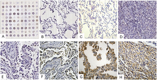
Figure 1. IHC analysis of stathmin in lung cancer and normal lung tissues (IHC×400).

A. tissue microarray construction; B. low staining of stathmin in normal lung tissues; C. moderate staining of stathmin in normal lung tissues; D. low staining of stathmin in well differentiated LSCC; E. low staining of stathmin in well differentiated LAC; F. moderate staining of stathmin in moderately differentiated LAC; G. high staining of stathmin in poorly differentiated LAC; H. moderate staining of stathmin in moderately differentiated LSCC; LAC, lung adenocarcinoma; LSCC, lung squamous cell carcinoma.
