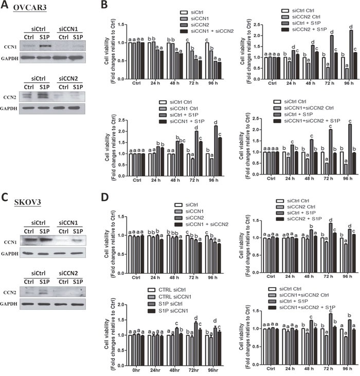
Figure 5. CCN1 and CCN2 mediate the S1P-induced cell proliferation in OVCAR3 and SKOV3 cells.

(A and C) OVCAR3 (A) or SKOV3 (C) cells were transfected for 48 h with 50 nM control siRNA (siCtrl), CCN1 siRNA (siCCN1) or CCN2 siRNA (siCCN2), and then treated with S1P for additional 2 h. The protein levels of CCN1 and CCN2 were analyzed using Western blot analysis. (B and D) OVCAR3 (B) or SKOV3 (D) cells were transfected for 48 h with 50 nM siCtrl, siCCN1, siCCN2 or combined siCCN1 and siCCN2 (siCCN1+siCCN2), and then treated with S1P for 0, 24, 48, 72 or 96 h. The cell viability was analyzed using an MTT assay. The results are expressed as the mean ± SEM from at least three independent experiments. All samples were compared using one-way ANOVA followed by Tukey's multiple comparison tests, and values without a common letter (a, b, c and d) are significantly different (P<0.05).
