

Abstract
Background
In recent years, a large number of neuroimaging studies found that the Cortico-Striato- Thalamo-Cortical circuit (CSTC), including the prefrontal lobe, a significant part of CSTC, has disturbance metabolically in patients with Obsessive-Compulsive Disorder (OCD).
Aim
Explore the correlation between the neuro-metabolic features and clinical characteristics of OCD patients using magnetic resonance spectroscopy technology.
Methods
88 patients with OCD who were not received medication and outpatient treatment for 8 weeks and 76 health controls were enrolled, there was no significant difference in gender, age or education level between the two groups. SIEMENS 3.0T MRI scanner was used to measure the spectral wave of Orbito Frontal Cortex (OFC) and Anterior Cingulate Cortex (ACC) of participants, setting mega-press sequences. Meanwhile, the concentrations of gamma-aminobutyric acid (GABA), glutamine/glutamate complex (Glx) and N-Acetyl Aspartate (NAA) were measured relative to concentration of water, on the ACC and OFC of participants, for statistical analysis via LC model version 6.3 software. The concentration of metabolic substances of the OCD group compared to the healthy control group was analyzed using two sample t-test. The correlation between substance concentration and scores on the scales, including Yale-Brown Obsessive Compulsive Scale (Y-BOCS), Hamilton Anxiety scale (HAMA) and Hamilton Depression scale (HAMD) was carried out using the Pearson correlation method.
Results
Compared with healthy controls, the GABA/W and NAA/W concentration in individuals with OCD are significantly decreased (p=0.031, t=2.193, p=0.002, t=3.223). Also, the concentration of GABA/W had a trend of decrease in the ACC. The GABA/W of the OFC had a negative correlation with Y-BOCS-O, Y-BOCS-C and Y-BOCS-T scores (p=0.037, r=0.221; p=0.007, r=0.283; p=0.014, r=0.259).
Conclusions
These results support that GABA concentration in the OFC area of patients with OCD is significantly decreased and the concentration in the ACC has a trend of decreasing. All of these indicate that there is a relationship between the GABA concentration and the psychopathology of OCD.
Key words: obsessive-compulsive disorder, magnetic resonance spectroscopy, orbitofrontal cortex, anterior cingulate cortex, gamma-aminobutyric acid, China
Abstract
背景
近年来, 大量的神经影像学研究发现强迫症(obsessive-compulsive disorder,OCD)在皮层- 纹状体-丘脑- 皮层(cortico-striato-thalamo-cortical,CSTC)环路存在代谢方面的异常,前额叶是CSTC 环路的重要脑区。
目的
本研究采用磁共振波谱技术研究OCD 患者前额叶神经代谢的特点及其与临床特征之间的关系。
方法
研究入组未用药(8 周及以上)的OCD 患者 78 例以及76 例健康对,性别、年龄及受教育年限无统计学差异。采用3.0 T 西门子磁共振扫描仪分别收集眶额回(orbito frontal cortex,OFC) 及前扣带回(anterior cingulate cortex,ACC)部位的磁共振波谱数据, 采集序列为mega-press 序列, 基于LCmodel 6.3 的分析方法得到OFC 及ACC 部位的γ- 氨基丁酸(gamma-aminobutyric acid,GABA)、谷氨酸复合物(glutamine/glutamate,Glx) 及N- 乙酰天门冬氨酸(NAA)比水的相对浓度进行统计分析;采用两样本t检验分析OCD 组与健康对照组代谢物质浓度的差异,基于Pearson 相关分析的方法,分析相关的代谢物质浓度与耶鲁- 布朗强迫量表(Y-BOCS),汉密尔顿焦虑、抑郁量表(HAMA、HAMD)得分的相关性。
结果
与健康对照组相比,OCD 患者OFC 部位的 GABA/W、NAA/W 显著减少(p=0.031,t=-2.193; p=0.002,t=-3.223);ACC 部位的GABA/W 有减少趋势(p=0.059,t=-1.921)。 OFC 部位的GABA/W 与耶鲁-布朗强迫思维、行为及强迫总分(Y-BOCS-O、Y-BOCS-C、 Y-BOCS-T) 负相关(p=0.037,r=-0.221;p=0.007, r=-0.283;p=0.014,r=-0.259)。
结论
OCD 患者OFC 部位的GABA 相对水平存在显著减少,ACC 存在减少趋势,且与临床症状相关,提示 GABA 与OCD 精神病理相关。
1. Introduction
The etiology of Obsessive-Compulsive Disorder (OCD) is still unclear. Existing neuro-imaging research shows that structural, functional and metabolic disturbance exists in the brain of OCD patients.[1-3] A currently accepted theory about etiological mechanism of OCD claims that the dysfunctions that exist on the Cortico- Striato-Thalamo-Cortical circuit (CSTC),[4-6]especially on Orbitofrontal Cortex (OFC),[5] Anterior Cingulate Cortex (ACC),[6] striatum,[4] thalamus[7] and other parts of the circuit, are crucial for the pathogenesis of OCD. The OFC is considered a functional area for sensation, perception, motion, association and memory in human beings[8,9] ; and the ACC is a subcortical structure on the frontal lobe, which probably plays a role of regulation for advanced brain functions, such as management of emotional information, behavior planning, and execution of actions. [10,11]After measuring the neurometabolism on white matters of the frontal lobe in OCD teenage patients via MR scanner, Weber and colleagues [12] reported that the decline of choline and N-Acetyl Aspartate (NAA) was found to be significant in these white matters, and the relationship between the decrease of substances and clinical symptoms of OCD was significant. Tukel and colleagues [13] found that NAA/Cr decreased significantly in the ACC of OCD patients, and was negatively correlated with the clinical scores within OCD patients.
Magnetic Resonance Spectroscopy (MRS) is a technological approach, which measures cerebral metabolic substances via magnetic resonance. Among the MRS methods, proton magnetic resonance spectroscopy (1H-MRS) is the most widely used. Currently, there are relatively few studies on gamma-aminobutyric acid (GABA) in OCD. Previous studies have reported that plasma levels of GABA decreased in patients with OCD.[14] Simpson[15] found that GABA/W was significantly decreased in the dorsal anterior medial pre-frontal cortex (MPFC)in patients with OCD, and the decrease is negatively associated with age of onset.
Because there are so few studies on Gamma-aminobutyric acid in OCD patients and the question of whether or not a dysfunction exists remains inconclusive, our study focuses on the change of GABA concentration in the frontal lobe of patients with OCD in order to provide further objective data in the exploration of the etiology of OCD.
2. Objectives and Methods
2.1 Objectives
The obsessive-compulsive disorder group (OCD group) was made up of patients being treated for OCD at the outpatient department of the Shanghai Mental Health Center from June 2014 to March 2016. Inclusion criteria were the following: (1) meeting diagnostic criteria of OCD according to the Diagnostic and Statistical Manual of Mental Disorders, Fourth Edition (DSM-IV); (2) patients were aged 18 to 54 years, were right-handed, both women and men were included, (3) score on the Yale-Brown Obsessive Compulsive Scale (Y-BOCS) was higher than 3 points at the beginning of the study, (4) education level of middle school or higher. Exclusion criteria were the following: (1) having a severe primary physical disease or metal implant, (2) having another diagnosis from axis I of DSM-IV (assessment via MINI). Patients enrolled were diagnosed and evaluated by two senior psychiatrists and provided written informed consent to participate in this study.
The healthy control group (HC group) was made up of volunteers recruited via public advertisement. Inclusion criteria were the following: (1) being assessed by researchers strained in the use of the Mini-international Neuropsychiatric Interview (MINI) (Chinese version) and having DSM-IV mental disorders excluded, (2) being aged 18 to 54 years, being right-handed, both women and men were included, (3) education level of middle school or higher, (4) no personal or family history of mental disorders. Those with severe primary physical diseases or metal implants were excluded.
88 unmedicated patients were enrolled into the OCD group (10 individuals declined to participate in the study and were dropped at the beginning). In the OCD group 46 patients were given magnetic resonance scanning of the OFC: 24 males and 22 females, mean (sd) age 29.1 (6.5) years and mean (sd) years of education 14.9 (1.8); 32 patients were given scanning of the ACC area: 17 males and 15 females, mean (sd) age 28.6 (5.4) years, mean (sd) years of education 14.7 (2.4).
76 individuals were enrolled in the HC group, including 44 including who accepted scanning for the OFC area: 23 males and 21 females, mean (sd) age 28.7 (6.5) years, mean (sd) years of education15.1 (1.7); 32 patients accepted scanning for the brain area of ACC: 18 males and 14 females, mean (sd) age 27.5 (5.4) years, mean (sd) years of education 14.8 (2.2).
There were no statistically significant differences found in gender, age and or level of education between the OCD group and control group or between the participants who were scanned in the OFC and ACC.
This study was approved by the IRB of the Shanghai Mental Health Center. All individuals provided written informed consent to participate in this study.
2.2 Methods
2.2.1 Questionnaires and Scales
The following questionnaires were used to gather patient data: (1) General information questionnaire: a self-created questionnaire including general items such as name, gender, age, years of education, age of onset for illness, circumstances surrounding disease onset, history of illness and others; (2) Yale-Brown Obsessive Compulsive Scales (Y-BOCS), which was designed by Goodman and colleagues.[16]On this questionnaire, which is widely used in clinical practice, one of the scales specifically assesses the severity of OCD symptoms. The reliability and validity of Y-BOCS Chinese edition has been demonstrated in a previous study by Xu and colleagues,[17] which showed that the internal consistency coefficient of the scale was 0.75. The Y-BOCS is a10-item scale, each item rated from 0 to 4. The obsessive symptoms are evaluated by items 1 to 5, and the compulsive symptoms are assessed by items 6 to 10. (3) The Hamilton Depression Scale (HAMD) was designed by Hamilton in 1960 [18], and is the most widely used scale for assessing the state of depression in clinical practice. The HAMD includes 17 items, each item is also rated from 0 to 4. A score of 0 to 6 is considered to be within normal limits, scores of 7 to 17 indicate that possible depression exists, scores of 18 to 24 indicate that depression is definitely present, and a score greater than 24 indicates presence of severe depression. (4)The Hamilton Anxiety Scale (HAMA) was designed by Hamilton in 1959[7], and includes 14 items, with each item also rated from 0 to 4. According to the information provided by the China cooperative group for the HAMA, a score higher than 29 indicates extremely severe anxiety, scores of 21 to 29 indicates presence of severe anxiety, score of 14 to 28 presence of some anxiety, scores of 7 to 27 indicate presence of possible anxiety, and a score less than 7 indicates no anxiety present.
2.2.2 Magnetic Resonance
Siemens Verio 3.0T magnetic resonance scanner with 32 channel array coil was used to perform the scanning in following steps: Firstly, a routine brain scan was performed on participants in order to make a T1-weighted image of entire 3D structure of the brain by Mega-Press sequence. Secondly, OFC area of patients was set as the volumes of interest (VOI, pic.A), setting voxel size at 30mm right-to-left, 30mm top-to-bottom, and 30mm prior-to-post, TR=1500ms, TE=68ms, scan time: 6m30s. Thirdly, the ACC was set as the VOI, where the anterior grim is bounded in the genu of corpus callosum, between the sulcus of corpus callosum and cingulate sulcus (pic.B); the brain was scanned with the following conditions: voxel size 20mm right-to-left, 20mm top-to-bottom, and 40mm prior-to-post, TR=1500ms, TE=68ms, scan time: 8m30s. The VOIs were set consistently by a trained researcher, who was blinded to the assessment of scales and diagnosis of participants. The processing of fluid attenuated inversion recovery (FLAIR) was used in the chemical shift selective saturation (CHESS) sequence, and was acquired individually using equipment hand-coded mean field protocol.
Picture A.
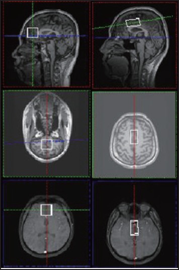
Position map of the orbitofrontal gyrus; Picture B. Position map of the anterior cingulate gyrus
2.3 Analysis of magnetic resonance imaging data
This study focused on the spectral peak at 3.0 ppm. According to the characteristics of GABA which has a low signal to noise ratio and overlapping spectral peak, software LC model 6.3 was used to analyze the absolute concentration and relative error of metabolic products of GABA, and data in which the estimation error was less than 20% was chosen for further statistical analysis.
2.4 Statistical Analysis
Statistical analysis was processed by SPSS software version 19.0. Participants’ age, years of education, Y-BOCS scores, HAMA scores and HAMD scores were analyzed using paired t-tests; gender was analyzed using chi-square tests; Pearson correlation analysis was used to analyze the relevance between abnormal substance concentration and clinical symptoms of OCD (as assessed by Y-BOCS, HAMD and HAMA). Statistical significance level was set at p<0.05.
3. Results
3.1 Analysis of substance concentration between groups
Compared with the control group, this study found that GABA/W and NAA/W were significantly decreased in the OFC of OCD patients (p=0.031, t=-2.193), no abnormal changes were found in Glx/W, and the GABA/W in ACC had a decreasing trend (see tables 1, 2).
Table 1.
Comparison of Substances Concentration in Orbitofrontal Cortex between OCD group and HC group
| Substance | OCD group | HC group | t | p |
|---|---|---|---|---|
| GABA/Wa | 0.0246(0.0075) | 0.0280(0.0072) | -2.193 | 0.031* |
| NAA/Wb | 0.0083(0.0024) | 0.0097(0.0017) | -3.223 | 0.002* |
| Glx/Wc | 0.0147(0.0029) | 0.0154(0.0031) | -1.160 | 0.249 |
*p<0.05 is statistically significant.
aGABA/W, concentration of gamma-aminobutyric acid
bNAA/W, concentration of N-acetyl-aspartate
cGlx/W, concentration of glutamate/glutamine complex
Table 2.
Comparison of Substances Concentration in Anterior Cingulate Cortex between OCD group and HC group
| Substance | OCD group | HC group | t | p |
|---|---|---|---|---|
| GABA/Wa | 0.0277(0.0062) | 0.0312(0.0083) | -1.921 | 0.059 |
| NAA/Wb | 0.0120(0.0036) | 0.0131(0.0036) | -1.151 | 0.255 |
| Glx/Wc | 0.0158(0.0027) | 0.0163(0.0027) | -0.680 | 0.499 |
*p<0.05 is statistically significant.
aGABA/W, concentration of gamma-aminobutyric acid
bNAA/W, concentration of N-acetyl-aspartate
cGlx/W, concentration of glutamate/glutamine complex
3.2 Correlational analysis between concentration of substances and clinical features
There was a significant negative correlation found between the concentration of GABA/W and scores on Y-BOCS (Y-BOCS-O, p=0.037, r=-0.221; Y-BOCS-C, p=0.007, r=-0.283; and Y-BOCS-T p=0.014, r=-0.259). The concentration of NAA/W was negatively correlated with the scores on Y-BOCS-O (p<0.001, r=-0.33), Y-BOCS-C (p<0.001, r=-0.38) and Y-BOCS-T (p<0.001. r=-0.36), as well as with the age of onset (p=0.01, r=-0.26), HAMD (p<0.001, r=-0.32) and HAMA scores (p<0.001, r=-0.35). (Table 3)
Table 3.
Comparative analysis between substances concentration and clinical data of participants with OCD.
| Substances | Age | Education years | Onset years | Entire OCD course | YBOCSc-O | YBOCS-C | YBOCS-T | HAMAd | HAMDe |
|---|---|---|---|---|---|---|---|---|---|
| GABA | -0.12 | -0.09 | -0.20 | -0.14 | -0.22 | -0.28 | -0.38 | -0.18 | -0.18 |
| /W r(p) | (0.24) | (0.36) | (0.05) | (0.18) | (0.03)* | (0.01)* | (0.00)** | (0.09) | (0.08) |
| NAA | -0.03 | -0.14 | -0.26 | -0.19 | -0.33 | -0.38 | -0.36 | -0.32 | -0.35 |
| /Wr(p) | (0.78) | (0.17) | (0.01)* | (0.07) | (0.00)** | (0.00)** | (0.00)** | (0.00)** | (0.00)** |
*p<0.05
**p<0.001 are statistically significant.
aGABA/W, concentration of gamma-aminobutyric acid
bNAA/W, concentration of N-acetyl-aspartate
cYale-Brown Obsessive Compulsive Scale
d Hamilton Anxiety scale
e Hamilton Depression scale
4. Discussion
4.1 Main findings
As we know, this study is the first to find that the concentration of GABA/W in the OFC in patients with OCD has a significant decrease. As well, our study also found that the Glx/W level was normal, and the GABA/W in the ACC area tended to decrease. These results indicate that dysfunction of GABAergic system is related with psychopathology in OCD, which is consistent with findings by Simpson and colleagues[15], which suggested that the concentration of GABA/W was decreased in the MPFC, but the change of the glutamate concentration level was not significant. GABA is mainly generated by glutamate via glutamic acid decarboxylase. Previous studies reported that glutamate is significantly increased in the cerebral spinal fluid of OCD patients.[19] Another study reported a change in glutamate concentration and abnormality in genes polymorphism, including the gene of glutamate-relative N- methyl-D-aspartic acid receptor (NMDA), NR2B, Grik2, and SLA1C1.Meanwhile the glutamate regulator riluzole was found to have a function of decreasing Y-BOCS scores for patients with OCD when applied as a synergistic agent.[20] As an excitatory neurotransmitter[21], the disturbance of glutamate also results in the abnormality of inhibitory neurotransmitter. The main symptom of OCD is the intrusive thoughts, which the individual is unable to control. The neurophysiological mechanism of the symptom as previously investigated includes disturbance of GABA and dysregulation of the NMDA receptor. Also, in previous studies, when researchers measured cortical inhibitory and facilitatory neurotransmitters via inhibition of short time interval (SICI), cortical cortex quiescent (CSP) and cortical facilitation (ICF), they found that patients with OCD have shorter CSP, increasing ICF and no significant SICI when compared with healthy controls. All these results suggest that injuries exist on signal pathways mediated by GABA-β receptor and NMDA receptor in patients with OCD, which is probably related to the etiological mechanism of OCD symptoms.[22]
The OFC is a classical brain area of CSTC. Previous studies reported that the CSTC has metabolic disturbance in OCD patients.[23] Our results, suggesting that metabolic dysfunctions exist in the OFC, are consistent with previous studies on GABA in OCD patients. Therefore, the results suggest that OFC is relative to psychopathology in OCD, lending crucial evidence supporting the conclusion of dysfunction in CSTC. Researchers have also reported that GABA decreases in the MPFC, which is related to clinical symptoms in OCD.[15]Zurowski and colleagues[23] attempt to predict the efficacy of cognitive-behavior treatment (CBT) in OCD treatment via exploration of neuro-metabolism in the OFC, the results suggested that the effect of CBT is significantly correlated with the (myo-) inositol (MI) in the OFC area, but the MI levels at baseline did not significantly differ between OCD and control groups. Another research compared the metabolism of head of caudate (HOC) nucleus and white matter of the OFC in OCD patients relative to healthy controls, and found that Glx/Cr and NAA/Cr levels were significantly increased in the OFC white matter of their right hemisphere and the MI/Cr level was decreased on caudate head of both sides.[24]Why are these results inconsistent to our findings? The reasons likely include the difference of equipment (study used 1.5T MRI), sample size, voxels and parameters. Whiteside and colleagues [25] compared patients with OCD and healthy controls, and found that NAA and Cr were significantly decreased in the Orbito Frontal White Matter (OFWM), and there was no significant difference of NAA and Glx in the caudate head at baseline. Moreover in their research, after behavior therapy, the NAA in the left HOC significantly increased. Another study by this group [26] found a significantly decreased NAA in the HOC in teenage OCD patients; these findings are consistent our results. NAA is thought to be related with the integrity and activity of neurons, therefore the dysfunction of NAA indicates abnormality of neuron integrity in the brain of individuals with OCD. This has a great potential to be regarded as a specific endophenotype for OCD.
ACC is also a typical brain area of the CSTC circuit. Previous studies reported that dysfunction of metabolism also exists in the ACC. [27]Tukel and colleagues[13] also found significant NAA/Cr loss in the ACC, and a trend of decreasing in the caudate nucleus and putamen. Also, after 12 weeks treatment of sertraline, the NAA/Cr of OCD group did not significantly differ from controls. Ortiz and colleagues [27] compared individuals with OCD and controls, and reported no significant difference in the glutamate complex, a result that is also consistent with our findings. Although all of these findings suggest that metabolic dysfunction exists in the ACC, there are still inconsistencies amongst results, which may require further studies with larger sample sizes to provide definitive evidence.
4.2 Limitations
There are several limitations in our study. First, the sample size is not sufficient to achieve acceptable reliability to reach a strong conclusion. Second, the characteristics of GABA, which have low signal to noise ratio and overlapping spectral peak, made the concentration itself difficult to measure precisely, a more rigorous method of analysis is required. Finally, OCD is a disease with heterogeneous features that may influence the results due to difference of subtypes. Therefore, more precise results require distinctly calculating for each OCD subtype, for which homogeneous OCD cases should been chosen as far as possible. In further research, we will expand the sample size and enroll OCD patients with only one prominent symptom (i.e. compulsions or obsessions) and analyze data according to subtype grouping.
4.3 Implication
Our research once again shows that metabolic dysfunctions exists on the CSTC circuit, and for the first time explored the GABA abnormality in the OFC within OCD patients, which is negatively correlated with clinical symptoms. These results indicate that GABA in OCD is related to the psychopathology of OCD, and further illuminate the etiology of OCD.
Figure 1.
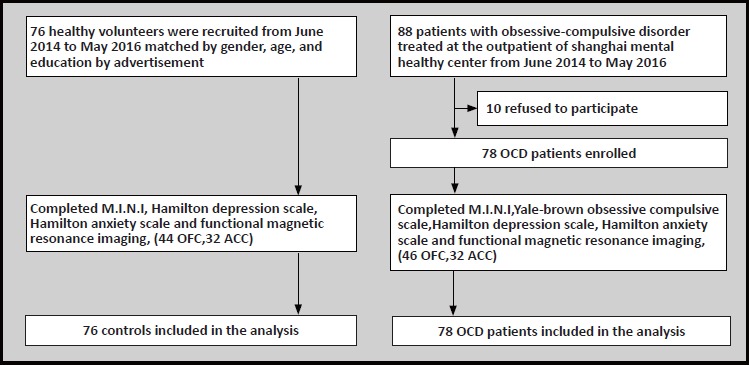
Enrollment of obsessive-compulsive disorder and healthy controls
Biography

Zongfeng Zhang graduated from Jining Medical University with a bachelor’s degree of Medicine in 2013, and she graduated from Shanghai Jiao Tong University Medical School with a master degree in 2016. She has been working as a graduate trainee at Shanghai Mental Health Center since 2014. Her current research interest is brain imaging of obsessive compulsive disorder.
Footnotes
Funding
This study was supported by the National Key Clinical Subject project-Shanghai Mental Health Center (2011-873, Health Administration, National Health and Family Planning Commission), by the National Natural Science Foundation of China (No. 8130115), by the Project Action for Scientific and Technological Innovation, Medical and Agricultural Science Supporting Subproject, Shanghai Science and Technology Committee, (No.15411950203) by the Key Disciplines, Shanghai Mental Health Center (No.2013-YJTSZK-02) and by the research fund of Shanghai Mental Health Center (NO.yj-2013-01).
Conflict of interest
The authors declare no conflict of interest.
Informed consent
Every individual provided written informed consent to participate in this study.
Ethics approval
This study was approved by the ethics committee of the Shanghai Mental Health Center.
Authors’ contributions
Dr. Zongfeng ZHANG was the principal investigator in charge of the overall design and analysis of the study, and also participated the evaluation, data collection and analysis. Prof. Qing FAN, Prof. Zhen WANG and Prof. Yanle BAI participated in the enrolling of participants, implementation, and analysis of the study. Prof. Haiyin ZHANG and Prof. Zeping XIAO helped in the composing of this article and manuscript-reviewing.
References
- 1.Yucel M, Wood SJ, Wellard RM, Harrison BJ, Fornito A, Pujol J, et al. Anterior cingulate glutamate-glutamine levels predict symptom severity in women with obsessive-compulsive disorder. Aust N Z J Psychiatry. 2008; 42(6): 467-477 [DOI] [PubMed] [Google Scholar]
- 2.Beucke JC, Sepulcre J, Eldaief MC, Sebold M, Kathmann N, Kaufmann C. Default mode network subsystem alterations in obsessive-compulsive disorder. Br J Psychiatry. 2014; 205(5): 376-382. doi: http://dx.doi.org/10.1192/bjp.bp.113.137380 [DOI] [PubMed] [Google Scholar]
- 3.Whiteside SP, Port JD, Abramowitz JS. A meta-analysis of functional neuroimaging in obsessive-compulsive disorder. Psychiatry Res. 2004; 132(1): 69-79. doi: http://dx.doi.org/10.1016/j.pscychresns.2004.07.001 [DOI] [PubMed] [Google Scholar]
- 4.Posner J, Marsh R, Maia TV, Peterson BS, Gruber A, Simpson HB. Reduced functional connectivity within the limbic cortico-striato-thalamo-cortical loop in unmedicated adults with obsessive-compulsive disorder. Hum Brain Mapp. 2014; 35(6): 2852-2860. doi: http://dx.doi.org/10.1002/hbm.22371 [DOI] [PMC free article] [PubMed] [Google Scholar]
- 5.Beucke JC, Sepulcre J, Talukdar T, Linnman C, Zschenderlein K, Endrass T, et al. Abnormally high degree connectivity of the orbitofrontal cortex in obsessive-compulsive disorder. JAMA Psychiatry. 2013; 70(6): 619-629. doi: http://dx.doi.org/10.1001/jamapsychiatry.2013.173 [DOI] [PubMed] [Google Scholar]
- 6.Norman LJ, Carlisi C, Lukito S, Hart H, Mataix-Cols D, Radua J, et al. Structural and functional brain abnormalities in attention-deficit/hyperactivity disorder and obsessive-compulsive disorder: A comparative meta-analysis. JAMA Psychiatry. 2016; 73(8):815-825. doi: http://dx.doi.org/10.1001/jamapsychiatry.2016.0700 [DOI] [PubMed] [Google Scholar]
- 7.Hamilton M. The assessment of anxiety states by rating. The British journal of medical psychology. 1959; 32(1): 50-55. doi: http://dx.doi.org/10.1111/j.2044-8341.1959.tb00467.x [DOI] [PubMed] [Google Scholar]
- 8.Stalnaker TA, Cooch NK, Schoenbaum G. What the orbitofrontal cortex does not do. Nat Neurosci. 2015; 18(5): 620-627. doi: http://dx.doi.org/10.1038/nn.3982 [DOI] [PMC free article] [PubMed] [Google Scholar]
- 9.Kwon JS, Jang JH, Choi JS, Kang DH. Neuroimaging in obsessive-compulsive disorder. Expert Rev Neurother. 2009; 9(2): 255-269. doi: http://dx.doi.org/10.1586/14737175.9.2.255 [DOI] [PubMed] [Google Scholar]
- 10.Palomero-Gallagher N, Mohlberg H, Zilles K, Vogt BA. Cytology and receptor architecture of human anterior cingulate cortex. J Comp Neurol. 2008; 508(6): 906-926. doi: http://dx.doi.org/10.1002/cne.21684 [DOI] [PMC free article] [PubMed] [Google Scholar]
- 11.Bush G, Luu P, Posner MI. Cognitive and emotional influences in anterior cingulate cortex. Trends Cogn Sci. 2000; 4(6): 215-222 [DOI] [PubMed] [Google Scholar]
- 12.Weber AM, Soreni N, Stanley JA, Greco A, Mendlowitz S, Szatmari P, et al. Proton magnetic resonance spectroscopy of prefrontal white matter in psychotropic naive children and adolescents with obsessive-compulsive disorder. Psychiatry Res. 2014; 222(1-2): 67-74. doi: http://dx.doi.org/10.1016/j.pscychresns.2014.02.004 [DOI] [PubMed] [Google Scholar]
- 13.Tukel R, Aydin K, Ertekin E, Ozyildirim SS, Taravari V. Proton magnetic resonance spectroscopy in obsessive-compulsive disorder: evidence for reduced neuronal integrity in the anterior cingulate. Psychiatry Res. 2014; 224(3): 275-280. doi: http://dx.doi.org/10.1016/j.pscychresns.2014.08.012 [DOI] [PubMed] [Google Scholar]
- 14.Russo AJ, Pietsch SC. Decreased Hepatocyte Growth Factor (HGF) and Gamma Aminobutyric Acid (GABA) in Individuals with Obsessive-Compulsive Disorder (OCD). Biomark Insights. 2013; 8: 107-114. doi: http://dx.doi.org/10.4137/BMI.S11931 [DOI] [PMC free article] [PubMed] [Google Scholar]
- 15.Simpson HB, Shungu DC, Jr JB, Mao X, Xu X, Slifstein M, et al. Investigation of cortical glutamate-glutamine and gamma-aminobutyric acid in obsessive-compulsive disorder by proton magnetic resonance spectroscopy. Neuropsychopharmacology. 2012; 37(12): 2684-2692 [DOI] [PMC free article] [PubMed] [Google Scholar]
- 16.Goodman WK, Price LH, Rasmussen SA, Mazure C, Fleischmann RL, Hill CL, et al. The Yale-Brown Obsessive Compulsive Scale. I. Development, use, and reliability. Arch Gen Psychiatry. 1989; 46(11): 1006-1011 [DOI] [PubMed] [Google Scholar]
- 17.Xu Y, Zhan HY. [The reliability and validity of the Chinese version of Yale-Brown obsessive-compulsive scale]. Shanghai Arch Psychiatry. 2006; 18(6): 321-323. Chinese. doi: http://dx.chinadoi.cn/10.3969/j.issn.1002-0829.2006.06.001 [Google Scholar]
- 18.Hamilton M. A rating scale for depression. Journal of Neurology, Neurosurgery, and Psychiatry. 1960; 23: 56-62 [DOI] [PMC free article] [PubMed] [Google Scholar]
- 19.Chakrabarty K, Bhattacharyya S, Christopher R, Khanna S. Glutamatergic dysfunction in OCD. Neuropsychopharmacology : official publication of the American College of Neuropsychopharmacology. 2005; 30(9): 1735-1740. [DOI] [PubMed] [Google Scholar]
- 20.Pittenger C, Bloch MH, Williams K. Glutamate abnormalities in obsessive compulsive disorder: neurobiology, pathophysiology, and treatment. Pharmacol Ther. 2011; 132(3): 314-332. doi: http://dx.doi.org/10.1016/j.pharmthera.2011.09.006 [DOI] [PMC free article] [PubMed] [Google Scholar]
- 21.Wu K, Hanna GL, Rosenberg DR, Arnold PD. The role of glutamate signaling in the pathogenesis and treatment of obsessive-compulsive disorder. Pharmacol Biochem Behav. 2012; 100(4): 726-735. doi: http://dx.doi.org/10.1016/j.pbb.2011.10.007 [DOI] [PMC free article] [PubMed] [Google Scholar]
- 22.Richter MA, de Jesus DR, Hoppenbrouwers S, Daigle M, Deluce J, Ravindran LN, et al. Evidence for cortical inhibitory and excitatory dysfunction in obsessive compulsive disorder. Neuropsychopharmacology. 2012; 37(5): 1144-1151. doi: http://dx.doi.org/10.1038/npp.2011.300 [DOI] [PMC free article] [PubMed] [Google Scholar]
- 23.Zurowski B, Kordon A, Weber-Fahr W, Voderholzer U, Kuelz AK, Freyer T, et al. Relevance of orbitofrontal neurochemistry for the outcome of cognitive-behavioural therapy in patients with obsessive-compulsive disorder. Eur Arch Psychiatry Clin Neurosci. 2012; 262(7): 617-624. doi: http://dx.doi.org/10.1007/s00406-012-0304-0 [DOI] [PubMed] [Google Scholar]
- 24.Whiteside SP, Port JD, Deacon BJ, Abramowitz JS. A magnetic resonance spectroscopy investigation of obsessive-compulsive disorder and anxiety. Psychiatry Res. 2006; 146(2): 137-147. doi: http://dx.doi.org/10.1016/j.pscychresns.2005.12.006 [DOI] [PubMed] [Google Scholar]
- 25.Whiteside SP, Abramowitz JS, Port JD. The effect of behavior therapy on caudate N-acetyl-l-aspartic acid in adults with obsessive-compulsive disorder. Psychiatry Res. 2012; 201(1):10-6. doi: http://dx.doi.org/10.1016/j.pscychresns.2011.04.004 [DOI] [PubMed] [Google Scholar]
- 26.Whiteside SP, Abramowitz JS, Port JD. Decreased caudate N-acetyl-l-aspartic acid in pediatric obsessive-compulsive disorder and the effects of behavior therapy. Psychiatry Res. 2012; 202(1): 53-59. doi: http://dx.doi.org/10.1016/j.pscychresns.2011.11.010 [DOI] [PubMed] [Google Scholar]
- 27.Ortiz AE, Ortiz AG, Falcon C, Morer A, Plana MT, Bargallo N, et al. 1H-MRS of the anterior cingulate cortex in childhood and adolescent obsessive-compulsive disorder: a case-control study. Eur Neuropsychopharmacol. 2015; 25(1): 60-68. doi: http://dx.doi.org/10.1016/j.euroneuro.2014.11.007 [DOI] [PubMed] [Google Scholar]