

Abstract
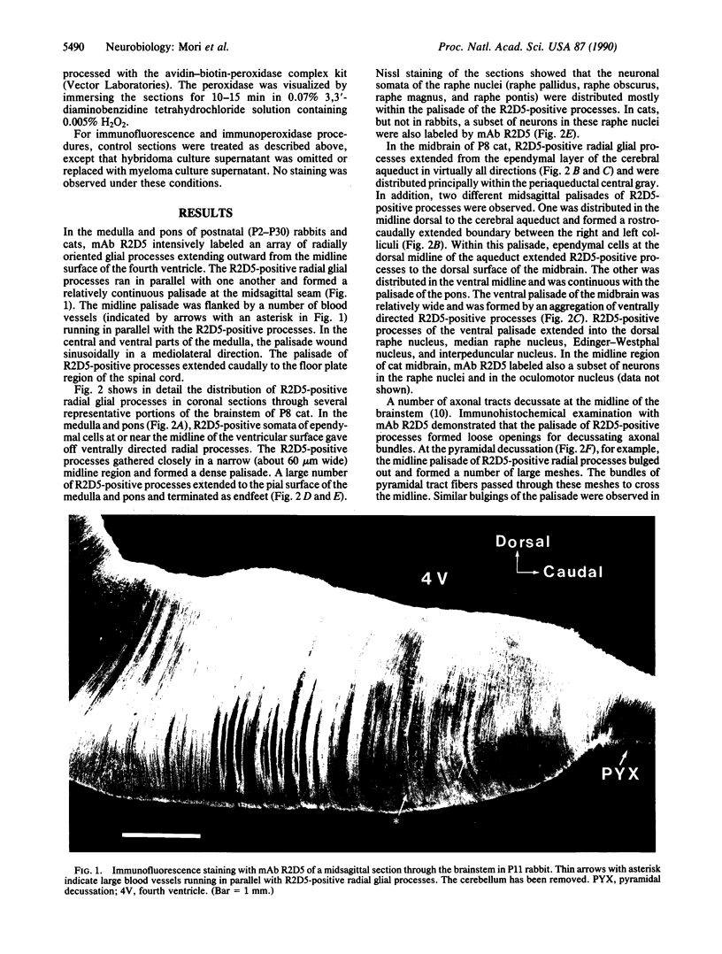
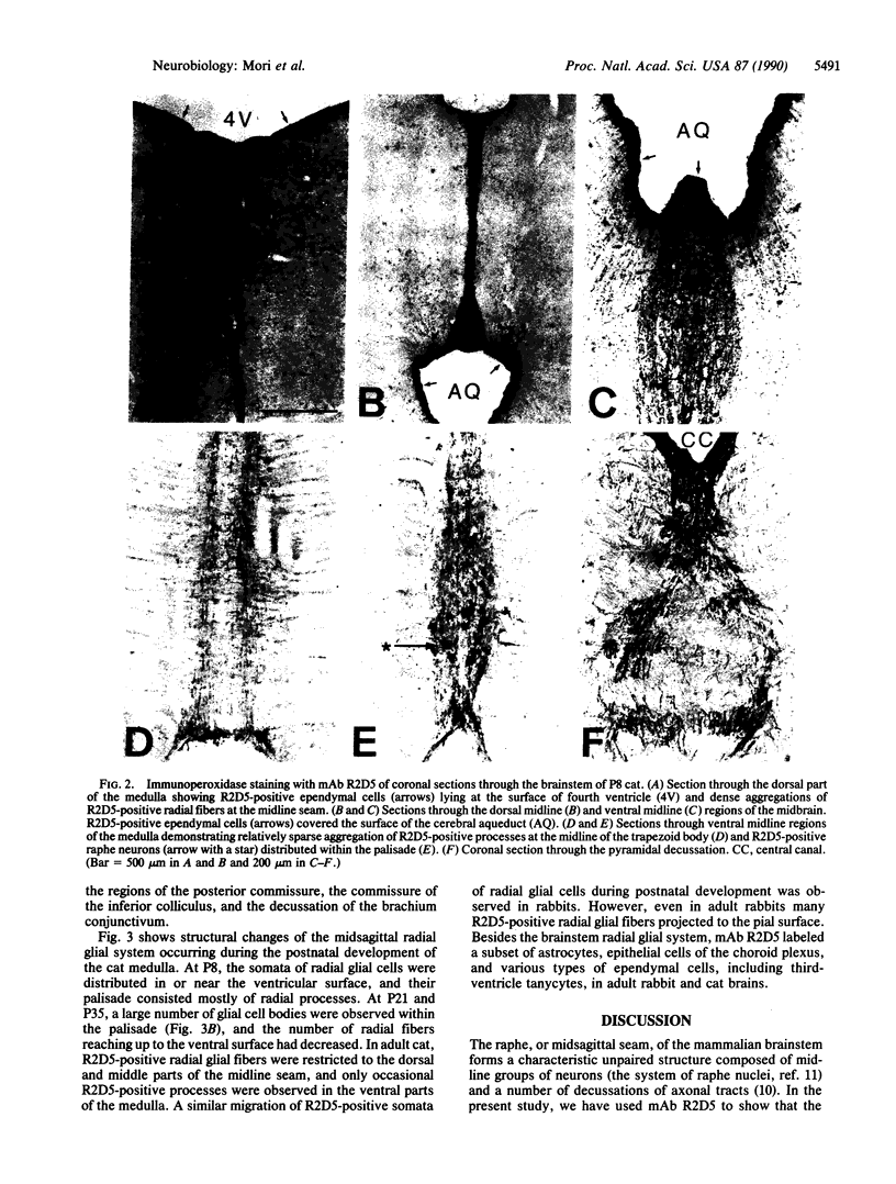
Radial glial cells and their processes play critical roles in organizing the spatial arrangement of the nervous system in the embryonic brain. It has been thought that following completion of their roles in the embryo, most of the radial glial processes disappear before or shortly after birth. Here we use R2D5, a monoclonal antibody to a soluble cytosolic protein, to demonstrate that a specific system of midsagittal radial glial cells persists in postnatal and adult brain. In the brainstem of postnatally developing and adult rabbits and cats, the R2D5-positive processes of radial glial cells were observed to be arranged in a precisely parallel array at the midsagittal seam. These radial glial processes formed a continuous palisade separating the right and left brainstem. In early postnatal animals, R2D5-positive radial processes were found to reach the pial surface and to cover the entire midsagittal seam of the brainstem. These processes embraced dendrites and somata of neurons in almost all of the midsagittal nuclei, including the raphe nuclei, suggesting that the radial glial cells may interact with the midsagittal groups of neurons. In addition, the palisade of R2D5-positive radial processes formed loose openings for crossing axonal bundles at the midline decussations of fiber tracts. In more mature brains, somata of R2D5-positive radial glial cells that had migrated ventrally were observed within the palisades, and in adult cats, most of the R2D5-positive radial processes were found to have retracted from the ventral parts of the midsagittal seam. The spatial arrangement of R2D5-positive processes suggests that they may have persistent functional roles as an interface between ventricular humoral signals and midsagittal groups of neurons in the postnatally developing brainstem and in the adult brainstem. The structure of the midline glial system suggests also that it plays a role in organizing the spatial arrangement of decussating axons during development.
Full text
PDF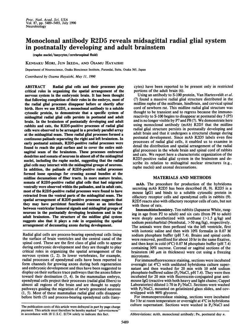
Images in this article
Selected References
These references are in PubMed. This may not be the complete list of references from this article.
- Basbaum A. I., Fields H. L. Endogenous pain control systems: brainstem spinal pathways and endorphin circuitry. Annu Rev Neurosci. 1984;7:309–338. doi: 10.1146/annurev.ne.07.030184.001521. [DOI] [PubMed] [Google Scholar]
- Burnett B. T., Felten D. L. Aqueductal tanycytes in the rabbit brain: A Golgi study. Anat Rec. 1981 Jul;200(3):337–347. doi: 10.1002/ar.1092000314. [DOI] [PubMed] [Google Scholar]
- Chase T. N., Murphy D. L. Serotonin and central nervous system function. Annu Rev Pharmacol. 1973;13:181–197. doi: 10.1146/annurev.pa.13.040173.001145. [DOI] [PubMed] [Google Scholar]
- Cowan W. M., Fawcett J. W., O'Leary D. D., Stanfield B. B. Regressive events in neurogenesis. Science. 1984 Sep 21;225(4668):1258–1265. doi: 10.1126/science.6474175. [DOI] [PubMed] [Google Scholar]
- Cummings J. P., Felten D. L. A raphe dendrite bundle in the rabbit medulla. J Comp Neurol. 1979 Jan 1;183(1):1–23. doi: 10.1002/cne.901830102. [DOI] [PubMed] [Google Scholar]
- Dodd J., Morton S. B., Karagogeos D., Yamamoto M., Jessell T. M. Spatial regulation of axonal glycoprotein expression on subsets of embryonic spinal neurons. Neuron. 1988 Apr;1(2):105–116. doi: 10.1016/0896-6273(88)90194-8. [DOI] [PubMed] [Google Scholar]
- Felten D. L., Harrigan P., Burnett B. T., Cummings J. P. Fourth ventricular tanycytes: a possible relationship with monoaminergic nuclei. Brain Res Bull. 1981 May;6(5):427–436. doi: 10.1016/s0361-9230(81)80013-5. [DOI] [PubMed] [Google Scholar]
- Flament-Durand J., Brion J. P. Tanycytes: morphology and functions: a review. Int Rev Cytol. 1985;96:121–155. doi: 10.1016/s0074-7696(08)60596-3. [DOI] [PubMed] [Google Scholar]
- Fujita S. C., Mori K., Imamura K., Obata K. Subclasses of olfactory receptor cells and their segregated central projections demonstrated by a monoclonal antibody. Brain Res. 1985 Feb 4;326(1):192–196. doi: 10.1016/0006-8993(85)91403-9. [DOI] [PubMed] [Google Scholar]
- Furley A. J., Morton S. B., Manalo D., Karagogeos D., Dodd J., Jessell T. M. The axonal glycoprotein TAG-1 is an immunoglobulin superfamily member with neurite outgrowth-promoting activity. Cell. 1990 Apr 6;61(1):157–170. doi: 10.1016/0092-8674(90)90223-2. [DOI] [PubMed] [Google Scholar]
- Hayaishi O. Sleep-wake regulation by prostaglandins D2 and E2. J Biol Chem. 1988 Oct 15;263(29):14593–14596. [PubMed] [Google Scholar]
- Levitt P., Rakic P. Immunoperoxidase localization of glial fibrillary acidic protein in radial glial cells and astrocytes of the developing rhesus monkey brain. J Comp Neurol. 1980 Oct 1;193(3):815–840. doi: 10.1002/cne.901930316. [DOI] [PubMed] [Google Scholar]
- Mori K., Fujita S. C., Imamura K., Obata K. Immunohistochemical study of subclasses of olfactory nerve fibers and their projections to the olfactory bulb in the rabbit. J Comp Neurol. 1985 Dec 8;242(2):214–229. doi: 10.1002/cne.902420205. [DOI] [PubMed] [Google Scholar]
- Nordlander R. H., Singer M. The role of ependyma in regeneration of the spinal cord in the urodele amphibian tail. J Comp Neurol. 1978 Jul 15;180(2):349–374. doi: 10.1002/cne.901800211. [DOI] [PubMed] [Google Scholar]
- Rakic P. Specification of cerebral cortical areas. Science. 1988 Jul 8;241(4862):170–176. doi: 10.1126/science.3291116. [DOI] [PubMed] [Google Scholar]
- Schoenenberger G. A., Monnier M. Characterization of a delta-electroencephalogram (-sleep)-inducing peptide. Proc Natl Acad Sci U S A. 1977 Mar;74(3):1282–1286. doi: 10.1073/pnas.74.3.1282. [DOI] [PMC free article] [PubMed] [Google Scholar]
- Singer M., Nordlander R. H., Egar M. Axonal guidance during embryogenesis and regeneration in the spinal cord of the newt: the blueprint hypothesis of neuronal pathway patterning. J Comp Neurol. 1979 May 1;185(1):1–21. doi: 10.1002/cne.901850102. [DOI] [PubMed] [Google Scholar]
- Snow D. M., Steindler D. A., Silver J. Molecular and cellular characterization of the glial roof plate of the spinal cord and optic tectum: a possible role for a proteoglycan in the development of an axon barrier. Dev Biol. 1990 Apr;138(2):359–376. doi: 10.1016/0012-1606(90)90203-u. [DOI] [PubMed] [Google Scholar]
- Steindler D. A., Cooper N. G., Faissner A., Schachner M. Boundaries defined by adhesion molecules during development of the cerebral cortex: the J1/tenascin glycoprotein in the mouse somatosensory cortical barrel field. Dev Biol. 1989 Jan;131(1):243–260. doi: 10.1016/s0012-1606(89)80056-9. [DOI] [PubMed] [Google Scholar]
- Tessier-Lavigne M., Placzek M., Lumsden A. G., Dodd J., Jessell T. M. Chemotropic guidance of developing axons in the mammalian central nervous system. Nature. 1988 Dec 22;336(6201):775–778. doi: 10.1038/336775a0. [DOI] [PubMed] [Google Scholar]
- Van Hartesveldt C., Moore B., Hartman B. K. Transient midline raphe glial structure in the developing rat. J Comp Neurol. 1986 Nov 8;253(2):174–184. doi: 10.1002/cne.902530205. [DOI] [PubMed] [Google Scholar]
- Wallace J. A., Lauder J. M. Development of the serotonergic system in the rat embryo: an immunocytochemical study. Brain Res Bull. 1983 Apr;10(4):459–479. doi: 10.1016/0361-9230(83)90144-2. [DOI] [PubMed] [Google Scholar]