Abstract
Epigallocatechin gallate (EGCG) is a main constituent of green tea polyphenols that are widely used as food preservatives and are considered to be safe for consumption. However, the underlying antimicrobial mechanism of EGCG and the bacterial response to EGCG are not clearly understood. In the present study, a genome-wide transcriptional analysis of a typical spoilage bacterium, Pseudomonas fluorescens that responded to EGCG was performed using RNA-seq technology. A total of 26,365,414 and 23,287,092 clean reads were generated from P. fluorescens treated with or without 1 mM EGCG and the clean reads were aligned to the reference genome. Differential expression analysis revealed 291 upregulated genes and 134 downregulated genes and the differentially expressed genes (DEGs) were verified using RT-qPCR. Most of the DGEs involved in iron uptake, antioxidation, DNA repair, efflux system, cell envelope and cell-surface component synthesis were significantly upregulated by EGCG treatment, while most genes associated with energy production were downregulated. These transcriptomic changes are likely to be adaptive responses of P. fluorescens to iron limitation and oxidative stress, as well as DNA and envelope damage caused by EGCG. The expression of specific genes encoding the extra-cytoplasmic function sigma factor (PvdS, RpoE and AlgU) and the two-component sensor histidine kinase (BaeS and RpfG) were markedly changed by EGCG treatment, which may play important roles in regulating the stress responses of P. fluorescens to EGCG. The present data provides important insights into the molecular action of EGCG and the possible cross-resistance mediated by EGCG on P. fluorescens, which may ultimately contribute to the optimal application of green tea polyphenols in food preservation.
Introduction
Contamination by pathogenic and spoilage bacteria is a major issue during food processing. To date, many synthetic and natural food additives have been used to control bacterial contamination and to extend the shelf-life of foods. With the increasing demands for safe and high-quality food, the development and utilization of effective natural preservatives are desirable. Green tea polyphenols (GTPs), key ingredients of green tea, have shown effective antimicrobial and antioxidant activities, and non-toxicity in the food industry [1]. In addition, GTPs are beneficial to health and are proposed to prevent cancer and several other chronic diseases [1–2]. Moreover, GTPs are ubiquity, abundance and low cost. So the application of GTPs in the food industry has attracted more interest, especially in fish or meat products. For examples, dip treatment with 0.2% GTP was effective in inhibiting spoilage bacteria growth and extending the ice storage life of fish samples to 35 days compared to 28 days for the control group [3]. The total viable counts reduced two orders of magnitude in Collichthys fish balls supplemented with 0.25 g/kg GTPs compared with the controls [4]. GTPs were also used combined with other natural preservatives. Application of GTPs (0.3% or 0.15%) in combination with 6-gingerol (0.3% or 0.15%) inhibited oxidation of protein and lipids, and reduced microorganism counts compared to control treatments during storage of shrimp paste [5]. The combination of nisin (0.625 g/L), GTPs (0.313 g/L) and chitosan (3.752 g/L) could be used as preservatives to efficiently inhibit the growth of spoilage microorganisms and pathogens in chilled mutton [6]. Although GTPs were considered to be a good choice of natural food preservatives, there were some challenges of the utilisation of GTPs in foods, such as the astringent and bitter taste [7], unstability during thermal processing and alkaline solutions [8–9], which may limit the application of GTPs in foods.
The most abundant components of GTPs are catechins, of which epigallocatechin gallate (EGCG) is the major one (50–80% of the total catechin content) [10]. GTPs and EGCG have a broad antimicrobial spectrum and inhibit the growth of many foodborne pathogenic and spoilage bacteria [4, 11]. Several studies have indicated that the antibacterial activity of EGCG is due to damage to the bacterial cell membrane, such as peptidoglycan [12], outer membrane proteins [13] and lipid bilayers [14]. Moreover, numerous key enzymes in bacterial cells have been suggested as the targets of EGCG, including FabG and FabI reductases [15], the DNA gyrase B subunit [16], thioredoxin, and thioredoxin reductase [17]. Instead of directly binding to protein targets, EGCG was reported to inhibit bacterial growth by producing H2O2 [18–19]. Most polyphenol compounds are effective metal chelators [20], and metal chelation may contribute to antibacterial activities [21]. In addition, green tea polyphenols and EGCG are able to disrupt biofilm formation and quorum sensing of bacteria [22–23]. Overall, the antibacterial action of EGCG may act in multiple ways and the complete mechanism has yet to be fully elucidated.
Modern food safety measures are designed only once there is a complete understanding of how foodborne microorganisms cope with stress conditions that are encountered in a variety of food products. Microorganisms can become resistant to otherwise lethal stresses due to exposure to sub-lethal stresses, especially when minimal food processing is involved, which creates challenges in developing new food processing techniques [24]. Although green tea polyphenols are widely used as food preservatives and are generally considered to be safe, cross-resistance can be induced by using green tea polyphenols and EGCG. Previously, EGCG-adapted strains of Staphylococcal have adapted increased resistance to antibiotics and heat stress [25]. Recently, short exposure of Pseudomonas aeruginosa to sub-lethal doses of green tea polyphenols or EGCG led to cross-resistance with some environmental stresses, including oxidants, various organic acids, salt, heat, ethanol and crystal violet [26]. Microorganisms often respond to environmental stress conditions by producing specific enzymes or proteins that aid in coping with various stresses [24]. Interestingly, expression of antioxidative genes is likely to be involved in the cross-resistance induced by green tea polyphenols in P. aeruginosa [19]. Proteomic analysis of Escherichia coli that has been exposed to green tea polyphenols indicates that several stress-related proteins were upregulated, such as the chaperone protein HSP60, RNA polymerase sigma factor RpoS, superoxide dismutase SodC and multidrug resistance efflux pump protein EmrK [27]. Additionally, short exposure to sublethal EGCG concentrations results in increased cell wall thickness, and the two-component VraSR system is suggested to be involved in modulating Staphylococcus aureus response to EGCG [25, 28]. Nevertheless, knowledge regarding how microorganisms respond to EGCG is still very limited.
Pseudomonas fluorescens is a common gram-negative spoilage bacterium that is widely found in food matrices, such as ready-prepared fresh vegetables [29], raw fish (especially sushi or sashimi) [30], meat and dairy products [31]. Due to its extreme adaptability, versatility and ability to replicate at refrigeration temperatures, long periods of shelf life can easily lead to increased P. fluorescens concentration in food products [4, 32]. Genome-wide transcriptomic analysis is an effective method in studying adaptive responses to the environment by identifying and linking associated transcriptional perturbations. However, there are currently few transcriptome analysis reports that describe the impact of polyphenols on bacteria [33]. In the present study, we exploited the global transcriptomic analysis of adaptive responses of P. fluorescens to EGCG by RNA-seq in order to further understand the antibacterial mechanisms of EGCG and the EGCG mediated cross-resistance in bacteria, which may ultimately contribute to the optimal application of green tea polyphenols in food preservation.
Materials and methods
Bacterial strains and media
The P. fluorescens strain ZJL511 was isolated previously from spoiled refrigerated turbot (Scophthalmus maximus). The bacterium was cultured in nutrient broth (NB; 1% peptone, 0.3% beef extract, 0.5% NaCl, pH 7.5) or plate count agar (PCA; 0.5% peptone, 0.25% yeast extract, 0.1% glucose, 1.55% agar, pH 7.0) at 30°C.
Antibacterial activity of EGCG to P. fluorescens
P. fluorescens was cultured in NB medium at 30°C for 16 h and the cell culture then was diluted 1000-fold with NB medium (approximately 106 CFU (colony forming units) /mL). The diluted P. fluorescens cell suspensions were cultivated in the presence (0.25, 0.5, 1, 2, and 4 mM) and absence of EGCG (Sigma, USA) at 30°C while shaking at 220 rpm. After 4 h of incubation, cells were diluted appropriately and poured in plate count agar. The cultures without EGCG treatment were used as controls. Viable cell numbers were determined by counting the colonies after 24–48 h of incubation at 30°C.
EGCG exposure and RNA extraction
One millilitre of an overnight P. fluorescens culture was inoculated into 100 mL fresh NB medium (pH 7.5) and was incubated at 30°C with shaking at 220 rpm. At an OD600 of 0.4 (logarithmic phase), the culture was immediately treated with 1 mM EGCG. Culture untreated with EGCG was used as the control. After 1 h of exposure, the cells were harvested by centrifugation at 7,000 g for 10 min and were frozen in liquid nitrogen prior to RNA isolation. Total RNA was isolated from the frozen cells using TRIzol® Plus RNA Purification Kit (Invitrogen, USA) according to the manufacturer’s instructions. Three independent biological replicates were performed for each treatment and the extracted RNA samples were mixed equally for each treatment. Total RNA was treated with RNase-Free DNase Set (Qiagen, Germany) to remove contaminating DNA. The quantity and quality of total RNA were assessed using a NanoDrop ND-2000 Spectrophotometer (Thermo, USA) and Agilent 2100 Bioanalyzer (Agilent, USA).
Library preparation and sequencing
Strand-specific RNA sequencing libraries were prepared by following a modified deoxy-UTP (dUTP) strand-marking protocol described previously [34]. Briefly, total RNA from each sample was depleted of rRNA using a Ribo-Zero rRNA removal kit for Gram-negative bacteria (Epicentre, USA) per the manufacturer’s protocol, followed by RNA fragmentation. First-strand cDNA was synthesized using a hexamer primer (Takara, Japan) and Superscript II reverse transcriptase (Invitrogen, USA). Double-stranded cDNA was synthesized with dUTP incorporation into the second strand. The double-stranded cDNA fragments were further processed to construct a sequencing library. Prior to final amplification, the dUTP-marked strand was selectively degraded with USER enzyme (NEB, USA) and the remaining strand was amplified to generate a cDNA library suitable for sequencing. Following validation with an ABI StepOne Plus Real-Time PCR System (ABI, USA), the cDNA library was sequenced on a flow cell using high-throughput 125-bp pair-end mode on an Illumina HiSeq 2500V4 platform (Illumina, USA).
Quality control and alignment
Clean data (clean reads) were obtained by removing adapter-containing, poly-N, and low-quality reads from the raw data (raw reads) using in-house Perl scripts. Meanwhile, the Q20, Q30 and GC content were calculated. All downstream analyses were based on clean data with high-quality. The genome sequence of P. fluorescens UK4 was used as the reference genome [35]. For each sample, sequence alignment with the reference genome sequences was performed using Tophat 1.3.1 [36]. The unique mapped reads were used in subsequent analyses.
Analysis of differentially expressed genes (DEGs)
For gene expression analysis, the python script rpkmforgenes.py (last modified 13 November, 2014) was used to estimate the expression level (relative abundance) of specific transcripts expressed using the RPKM (Reads Per Kilobase per Million reads mapped) method [37]. The RPKM value was directly used for comparing differences in gene expression among samples, as this method eliminates influences due to different gene lengths and sequencing discrepancies in calculating gene expression. The software package edgeR 3.12.0 was used to calculate the fold-change of transcripts and to screen all differentially expressed genes (DEGs). The criteria of significant difference expression were |log2 fold change| ≥ 1 and False Discovery Rate (FDR) ≤ 0.05 [38–39].
Cluster of orthologous group (COG) analysis for DEGs
DEGs functional description was performed using a P. fluorescens UK4 genome annotation and GO database. COG functional categories for the DEGs were obtained from the NCBI COG database (http://www.ncbi.nlm.nih.gov/COG/).
DEG verification using RT-qPCR
In order to confirm the RNA-seq results, 17 upregulated and 5 downregulated genes from the RNA-seq analysis were selected and RT-qPCR was performed to confirm the gene expression changes of these 22 genes based on functional categories. P. fluorescens was treated with 1 mM EGCG and three biological replicates were collected. In addition, the expression levels of 4 representative upregulated genes were detected by RT-qPCR after P. fluorescens was treated with 0.5, 1 and 2 mM EGCG. Total RNA was isolated from each sample and reversely transcribed with a hexamer primer and SuperScript™ III First-Strand Synthesis SuperMix for RT-qPCR (Invitrogen, USA) according to the manufacturer’s instructions. The resulting cDNA was then used as the template for qPCR. Gene-specific primers were designed using the Primer Premier 6.0 and Beacon designer 7.8 software, while the 16S rRNA gene was used as the internal control (S3 Table). The reactions were performed using a CFX384 Touch™ Real-Time PCR Detection System (Bio-Rad, USA) with Power SYBR® Green PCR Master Mix (Applied Biosystems, USA). The two-step qPCR program began at 95°C for 1 min, followed by 40 cycles of 95°C for 15 s and 63°C for 25 s. Fluorescent signals were collected at each polymerization step. Melting curve analysis of amplification products was performed at the end of each PCR program to confirm that a single PCR product was detected. Samples were assayed in triplicate and PCR reactions without the template were performed as negative controls. The comparative Ct method for relative quantification (ΔΔCt method) was used to analyze the data [40].
Results and discussion
Global overview of the RNA-seq data
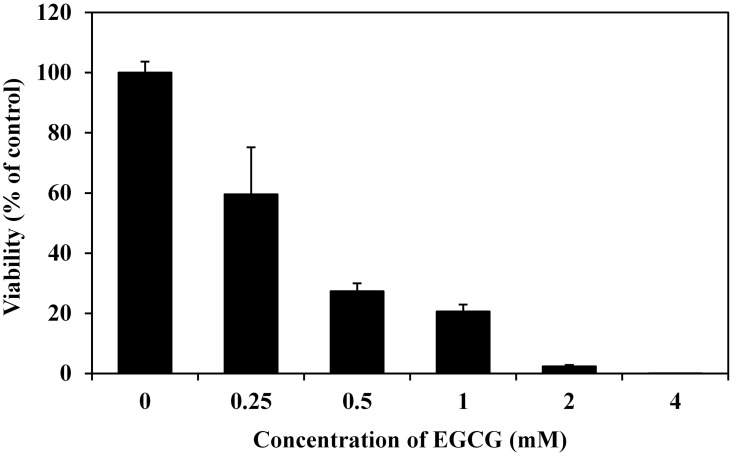
Prior to RNA-seq, the antibacterial action of EGCG against P. fluorescens was tested and EGCG had significant antibacterial activity that was dose dependent (Fig 1). Treatments of P. fluorescens with 0.25, 0.5, 1, 2 and 4 mM EGCG for 4 h significantly decreased survival rates (59.60%, 27.34%, 20.65%, 2.35% and 0.01% vs. control, respectively), demonstrating comparable antibacterial activity against P. aeruginosa as previously reported [19]. In order to avoid complete suppression of cellular metabolism, a moderate inhibiting concentration (1 mM) was selected for treatment in investigating the transcriptomic response of P. fluorescens to EGCG.
Fig 1. The antimicrobial action of EGCG against P. fluorescens.
Different EGCG concentrations were supplemented to P. fluorescens cell suspensions (~106 CFU/mL in NB medium, pH 7.5). After incubation at 30°C for 4 h with shaking at 220 rpm, cell viability was determined. Cell viability is expressed as a percentage of the control. Data are mean ± standard deviation from three independent experiments.
RNA-seq is an attractive method for monitoring global transcriptomic changes and overcomes many of the defects related to traditional DNA microarrays [41]. RNA-seq using the Illumina paired-end sequencing technology was used to examine the effect of EGCG on the P. fluorescens transcriptome. After filtering through the raw reads, there were 26,365,414 clean reads for the control sample and 23,287,092 for the EGCG-treated sample, giving rise to total clean bases of 3.30 G and 2.92 G, respectively (S1 Table). The clean sequencing data were deposited to the Short Reads Archive of NCBI with accession numbers SAMN05559903, SAMN05559904. Of the clean reads, the Q20 and Q30 values were > 92.68% and 85.68%, respectively, indicating that the data were of high quality. Among the P. fluorescens strains with complete genome sequence in GenBank, UK4 strain had the highest 16S gene homology with the test strain ZJL511 (data not shown), and thus the P. fluorescens UK4 strain sequence was used as the reference genome [35]. The clean reads of P. fluorescens transcriptome were compared to the reference genome sequence and the total mapped rates of the reads to the reference genome were 57.00% in the control sample and 58.00% in the EGCG group. There were 61.41% of the total genes in the reference genome detected (read number ≥ 1) by RNA-seq in the control, while there were 62.57% detected in the treated sample (S2 Table). The mapping rates were not high, which was likely due to high variability among the P. fluorescens genomes isolated [42]. For example, comparisons of three P. fluorescens genomes (SBW25, Pf0-1, Pf-5) revealed considerable divergence, showing that only 61% of genes were shared [43].
Analyses of DEGs
A total of 425 DEGs were identified using the gene expression levels calculated by RPKM and the statistical criteria (|log2 fold change| ≥ 1, FDR ≤ 0.05). Among these genes, 291 genes were markedly upregulated and 134 genes were markedly downregulated in P. fluorescens following EGCG treatment (S1 Fig). The gene locus tags, log2 FC (fold change), FDR, RPKM, COG categories, gene product description and other data of the DGEs are provided in S4 and S5 Tables.
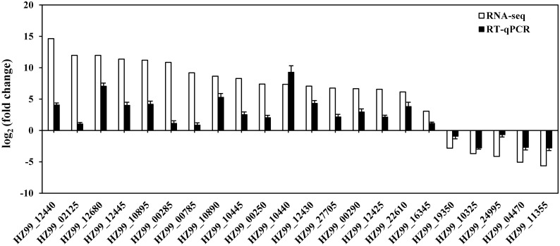
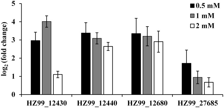
In order to validate the data generated from the RNA-seq experiment, 17 upregulated and 5 downregulated genes were selected from DGEs based on functional categories to verify their expression by RT-qPCR. All of the genes examined were consistent with the RNA-seq results (Fig 2), although the observed fold changes differed between qRT-PCR and RNA-seq data, which may reflect differences in the sensitivity and specificity between RT-qPCR and high-throughput sequencing technology. In addition, the expression levels of 4 representative upregulated genes were detected after 0.5, 1 and 2 mM EGCG treatment by RT-qPCR (Fig 3). The expressions of the 4 genes could be upregulated by each of the EGCG concentrations. These results suggested that the RNA-seq results were generally reliable.
Fig 2. Validation of RNA-seq data using RT-qPCR.
White bars represent RNA-seq data, while the black bars represent mean values of log2 (fold change) observed for EGCG-treated samples vs controls. Three biological replicates were performed for qRT-PCR. Error bars represent standard deviation.
Fig 3. Expression analysis of DGEs after treatments with different concentrations of EGCG in P. fluorescens.
White, gray and white bars represent 0.5, 1 and 2 mM EGCG treatment, respectively. The Y-axis shows values of log2 (fold change) observed for EGCG-treated samples vs controls. Three biological replicates were performed for qRT-PCR. Error bars represent standard deviation.
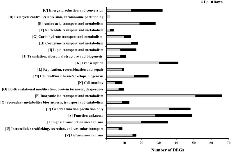
The 425 DGEs were classified using the known function of the gene products or through homology with protein functions determined from the COG database (Fig 4). COG functional analysis assigned DEGs to 20 predicted pathways, most of which belonged to the following groups: inorganic ion transport and metabolism (COG P; 15.53%), function unknown (COG S; 11.53%), general function prediction only (COG R; 11.29%), transcription (COG K; 9.65%), signal transduction mechanisms (COG T; 8.24%), energy production and conversion (COG C; 7.53%), amino acid transport and metabolism (COG E; 6.59%), cell wall/membrane/envelope biogenesis (COG M; 5.65%), coenzyme transport and metabolism (COG H; 4.24%), lipid transport and metabolism (COG I; 4.00%), and defense mechanisms (COG V; 4.00%). Moreover, in most of the COGs, a significantly larger number of the genes were upregulated rather than downegulated in the EGCG-treated sample versus the control (Fig 4). Only in COG C and COG I were more genes downregulated, and this was especially notable in COG I.
Fig 4. Classification of the upregulated and downregulated genes according to the COGs.
DEGs related to iron uptake
Among the DGEs classified in COG P, a majority (38/66) of the genes were related to iron uptake, including 35 upregulated genes and 3 downregulated genes (Table 1). The 35 upregulated genes contained 9 genes encoding the TonB-dependent siderophore receptor, 6 genes encoding the TonB-dependent heme receptor, 5 genes encoding the TonB-ExbBD complex, 6 genes encoding proteins involved in biosynthesis of pyoverdine (the high-affinity siderophore of P. fluorescens) and some other genes related to heme uptake—such as heme receptor genes and the heme oxygenase gene hemO (HZ99_10895). HemO catalyzes the rate-limiting step in degrading heme to bilirubin and is essential for recycling iron from heme [44]. In addition, three genes that code for bacterioferritin were significantly downregulated. Bacterioferritins convert excess cellular iron (II) to iron (III), which is then stored as ferric-oxy-hydroxide-phosphate complexes [45]. According to the RNA-seq results, most of the genes in iron transport and metabolism pathways were significantly changed after EGCG exposure. The genes related to iron uptake can be induced by a series of environmental stresses, such as iron limitation [46], H2O2 stress [47], cold shock [48] or arsenic exposure [49]. Polyphenols are easily deprotonated at or below physiological pH (e.g., 5.0–8.0) in the presence of iron (III) and form complexes with stability constants comparable to iron-siderophore complexes [20, 50]. As P. fluorescens produces high-affinity siderophore pyoverdines in order to acquire iron, EGCG may play a role in inhibiting the pyoverdine-dependent transport of iron by competitive binding and may result in iron starvation. Moreover, some reports have indicated that green tea polyphenols or EGCG result in oxidative stresses in bacteria that depend on H2O2 release [19, 51]. For human tumor cell lines, the induction of apoptosis by EGCG was either partially or completely mediated by H2O2 [52]. In Escherichia coli H2O2 may antagonize the function of Fur protein, a major transcriptional repressor that controls ferric uptake, by oxidizing the Fur-Fe2+ complex and inactivating its repressor function [53], which then results in up regulation of these iron acquisition-related genes.
Table 1. DEGs related to iron uptake.
| Gene | Log2FC | FDR | Gene product description |
|---|---|---|---|
| Siderophore receptor | |||
| HZ99_12680 | 11.97 | 2.80E-13 | TonB dependent siderophore (ferric enterobactin) receptor FepA |
| HZ99_15300 | 8.81 | 1.33E-05 | TonB-dependent siderophore (ferrichrome) receptor |
| HZ99_00225 | 8.15 | 2.29E-04 | TonB-dependent siderophore (ferric coprogen) receptor FhuE |
| HZ99_26610 | 7.87 | 7.03E-04 | TonB-dependent siderophore (ferric coprogen) receptor FhuE |
| HZ99_18870 | 6.70 | 2.58E-02 | TonB-dependent catecholate siderophore receptor CirA |
| HZ99_22610 | 6.14 | 2.94E-09 | TonB-dependent ferric enterochelin receptor FepA |
| HZ99_18545 | 4.40 | 4.06E-05 | TonB-dependent ferrichrome receptor |
| HZ99_00065 | 3.71 | 1.74E-04 | ferric-pseudobactin BN7/BN8 receptor |
| HZ99_00780 | 2.64 | 2.08E-02 | TonB-dependent ferric coprogen receptor FhuE |
| Heme receptor | |||
| HZ99_10890 | 8.64 | 2.92E-16 | TonB-dependent heme/hemoglobin receptor |
| HZ99_24755 | 4.25 | 1.76E-05 | TonB-dependent heme/hemoglobin receptor |
| HZ99_17300 | 2.96 | 7.48E-03 | TonB-dependent heme/hemoglobin receptor |
| HZ99_13170 | 2.91 | 1.22E-03 | TonB-dependent heme/hemoglobin receptor |
| HZ99_11600 | 2.72 | 2.40E-02 | TonB-dependent heme/hemoglobin receptor |
| HZ99_02135 | 2.67 | 2.22E-02 | TonB-dependent heme/hemoglobin receptor |
| TonB-ExbBD complex | |||
| HZ99_01780 | 5.05 | 2.27E-07 | Energy transducer TonB |
| HZ99_17025 | 4.74 | 8.96E-08 | biopolymer transporter ExbD |
| HZ99_17030 | 4.65 | 1.47E-07 | Energy transducer TonB |
| HZ99_17020 | 4.32 | 8.42E-07 | TonB-system energizer ExbB |
| HZ99_18895 | 2.54 | 1.82E-02 | Biopolymer transporter ExbB |
| Pyoverdine biosynthesis | |||
| HZ99_27755 | 9.55 | 2.66E-07 | diaminobutyrate—2-oxoglutarate aminotransferase PvdH |
| HZ99_00255 | 9.05 | 3.50E-06 | Chromophore maturation protein PvdO |
| HZ99_02660 | 8.69 | 2.23E-05 | Acyl-homoserine lactone acylase PvdQ |
| HZ99_00250 | 7.39 | 4.21E-13 | Pyoverdine ABC transporter, permease/ATP-binding protein PvdE |
| HZ99_27705 | 6.77 | 4.14E-13 | Non-ribosomal peptide synthetase PvdL |
| HZ99_00290 | 6.66 | 4.01E-11 | L-ornithine 5-monooxygenase PvdA |
| HZ99_00220 | 6.10 | 2.40E-11 | Pyoverdine sidechain peptide synthetase IV |
| Other iron uptake related genes | |||
| HZ99_10895 | 11.21 | 2.08E-11 | Heme oxygenase HemO |
| HZ99_27680 | 10.23 | 6.28E-09 | Acetyltransferase, siderophore biosynthesis protein |
| HZ99_22370 | 8.15 | 2.29E-04 | Substrate-binding protein function in the ABC transport of ferric siderophores |
| HZ99_02655 | 7.28 | 5.74E-03 | Fe2+ Zn2+ uptake regulation protein |
| HZ99_10340 | 4.14 | 2.23E-06 | Imelysin-like iron-regulated protein A-like |
| HZ99_16430 | 2.14 | 2.08E-02 | Iron ABC transporter substrate-binding protein |
| HZ99_23135 | 2.12 | 2.56E-02 | TonB-dependent ferric citrate transporter FecA |
| Iron storage | |||
| HZ99_11350 | 3.66 | 4.06E-05 | (2Fe-2S)-binding protein |
| HZ99_11355 | -5.86 | 7.06E-11 | Bacterioferritin |
| HZ99_20890 | -3.41 | 1.96E-04 | Bacterioferritin |
| HZ99_14300 | -2.96 | 8.43E-04 | Bacterioferritin |
DEGs related to antioxidation and DNA repair
Transcriptional changes were also observed for genes that encode oxidative stress response proteins, such as superoxide dismutases, which counter reactive oxygen species by converting O2- to H2O2 (Table 2). The sodA gene (HZ99_12430) that encodes manganese-based superoxide dismutase was markedly upregulated after EGCG exposure. sodA is part of the HZ99_12445-fumC- HZ99_12435-sodA operon, which is inducible by treatment with H2O2 or iron starvation, as well as Fur repression [46–47]. Based on the RNA-seq data, the other two genes in the operon were also significantly upregulated (S4 Table). Conversely, the sodB gene (HZ99_10325), which codes for a superoxide dismutase that utilizes iron as cofactor, was significantly downregulated after EGCG treatment (Table 2), presumably due to reduced iron availability in the cells after EGCG treatment. Other relevant for oxidative stress adaptation genes were also upregulated, including genes coding for alkylhydroperoxidase, peroxiredoxin, and AhpF (alkylhydroperoxide reductase subunit F) (Table 2). The ahpF gene has been induced by treatment with H2O2 [47, 54]. These genes are important to bacterial defense against toxic peroxides and were significantly upregulated in P. fluorescens after EGCG treatment, suggesting a cell response mediated by EGCG-induced oxidative stress.
Table 2. DEGs related to antioxidation and DNA repair.
| Gene | Log2FC | FDR | Gene product description |
|---|---|---|---|
| Antioxidation | |||
| HZ99_12430 | 7.07 | 1.49E-12 | superoxide dismutase SodA |
| HZ99_06790 | 6.44 | 4.74E-02 | alkylhydroperoxidase |
| HZ99_27730 | 2.43 | 1.51E-02 | peroxiredoxin |
| HZ99_02330 | 2.21 | 2.80E-02 | alkylhydroperoxide reductase subunit F, AhpF |
| HZ99_10325 | -3.91 | 5.61E-06 | superoxide dismutase SodB |
| DNA repair | |||
| HZ99_25720 | 7.94 | 5.77E-04 | ATP-dependent DNA helicase RecQ |
| HZ99_25700 | 7.28 | 5.74E-03 | recombinase RmuC |
| HZ99_11700 | 7.20 | 7.42E-03 | DNA repair protein RecO |
| HZ99_18100 | 6.58 | 3.49E-02 | DNA repair photolyase SplB |
| HZ99_13290 | 4.21 | 4.29E-05 | endonuclease |
| HZ99_01050 | 3.29 | 1.36E-02 | exonuclease subunit SbcC |
| HZ99_24920 | 2.44 | 1.44E-02 | uracil-DNA glycosylase UDG |
| HZ99_16060 | 2.17 | 2.89E-02 | DNA-formamidopyrimidine glycosylase MutM |
| HZ99_22865 | -7.61 | 1.87E-03 | deoxyribodipyrimidine photolyase Phr |
In addition to impact of EGCG on the gene transcription of antioxidant enzymes, there was an increase in mRNA levels of 8 genes relevant for DNA repair in EGCG-treated cells (Table 2). Among these upregulated genes, the recQ (HZ99_25720) and rmuC (HZ99_25700) were observed to belong to the SOS regulon in E. coli [55–56]. The SOS response is induced by DNA-damaging factors, such as UV, oxidants and antibiotics [57]. The recO gene encodes a gap repair protein and is essential for processing DNA damage prior to SOS induction in E. coli [58]. Consistent with the present results, previous research has demonstrated that green tea polyphenols also induced two DNA repair-related genes, lexA and recN, in P. aeruginosa [19]. Increased expression of genes related to DNA repair implies that EGCG caused DNA damage and induced the DNA damage response in P. fluorescens.
DEGs related to cell envelope and cell-surface component synthesis
Several genes involved in cell envelope biogenesis and polysaccharide synthesis—in addition to other surface components—were differentially expressed in P. fluorescens after EGCG exposure (Table 3). Among the 24 DEGs that encode proteins in group COG M, 7 upregulated genes were related to lipopolysaccharide biosynthesis and 4 upregulated genes were related to peptidoglycan biosynthesis (Table 3). For example, the wzzB gene (HZ99_27115) encodes a lipopolysaccharide O-antigen chain length determinant protein which confers resistance to colicin E2 and the serum complement [59–60]. The rfbC gene (HZ99_27125) codes for an enzyme that reduces the lethal effects of antimicrobial agents and environmental stressors in E. coli [61]. The peptidoglycan biosynthesis protein MviN, encoded by HZ99_00070, is required for cell viability and maintenance of cellular shape and integrity [62–63]. The expression of murF (HZ99_12130), which encodes for an enzyme that catalyzes the last cytoplasmic step in peptidoglycan biosynthesis, was upregulated in response to subinhibitory doses of EGCG in Staphylococcus aureus [28]. In addition to DGEs related to lipopolysaccharide and peptidoglycan biosynthesis, a number of genes related to envelope lipoproteins (MlaA, RlpA), outer membrane porins (OprB, OprD), and exopolysaccharides (PgaB, WcaA, AlgL) were also differentially expressed after EGCG exposure. The expression of genes with functions related to cell wall synthesis and maintenance was affected by external stresses that damaged cell envelope, such as antibiotics that specifically target the cell envelope [64]. Some reports have demonstrated that catechins inhibit bacterial growth by affecting the structure and function of the cell wall [14, 65]. In this study, EGCG acted as a cell wall stress signal that induced the transcription of genes that encode cell wall repair enzymes.
Table 3. DEGs related to cell envelope and cell-surface component synthesis.
| Gene | Log2FC | FDR | Gene product description |
|---|---|---|---|
| Lipopolysaccharide | |||
| HZ99_27115 | 7.81 | 8.42E-04 | LPS O-antigen chain length determinant protein WzzB |
| HZ99_25755 | 6.92 | 1.53E-02 | UDP-2,3-diacylglucosamine hydrolase LpxH |
| HZ99_27125 | 6.70 | 2.58E-02 | dTDP-4-dehydrorhamnose 3,5-epimerase RfbC |
| HZ99_19685 | 3.43 | 1.18E-03 | 4-amino-4-deoxy-L-arabinose transferase ArnT |
| HZ99_00100 | 3.57 | 7.76E-04 | glycosyl transferase family 1 RfaB |
| HZ99_00085 | 2.50 | 3.07E-02 | glycosyl transferase family 1 RfaB |
| HZ99_12380 | 2.24 | 3.63E-02 | 3-deoxy-D-manno-octulosonate 8-phosphate phosphatase KdsC |
| HZ99_22965 | -3.22 | 3.05E-03 | glycoside hydrolase RfaB |
| Peptidoglycan | |||
| HZ99_00070 | 7.32 | 4.53E-03 | peptidoglycan biosynthesis protein MviN |
| HZ99_13545 | 3.56 | 1.05E-02 | LD-carboxypeptidase |
| HZ99_17745 | 2.42 | 7.97E-03 | glucosamine fructose-6-phosphate aminotransferase GlmS |
| HZ99_12130 | 1.98 | 4.14E-02 | UDP-N-acetylmuramoyl-tripeptide—D-alanyl-D-alanine ligase MurF |
| HZ99_06200 | -2.36 | 2.52E-02 | D-alanyl-D-alanine endopeptidase PBP7 |
| Cell wall related proteins and exopolysaccharides | |||
| HZ99_06855 | 8.84 | 1.03E-05 | porin OprD |
| HZ99_18605 | 7.28 | 5.74E-03 | poly-beta-1,6-N-acetyl-D-glucosamine N-deacetylase PgaB |
| HZ99_20290 | 7.11 | 9.43E-03 | glycosyl transferase WcaA |
| HZ99_16630 | 6.70 | 2.58E-02 | small-conductance mechanosensitive channel MscS |
| HZ99_08015 | 5.00 | 1.44E-03 | lipoprotein component MlaA |
| HZ99_12515 | -2.53 | 1.87E-02 | rare lipoprotein A RlpA |
| HZ99_17495 | -3.02 | 1.84E-03 | glycosyl transferase WcaA |
| HZ99_04930 | -6.50 | 4.74E-02 | mechanosensitive ion channel protein MscS |
| HZ99_11950 | -8.10 | 2.78E-04 | poly(beta-D-mannuronate) lyase AlgL |
| HZ99_11605 | -8.46 | 5.54E-05 | porin OprB |
| HZ99_19350 | -8.94 | 6.09E-06 | choline transporter |
DGEs related to efflux
In the functional group COG V, there were 14 genes (82.35%) that encode efflux system proteins, including 13 upregulated genes and 1 downregulated gene (Table 4). Efflux is a mechanism that is responsible for bacterial antibiotic resistance. Active drug efflux is ascribed to low intrinsic susceptibility, cross-resistance to chemically unrelated classes of molecules, and selection/acquisition of additional resistance mechanisms [66]. Among the 12 upregulated efflux transporters, there were 6 Resistance-Nodulation-Cell Division (RND)-type multidrug efflux pumps. The expression of RND pumps is regulated in response to external stress factors—such as reactive oxygen species—or other agents that impose stress to the bacterial cell, like membrane damaging agents or ribosome blocking substances [67]. Thus, efflux pumps may be a component of a versatile protection mechanism against cellular stresses from EGCG exposure.
Table 4. DGEs related to efflux system.
| Gene | Log2FC | FDR | Gene product description |
|---|---|---|---|
| Efflux system | |||
| HZ99_18530 | 8.02 | 3.99E-04 | RND efflux transporter NodT |
| HZ99_18585 | 5.34 | 3.62E-08 | RND efflux transporter NodT |
| HZ99_03985 | 5.17 | 7.91E-04 | MexE family multidrug efflux RND transporter periplasmic adaptor subunit |
| HZ99_03845 | 4.43 | 4.88E-06 | RND efflux transporter NodT |
| HZ99_12050 | 3.88 | 3.06E-03 | RND efflux transporter MFP subunit |
| HZ99_00280 | 3.48 | 1.66E-04 | MacB family efflux pump subunit |
| HZ99_10930 | 3.45 | 1.09E-03 | MexH family multidrug efflux RND transporter periplasmic adaptor subunit |
| HZ99_03855 | 2.86 | 1.79E-03 | multidrug efflux system subunit MdtB |
| HZ99_08195 | 2.69 | 1.98E-02 | MATE multidrug family efflux transporter |
| HZ99_05915 | 2.44 | 8.34E-03 | MFS efflux transporter EmrB |
| HZ99_03850 | 2.35 | 1.22E-02 | multidrug efflux system subunit MdtC |
| HZ99_24615 | 2.24 | 1.26E-02 | multidrug efflux system transporter AcrA |
| HZ99_24610 | 1.92 | 3.77E-02 | multidrug efflux RND transporter permease subunit |
| HZ99_18980 | -2.71 | 2.90E-03 | Multidrug efflux pump subunit AcrB |
DEGs related to metabolism and energy production
EGCG exposure in P. fluorescens caused the expression of a large group of genes that are related to bacterial metabolism (S4 and S5 Tables). This was especially notable in genes classified to COGs E (26 genes), G (19 genes), I (18 genes) and C (32 genes). Many group E and G genes encode enzymes and components of various transport systems. In group E, a majority of the genes (20/29) associated with amino acid transport and metabolism were upregulated, and, among them, there were 4 genes (HZ99_19340, HZ99_24080, HZ99_19490 and HZ99_18995) involved in the transport and metabolism of sulfur amino acids, cysteine and methionine. In the case of group G, the expression of some genes for sugar transport systems was significantly upregulated after EGCG exposure, such as HZ99_03895 that codes for glycerol-3-phosphate transporter UgpC and HZ99_20215 that codes for predicted arabinose efflux permease AraJ. Interestingly, in group I two fabG genes (HZ99_21295 and HZ99_02630) that encode type II fatty-acid synthases were remarkably upregulated. The fabG gene was observed to be induced by heat, oxidative damage, hypoxia and/or nutrient starvation stressors [68–69]. Among COG C, a majority of the genes (18/32) were downregulated, including nine respiratory chain genes related to cytochrome c oxidase and 3 genes related to nitrate reductase. Similar expression changes were also observed when P. fluorescens Pf-5 was under limiting iron conditions [46]. The changes observed in the expression of groups G, E, I and C may suggest metabolism response and adjustment to EGCG stress.
DGEs related to transcription and signal transduction mechanisms
Bacterial adaptation to environmental stimuli is mediated primarily through transcription and signal transduction. Based on the RNA-seq results, the functional group COG K was also highly represented in P. fluorescens after EGCG exposure (Fig 4), while a significant majority (30/41) of the genes from COG K were upregulated. These DGEs encoded transcriptional factors from various families, including 10 alternative sigma factors (Table 5), 4 anti-sigma factors, 6 LysR family transcriptional regulators, 2 XRE family transcriptional regulators, 4 OmpR family DNA-binding response regulators, 3 MarR family transcriptional regulators, 3 AraC family transcriptional regulators, 1 TetR family transcriptional regulator, and 1 Fis family transcriptional regulator (S4 and S5 Tables). Alternative sigma factors control a wide variety of adaptive responses to environmental stresses [70]. Putative functions were assigned for HZ99_27685, HZ99_23165, HZ99_17290 and HZ99_24995 based on sequence conservation. HZ99_27685 encodes the extra-cytoplasmic function (ECF) sigma factor PvdS, a transcriptional regulator of pyoverdine biosynthesis genes [71]. The upregulation of pvdS is in agreement with increased expression of some pyoverdine biosynthesis genes (Table 1). PvdS has considerable similarity to the E. coli sigma factor FecI, a positive regulator of genes involved in ferric citrate transport, and FecI is controlled by the anti-sigma factor FecR, an inner-membrane sensor that transducers signals to FecI [72]. Accordingly, the RNA-seq data indicated that 5 genes coding for FecR were also highly upregulated. It was reported that pvdS and fecR were regulated by the Fur repressor [47, 73], suggesting adaptation responses to the iron limitation or oxidative stresses caused by EGCG. In addition, two rpoE genes (HZ99_23165 and HZ99_17290) were significantly upregulated by EGCG treatment, which was consistent with previous reports that antibiotics targeting the cell envelope induce a generalized response that involves activation of transcription of RpoE, as well as elements of heat, osmotic and oxidative stress regulons [64]. The RpoE regulon contains cell wall biogenesis genes, lipid detoxification gene ahpF and DNA repair related genes in E. coli K-12 [74] and RpoE has been characterized to be an important regulatory factors in response to environmental stress, especially envelope stress [70]. Thus, the two rpoE genes of P. fluorescens might be induced by envelope stress that is caused by EGCG. As shown in Tables 2 and 3, the expression of genes involved in cell wall biogenesis, oxidative stress and DNA damage was altered after EGCG exposure, which may be dependent on RpoE. Among the ten sigma factor genes, only the gene HZ99_24995, which encodes ECF sigma factor AlgU, was significantly downregulated after EGCG exposure, which mainly controls expression of genes for alginate biosynthesis. The decreased expression of algU was consistent with the observation that algL expression (HZ99_11950), an alginate biosynthesis related gene, was obviously downregulated after EGCG treatment (Table 3). In addition to the alternative sigma factors, many other genes that encode transcriptional regulators in different families also responded to EGCG exposure. However, a majority of these regulators were putative DNA-binding transcriptional regulators with undetermined functions.
Table 5. DGEs related to alternative sigma factors and two-component system histidine kinases.
| Gene | Log2FC | FDR | Gene product description |
|---|---|---|---|
| Alternative sigma factors | |||
| HZ99_02125 | 11.97 | 2.80E-13 | RNA polymerase sigma factor |
| HZ99_18535 | 7.63 | 8.90E-12 | RNA polymerase sigma factor |
| HZ99_27685 | 7.33 | 2.63E-13 | RNA polymerase sigma factor, ECF subfamily, PvdS |
| HZ99_23165 | 5.35 | 7.59E-08 | RNA polymerase subunit sigma-24, ECF subfamily, RpoE |
| HZ99_15290 | 4.92 | 3.17E-07 | RNA polymerase sigma factor |
| HZ99_23145 | 4.75 | 1.96E-06 | RNA polymerase sigma factor |
| HZ99_13180 | 3.71 | 5.54E-05 | RNA polymerase sigma, ECF subfamily |
| HZ99_17290 | 2.87 | 8.65E-03 | RNA polymerase sigma-24 factor, ECF subfamily, RpoE |
| HZ99_24765 | 2.64 | 4.90E-03 | RNA polymerase sigma factor |
| HZ99_24995 | -4.31 | 9.67E-05 | RNA polymerase sigma-H factor AlgU, ECF subfamily |
| Two-component system histidine kinase | |||
| HZ99_10445 | 8.29 | 5.53E-11 | two-component sensor histidine kinase BaeS |
| HZ99_14705 | 7.87 | 7.03E-04 | histidine kinase |
| HZ99_01210 | 7.36 | 4.53E-03 | two-component sensor histidine kinase RstB |
| HZ99_27235 | 6.92 | 1.53E-02 | K+-sensing histidine kinase KdpD |
| HZ99_22560 | 2.87 | 1.36E-02 | two-component sensor histidine kinase |
| HZ99_23875 | 2.24 | 4.64E-02 | histidine kinase EvgS |
| HZ99_25705 | 2.24 | 4.64E-02 | histidine kinase |
| HZ99_23310 | -4.40 | 2.14E-05 | histidine kinase |
| HZ99_15325 | -4.55 | 4.46E-02 | two-component system sensor histidine kinase CreC |
| HZ99_22295 | -5.60 | 5.54E-05 | histidine kinase |
| HZ99_02845 | -6.72 | 2.58E-02 | two-component sensor histidine kinase |
| HZ99_00900 | -7.30 | 4.53E-03 | hybrid sensor histidine kinase/response regulator BaeS |
| HZ99_11515 | -8.23 | 1.42E-04 | two-component system response regulator RpfG |
Importantly, in the COG T group, there were 35 genes differentially expressed after EGCG treatment, including two-component system genes, second messenger system genes and chemotaxis protein (S4 and S5 Tables). Among these DGEs, there were 13 genes that coded for two-component system histidine kinases (Table 5), which play a significant role in signaling transduction with transcriptional factors. For example, the highly upregulated gene HZ99_10445 encodes the two-component sensor histidine kinase BaeS, which is involved in envelope stress response in E. coli, while the main function of the Bae response is to upregulate efflux pump expression in response to specific envelope-damaging agents [75]. HZ99_10440, a COG R gene that codes for a LTXXQ domain protein, was adjacent to HZ99_10445 and was also highly upregulated after EGCG exposure (S4 Table). The two genes may be transcribed from the same operon, but the function of HZ99_10440 is still undetermined. HZ99_11515 codes for the two-component system response regulator RpfG, which was significantly downregulated. Mutation of rpfG greatly upregulates the genes related to extracellular polysaccharide production, such as pgaB in Xanthomonas oryzae pv. Oryzicola [76]. In this study, the pgaB gene (HZ99_18605) was observed to be upregulated (Table 3), which is in agreement with the downregulation of rpfG.
Conclusions
In this study, for the first time transcriptomic analysis of the responses of P. fluorescens to EGCG was performed using RNA-seq technology. Expression of most DGEs involved in iron uptake, antioxidation, DNA repair, cell envelope and cell-surface component synthesis, and efflux system were significantly upregulated after EGCG exposure, while most genes associated with energy production were downregulated. These transcriptomic changes could be adaptive responses of P. fluorescens to iron limitation, oxidative stress, DNA and envelope damage that are caused by EGCG. In addition, many of the observed DGEs were ascribed to transcription and signal transduction mechanisms. Among them, the expression of specific genes encoding ECF sigma factor (PvdS, RpoE and AlgU) and two-component sensor histidine kinase (BaeS and RpfG) were strongly changed by EGCG treatment, and the related gene products may play important roles in regulating the stress responses of P. fluorescens to EGCG. The data presented in this study provides interesting insights into the molecular action related to the possible cross-resistance mediated by EGCG on the typical spoilage bacterium, P. fluorescens.
Supporting information
Red dots indicate differentially-expressed genes, while black dots represent genes that are not differentially expressed. The X-axis represents the average count of reads per million reads based on a log2 scale, while the Y-axis shows the fold-change values between the control and EGCG group based on a log2 scale. The blue horizontal line indicates the location at which the fold change is 2.
(TIF)
(DOCX)
(DOCX)
(DOCX)
(XLSX)
(XLSX)
Acknowledgments
We thank LetPub for its linguistic assistance during the preparation of this manuscript.
Data Availability
All relevant data are within the paper and its Supporting Information files.
Funding Statement
This work was supported by Grants from the National Natural Sciences Foundation of China (31501581, http://www.nsfc.gov.cn/) to XL, and Zhejiang Provincial Program for the Cultivation of High-level Innovative Health Talents (http://325rc.zjwjw.gov.cn:8081/zjmedrc/html/index.htm) to XL and AS, and the National Natural Sciences Foundation of China (31471639 and 31401500, http://www.nsfc.gov.cn/) to JL and NW. The funders had no role in study design, data collection and analysis, decision to publish, or preparation of the manuscript.
References
- 1.Vuong QV, Stathopoulos CE, Nguyen MH, Golding JB, Roach PD. Isolation of green tea catechins and their utilization in the food industry. Food Rev Int. 2011;27: 227–247. [Google Scholar]
- 2.Zhou H, Manthey J, Lioutikova E, Yang W, Yoshigoe K, Yang MQ, et al. The up-regulation of Myb may help mediate EGCG inhibition effect on mouse lung adenocarcinoma. Hum Genomics. 2016;10 Suppl 2: 19. [DOI] [PMC free article] [PubMed] [Google Scholar]
- 3.Fan W, Chi Y, Zhang S. The use of a tea polyphenol dip to extend the shelf life of silver carp (Hypophthalmicthys molitrix) during storage in ice. Food Chem. 2008;108: 148–153. [Google Scholar]
- 4.Yi S, Li J, Zhu J, Lin Y, Fu L, Chen W, Li X. Effect of tea polyphenols on microbiological and biochemical quality of Collichthys fish ball. J Sci Food Agric. 2011;91: 1591–1597. 10.1002/jsfa.4352 [DOI] [PubMed] [Google Scholar]
- 5.Cai L, Liu S, Sun L, Wang Y, Ji H, Li J. Application of tea polyphenols in combination with 6-gingerol on shrimp paste of during storage: biogenic amines formation and quality determination. Front Microbiol. 2015;6: 981 10.3389/fmicb.2015.00981 [DOI] [PMC free article] [PubMed] [Google Scholar]
- 6.He L, Zou L, Yang Q, Xia J, Zhou K, Zhu Y, et al. Antimicrobial activities of nisin, tea polyphenols, and chitosan and their combinations in chilled mutton. J Food Sci. 2016;81(6): M1466–1471. 10.1111/1750-3841.13312 [DOI] [PubMed] [Google Scholar]
- 7.Drewnowski A, Gomez-Carneros C. Bitter taste, phytonutrients, and the consumer: a review. Am J Clin Nutr. 2000;72: 1424–1435. [DOI] [PubMed] [Google Scholar]
- 8.Zhu QY, Zhang A, Tsang D, Huang Y, Chen Z. Stability of green tea catechins. J Agric Food Chem. 1997;45: 4624–4628. [Google Scholar]
- 9.Wang R, Zhou W, Jiang X. Mathematical modeling of the stability of green tea catechin epigallocatechin gallate (EGCG) during bread baking. J Food Eng. 2008;87: 505–513. [Google Scholar]
- 10.Khan N, Mukhtar H. Tea polyphenols for health promotion. Life Sci. 2007;81: 519–533. 10.1016/j.lfs.2007.06.011 [DOI] [PMC free article] [PubMed] [Google Scholar]
- 11.Friedman M. Overview of antibacterial, antitoxin, antiviral, and antifungal activities of tea flavonoids and teas. Mol Nutr Food Res. 2007;51: 116–134. 10.1002/mnfr.200600173 [DOI] [PMC free article] [PubMed] [Google Scholar]
- 12.Yoda Y, Hu ZQ, Zhao WH, Shimamura T. Different susceptibilities of Staphylococcus and Gram-negative rods to epigallocatechin gallate. J Infect Chemother. 2004;10: 55e58. [DOI] [PubMed] [Google Scholar]
- 13.Nakayama M, Shimatani K, Ozawa T, Shigemune N, Tomiyama D, Yui K, et al. Mechanism for the antibacterial action of epigallocatechin gallate (EGCg) on Bacillus subtilis. Biosci Biotechnol Biochem. 2015;79: 845–854. 10.1080/09168451.2014.993356 [DOI] [PubMed] [Google Scholar]
- 14.Abram V, Berlec B, Ota A, Šentjurc M, Blatnik P, Ulrih NP. Effect of flavonoid structure on the fluidity of model lipid membranes. Food Chem. 2013;139: 804–813. 10.1016/j.foodchem.2013.01.100 [DOI] [PubMed] [Google Scholar]
- 15.Zhang YM, Rock CO. Evaluation of epigallocatechin gallate and related plant polyphenols as inhibitors of the FabG and FabI reductases of bacterial type II fatty acid synthase. J Biol Chem. 2004;279: 30994–31001. 10.1074/jbc.M403697200 [DOI] [PubMed] [Google Scholar]
- 16.Gradisar H, Pristovsek P, Plaper A, Jerala R. Green tea catechins inhibit bacterial DNA gyrase by interaction with its ATP binding site. J Med Chem. 2007;50: 264–271. 10.1021/jm060817o [DOI] [PubMed] [Google Scholar]
- 17.Liang W, Fernandes AP, Holmgren A, Li X, Zhong L. Bacterial thioredoxin and thioredoxin reductase as mediators for epigallocatechin 3-gallate-induced antimicrobial action. FEBS J. 2016;283: 446–458. 10.1111/febs.13587 [DOI] [PubMed] [Google Scholar]
- 18.Arakawa H, Maeda M, Okubo S, Shimamura T. Role of hydrogen peroxide in bactericidal action of Catechin. Biol Pharm Bull. 2004;27: 277e281. [DOI] [PubMed] [Google Scholar]
- 19.Liu X, Li J, Wang Y, Li T, Zhao J, Zhang C. Green tea polyphenols function as prooxidants to inhibit Pseudomonas aeruginosa and induce the expression of oxidative stress-related genes. Folia Microbiol (Praha), 2013;58: 211–217. [DOI] [PubMed] [Google Scholar]
- 20.Hider RC, Liu ZD, Khodr HH. Metal chelation of polyphenols. Methods Enzymol. 2001;335: 190–203. [DOI] [PubMed] [Google Scholar]
- 21.Thode SK, Kahlke T, Robertsen EM, Hansen H, Haugen P. The immediate global responses of Aliivibrio salmonicida to iron limitations. BMC Microbiol. 2015;15: 9 10.1186/s12866-015-0342-7 [DOI] [PMC free article] [PubMed] [Google Scholar]
- 22.Zhu J, Huang X, Zhang F, Feng L, Li J. Inhibition of quorum sensing, biofilm, and spoilage potential in Shewanella baltica by green tea polyphenols. J Microbiol. 2015; 53: 829–836. 10.1007/s12275-015-5123-3 [DOI] [PubMed] [Google Scholar]
- 23.Serra DO, Mika F, Richter AM, Hengge R. The green tea polyphenol EGCG inhibits E. coli biofilm formation by impairing amyloid curli fibre assembly and downregulating the biofilm regulator CsgD via the σ(E)-dependent sRNA RybB. Mol Microbiol. 2016;101: 136–151. 10.1111/mmi.13379 [DOI] [PubMed] [Google Scholar]
- 24.den Besten HM, Arvind A, Gaballo HM, Moezelaar R, Zwietering MH, Abee T. Short- and long-term biomarkers for bacterial robustness: a framework for quantifying correlations between cellular indicators and adaptive behavior. PLoS One. 2010; 5: e13746 10.1371/journal.pone.0013746 [DOI] [PMC free article] [PubMed] [Google Scholar]
- 25.Bikels-Goshen T, Landau E, Saguy S, Shapira R. Staphylococcal strains adapted to epigallocathechin gallate (EGCG) show reduced susceptibility to vancomycin, oxacillin and ampicillin, increased heat tolerance, and altered cell morphology. Int J Food Microbiol. 2010;138: 26–31. 10.1016/j.ijfoodmicro.2010.01.011 [DOI] [PubMed] [Google Scholar]
- 26.Liu X, Li J, Yang Y, Chen X. Exposure of Pseudomonas aeruginosa to green tea polyphenols enhances the tolerance to various environmental stresses. World J Microbiol Biotechnol. 2012;28: 3373–3380. 10.1007/s11274-012-1149-4 [DOI] [PubMed] [Google Scholar]
- 27.Cho YS, Schiller NL, Kahng HY, Oh KH. Cellular responses and proteomic analysis of Escherichia coli exposed to green tea polyphenols. Curr Microbiol. 2007;55: 501–506. 10.1007/s00284-007-9021-8 [DOI] [PubMed] [Google Scholar]
- 28.Levinger O, Bikels-Goshen T, Landau E, Fichman M, Shapira R. Epigallocatechin gallate induces upregulation of the two-component VraSR system by evoking a cell wall stress response in Staphylococcus aureus. Appl Environ Microbiol. 2012; 78: 7954–7959. 10.1128/AEM.02253-12 [DOI] [PMC free article] [PubMed] [Google Scholar]
- 29.Caldera L, Franzetti L. Effect of storage temperature on the microbial composition of ready-to-use vegetables. Curr Microbiol. 2014; 68: 133–139. 10.1007/s00284-013-0430-6 [DOI] [PubMed] [Google Scholar]
- 30.Shen Q, Yang Q, Cheung HY. Hydrophilic interaction chromatography based solid-phase extraction and MALDI TOF mass spectrometry for revealing the influence of Pseudomonas fluorescens on phospholipids in salmon fillet. Anal Bioanal Chem. 2015, 407: 1475–1484. 10.1007/s00216-014-8365-8 [DOI] [PubMed] [Google Scholar]
- 31.Andreani NA, Carraro L, Martino ME, Fondi M, Fasolato L, Miotto G, et al. A genomic and transcriptomic approach to investigate the blue pigment phenotype in Pseudomonas fluorescens. Int J Food Microbiol. 2015, 213: 88–98. 10.1016/j.ijfoodmicro.2015.05.024 [DOI] [PubMed] [Google Scholar]
- 32.Decimo M, Morandi S, Silvetti T, Brasca M. Characterization of Gram-negative psychrotrophic bacteria isolated from Italian bulk tank milk. J Food Sci. 2014;79: M2081–M2090. 10.1111/1750-3841.12645 [DOI] [PubMed] [Google Scholar]
- 33.Carraro L, Fasolato L, Montemurro F, Martino ME, Balzan S, Servili M, et al. Polyphenols from olive mill waste affect biofilm formation and motility in Escherichia coli K-12. Microb Biotechnol. 2014;7: 265–275. 10.1111/1751-7915.12119 [DOI] [PMC free article] [PubMed] [Google Scholar]
- 34.Borodina T, Adjaye J, Sultan M. A strand-specific library preparation protocol for RNA sequencing. Methods Enzymol. 2011;500: 79–98. 10.1016/B978-0-12-385118-5.00005-0 [DOI] [PubMed] [Google Scholar]
- 35.Dueholm MS, Danielsen HN, Nielsen PH. Complete genome sequence of Pseudomonas sp. UK4, a model organism for studies of functional amyloids in Pseudomonas. Genome Announc. 2014;2: e00898–14. 10.1128/genomeA.00898-14 [DOI] [PMC free article] [PubMed] [Google Scholar]
- 36.Trapnell C, Pachter L, Salzberg SL. TopHat: discovering splice junctions with RNA-Seq. Bioinformatics. 2009;25: 1105–1111. 10.1093/bioinformatics/btp120 [DOI] [PMC free article] [PubMed] [Google Scholar]
- 37.Trapnell C, Williams BA, Pertea G, Mortazavi A, Kwan G, Van Baren M, et al. Transcript assembly and quantification by RNA-Seq reveals unannotated transcripts and isoform switching during cell differentiation. Nat Biotechnol. 2010;28: 511–515. 10.1038/nbt.1621 [DOI] [PMC free article] [PubMed] [Google Scholar]
- 38.Wang L, Feng Z, Wang X, Wang X, Zhang X. DEGseq: an R package for identifying differentially expressed genes from RNA-seq data. Bioinformatics. 2010;26: 136–138. 10.1093/bioinformatics/btp612 [DOI] [PubMed] [Google Scholar]
- 39.Robinson MD, Mccarthy DJ, Smyth GK. edgeR: a Bioconductor package for differential expression analysis of digital gene expression data. Bioinformatics. 2010;26: 139–140. 10.1093/bioinformatics/btp616 [DOI] [PMC free article] [PubMed] [Google Scholar]
- 40.Livak KJ, Schmittgen TD. Analysis of relative gene expression data using real-time quantitative PCR and the 2(-Delta Delta C(T)) Method. Methods. 2001;25: 402–408. 10.1006/meth.2001.1262 [DOI] [PubMed] [Google Scholar]
- 41.Marioni JC, Mason CE, Mane SM, Stephens M, Gilad Y. RNA-seq: an assessment of technical reproducibility and comparison with gene expression arrays. Genome Res. 2008;18: 1509–1517. 10.1101/gr.079558.108 [DOI] [PMC free article] [PubMed] [Google Scholar]
- 42.Silby MW, Winstanley C, Godfrey SA, Levy SB, Jackson RW. Pseudomonas genomes: diverse and adaptable. FEMS Microbiol Rev. 2011;35: 652–680. 10.1111/j.1574-6976.2011.00269.x [DOI] [PubMed] [Google Scholar]
- 43.Silby MW, Cerdeño-Tárraga AM, Vernikos GS, Giddens SR, Jackson RW, Preston GM et al. Genomic and genetic analyses of diversity and plant interactions of Pseudomonas fluorescens. Genome Biol. 2009;10(5):R51 10.1186/gb-2009-10-5-r51 [DOI] [PMC free article] [PubMed] [Google Scholar]
- 44.Wegele R, Tasler R, Zeng Y, Rivera M, Frankenberg-Dinkel N. The heme oxygenase(s)-phytochrome system of Pseudomonas aeruginosa. J Biol Chem. 2004;279: 45791–45802. 10.1074/jbc.M408303200 [DOI] [PubMed] [Google Scholar]
- 45.Ma JF, Ochsner UA, Klotz MG, Nanayakkara VK, Howell ML, Johnson Z, et al. Bacterioferritin A modulates catalase A (KatA) activity and resistance to hydrogen peroxide in Pseudomonas aeruginosa. J Bacteriol. 1999;181: 3730–3742. [DOI] [PMC free article] [PubMed] [Google Scholar]
- 46.Lim CK, Hassan KA, Tetu SG, Loper JE, Paulsen IT. The effect of iron limitation on the transcriptome and proteome of Pseudomonas fluorescens Pf-5. PLoS One. 2012;7: e39139 10.1371/journal.pone.0039139 [DOI] [PMC free article] [PubMed] [Google Scholar]
- 47.Palma M, DeLuca D, Worgall S, Quadri LE. Transcriptome analysis of the response of Pseudomonas aeruginosa to hydrogen peroxide. J Bacteriol. 2004;186: 248–252. 10.1128/JB.186.1.248-252.2004 [DOI] [PMC free article] [PubMed] [Google Scholar]
- 48.Dahlsten E, Isokallio M, Somervuo P, Lindström M, Korkeala H. Transcriptomic analysis of (group I) Clostridium botulinum ATCC 3502 cold shock response. PLoS One. 2014;9: e89958 10.1371/journal.pone.0089958 [DOI] [PMC free article] [PubMed] [Google Scholar]
- 49.Zhang Y, Chen S, Hao X, Su JQ, Xue X, Yan Y, et al. Transcriptomic analysis reveals adaptive responses of an Enterobacteriaceae Strain LSJC7 to arsenic exposure. Front Microbiol. 2016;7: 636 10.3389/fmicb.2016.00636 [DOI] [PMC free article] [PubMed] [Google Scholar]
- 50.Elhabiri M, Carrer C, Marmolle F, Traboulsi H. Complexation of iron(III) by catecholate-type polyphenols. Inorg Chim Acta. 2007;360: 353–359. [Google Scholar]
- 51.Cui Y, Oh YJ, Lim J, Youn M, Lee I, Pak HK, et al. AFM study of the differential inhibitory effects of the green tea polyphenol (-)-epigallocatechin-3-gallate (EGCG) against Gram-positive and Gram-negative bacteria. Food Microbiol. 2012;29: 80–87. 10.1016/j.fm.2011.08.019 [DOI] [PubMed] [Google Scholar]
- 52.Vittal R, Selvanayagam ZE, Sun Y, Hong J, Liu F, Chin KV, et al. Gene expression changes induced by green tea polyphenol (-)-epigallocatechin-3-gallate in human bronchial epithelial 21BES cells analyzed by DNA microarray. Mol Cancer Ther. 2004;3: 1091–1099. [PubMed] [Google Scholar]
- 53.Varghese S, Wu A, Park S, Imlay KR, Imlay JA. Submicromolar hydrogen peroxide disrupts the ability of Fur protein to control free-iron levels in Escherichia coli. Mol Microbiol. 2007;64: 822–830. 10.1111/j.1365-2958.2007.05701.x [DOI] [PMC free article] [PubMed] [Google Scholar]
- 54.Ravindra Kumar S, Imlay JA. How Escherichia coli tolerates profuse hydrogen peroxide formation by a catabolic pathway. J Bacteriol. 2013;195: 4569–4579. 10.1128/JB.00737-13 [DOI] [PMC free article] [PubMed] [Google Scholar]
- 55.Irino N, Nakayama K, Nakayama H. The recQ gene of Escherichia coli K12: primary structure and evidence for SOS regulation. Mol Gen Genet. 1986;205: 298–304. [DOI] [PubMed] [Google Scholar]
- 56.Lewis LK, Harlow GR, Gregg-Jolly LA, Mount DW. Identification of high affinity binding sites for LexA which define new DNA damage-inducible genes in Escherichia coli. J Mol Biol. 1994;241: 507–523. 10.1006/jmbi.1994.1528 [DOI] [PubMed] [Google Scholar]
- 57.Qin TT, Kang HQ, Ma P, Li PP, Huang LY, Gu B. SOS response and its regulation on the fluoroquinolone resistance. Ann Transl Med. 2015;3: 358–374. 10.3978/j.issn.2305-5839.2015.12.09 [DOI] [PMC free article] [PubMed] [Google Scholar]
- 58.Prada Medina CA, Aristizabal Tessmer ET, Quintero Ruiz N, Serment-Guerrero J, Fuentes JL. Survival and SOS response induction in ultraviolet B irradiated Escherichia coli cells with defective repair mechanisms. Int J Radiat Biol. 2016;92: 321–328. 10.3109/09553002.2016.1152412 [DOI] [PubMed] [Google Scholar]
- 59.Osawa K, Shigemura K, Iguchi A, Shirai H, Imayama T, Seto K, et al. Modulation of O-antigen chain length by the wzz gene in Escherichia coli O157 influences its sensitivities to serum complement. Microbiol Immunol. 2013;57: 616–623. 10.1111/1348-0421.12084 [DOI] [PubMed] [Google Scholar]
- 60.Tran EN, Papadopoulos M, Morona R. Relationship between O-antigen chain length and resistance to colicin E2 in Shigella flexneri. Microbiology. 2014;160: 589–601. 10.1099/mic.0.074955-0 [DOI] [PubMed] [Google Scholar]
- 61.Han X, Dorsey-Oresto A, Malik M, Wang JY, Drlica K, Zhao X, et al. Escherichia coli genes that reduce the lethal effects of stress. BMC Microbiol. 2010;10: 35–43. 10.1186/1471-2180-10-35 [DOI] [PMC free article] [PubMed] [Google Scholar]
- 62.Ruiz N. Bioinformatics identification of MurJ (MviN) as the peptidoglycan lipid II flippase in Escherichia coli. Proc Natl Acad Sci U S A. 2008;105: 15553–15557. 10.1073/pnas.0808352105 [DOI] [PMC free article] [PubMed] [Google Scholar]
- 63.Inoue A, Murata Y, Takahashi H, Tsuji N, Fujisaki S, Kato J, et al. Involvement of an essential gene, mviN, in murein synthesis in Escherichia coli. J Bacteriol. 2008;190: 7298–7301. 10.1128/JB.00551-08 [DOI] [PMC free article] [PubMed] [Google Scholar]
- 64.Hesketh A, Hill C, Mokhtar J, Novotna G, Tran N, Bibb M, et al. Genome-wide dynamics of a bacterial response to antibiotics that target the cell envelope. BMC Genomics. 2011;12: 226 10.1186/1471-2164-12-226 [DOI] [PMC free article] [PubMed] [Google Scholar]
- 65.Yi SM, Zhu JL, Fu LL, Li JR. Tea polyphenols inhibit Pseudomonas aeruginosa through damage to the cell membrane. Int J Food Microbiol. 2010;144: 111–117. 10.1016/j.ijfoodmicro.2010.09.005 [DOI] [PubMed] [Google Scholar]
- 66.Mahamoud A, Chevalier J, Alibert-Franco S, Kern WV, Pagès JM. Antibiotic efflux pumps in Gram-negative bacteria: the inhibitor response strategy. J Antimicrob Chemother. 2007;59: 1223–1229. 10.1093/jac/dkl493 [DOI] [PubMed] [Google Scholar]
- 67.Dreier J, Ruggerone P. Interaction of antibacterial compounds with RND efflux pumps in Pseudomonas aeruginosa. Front Microbiol. 2015;6: 660 10.3389/fmicb.2015.00660 [DOI] [PMC free article] [PubMed] [Google Scholar]
- 68.Kawaji S, Zhong L, Whittington RJ. Partial proteome of Mycobacterium avium subsp. paratuberculosis under oxidative and nitrosative stress. Vet Microbiol. 2010;145: 252–264. 10.1016/j.vetmic.2010.03.025 [DOI] [PubMed] [Google Scholar]
- 69.Liang WD, Bi YT, Wang HY, Dong S, Li KS, Li JS. Gene expression profiling of Clostridium botulinum under heat shock stress. Biomed Res Int. 2013;2013: 760904 10.1155/2013/760904 [DOI] [PMC free article] [PubMed] [Google Scholar]
- 70.Paget MS. Bacterial sigma factors and anti-sigma factors: structure, function and distribution. Biomolecules. 2015;5: 1245–1265. 10.3390/biom5031245 [DOI] [PMC free article] [PubMed] [Google Scholar]
- 71.Cornelis P. Iron uptake and metabolism in pseudomonads. Appl Microbiol Biotechnol. 2010;86: 1637–1645. 10.1007/s00253-010-2550-2 [DOI] [PubMed] [Google Scholar]
- 72.Potvin E, Sanschagrin F, Levesque RC. Sigma factors in Pseudomonas aeruginosa. FEMS Microbiol Rev. 2008;32: 38–55. 10.1111/j.1574-6976.2007.00092.x [DOI] [PubMed] [Google Scholar]
- 73.Braun V, Mahren S, Ogierman M. Regulation of the FecI-type ECF sigma factor by transmembrane signalling. Curr Opin Microbiol. 2003;6: 173–180. [DOI] [PubMed] [Google Scholar]
- 74.Rhodius VA, Suh WC, Nonaka G, West J, Gross CA. Conserved and variable functions of the sigmaE stress response in related genomes. PLoS Biol. 2006;4(1): e2 10.1371/journal.pbio.0040002 [DOI] [PMC free article] [PubMed] [Google Scholar]
- 75.Leblanc SK, Oates CW, Raivio TL. Characterization of the induction and cellular role of the BaeSR two-component envelope stress response of Escherichia coli. J Bacteriol. 2011;193: 3367–3375. 10.1128/JB.01534-10 [DOI] [PMC free article] [PubMed] [Google Scholar]
- 76.Zhang Y, Wei C, Jiang W, Wang L, Li C, Wang Y, et al. The HD-GYP domain protein RpfG of Xanthomonas oryzae pv. oryzicola regulates synthesis of extracellular polysaccharides that contribute to biofilm formation and virulence on rice. PLoS One. 2013;8: e59428 10.1371/journal.pone.0059428 [DOI] [PMC free article] [PubMed] [Google Scholar]
Associated Data
This section collects any data citations, data availability statements, or supplementary materials included in this article.
Supplementary Materials
Red dots indicate differentially-expressed genes, while black dots represent genes that are not differentially expressed. The X-axis represents the average count of reads per million reads based on a log2 scale, while the Y-axis shows the fold-change values between the control and EGCG group based on a log2 scale. The blue horizontal line indicates the location at which the fold change is 2.
(TIF)
(DOCX)
(DOCX)
(DOCX)
(XLSX)
(XLSX)
Data Availability Statement
All relevant data are within the paper and its Supporting Information files.


