Summary
Adverse outcome pathways (AOPs) are novel tools in toxicology and human risk assessment with broad potential. AOPs are designed to provide a clear-cut mechanistic representation of toxicological effects that span over different layers of biological organization. AOPs share a common structure consisting of a molecular initiating event, a series of key events connected by key event relationships and an adverse outcome. Development and evaluation of AOPs ideally complies with OECD guidelines. AOP frameworks have yet been proposed for major types of drug-induced injury, especially in the liver, including steatosis, fibrosis and cholestasis. These newly postulated AOPs can serve a number of purposes pertinent to safety assessment of drugs, in particular the establishment of quantitative structure-activity relationships, the development of novel in vitro toxicity screening tests and the elaboration of prioritization strategies.
Keywords: AOP, drug safety, steatosis, fibrosis, cholestasis
1. Introduction
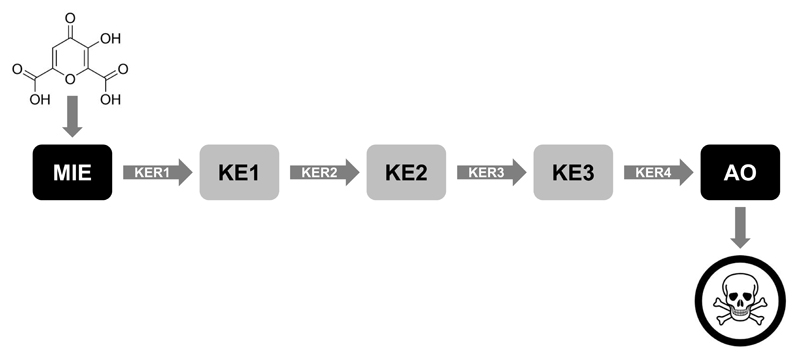
Predictive toxicology, based upon mechanistic information, has become a critical aspect of human risk assessment in the last decade. A major step in this direction came with the introduction of the mode-of-action concept, which relates to a series of key events (KEs) along a biological pathway from the initial chemical interaction to the adverse outcome (AO) (1). The mode-of-action concept was originally used by the US Environmental Protection Agency (EPA) in the cancer field (2), but seemed equally exploitable for non-cancer points (3–6). Another milestone was the well-known report published by the US National Academy of Science in 2007, outlining a vision on toxicology in the twenty-first century and placing toxicity pathways on the foreground (7). These toxicity pathways denote cellular pathways that, when disturbed, can lead to adverse health effects (1). Toxicity pathways align with adverse outcome pathways (AOPs), which have their roots in the area of ecotoxicology. An AOP refers to a conceptual construct that portrays existing knowledge concerning the linkage between a direct molecular initiating event (MIE) and an AO at a biological level of organization relevant to risk assessment (Fig. 1) (1, 8). In comparison with the mode-of-action, the scope of an AOP is broader, as it starts with the exposure and can go up to the population level. Thus far, AOPs have been designed for a number of different human-relevant toxicological endpoints. In response to the increasing use of AOPs, the Organization for Economic Cooperation and Development (OECD) together with the US EPA, the US Army Engineer Research and Development Center and the European Joint Research Center has initiated a project to facilitate the use of AOPs in assessing the safety of chemicals, called the AOP Knowledge Base (AOP-KB). The AOP-KB consists of four modules, namely the AOP Xplorer, Effectopedia, the Intermediate Effects Database and the AOP Wiki. The AOP Xplorer is a computational tool that enables automated graphical representation of AOPs and networks among them. Effectopedia is a modeling platform designed for collaborative development and utilization of AOPs. The Intermediate Effects Database hosts chemical-related data derived from non-apical endpoint methods and informs how individual compounds trigger MIEs and KEs. The AOP Wiki is a module of the AOP-KB that provides an open-source interface for rapid, widely accessible and collaborative sharing of established AOPs and building new AOPs (9). The AOP Wiki was launched in late 2014 and yet contains about fifty AOPs for several human-relevant toxicological endpoints, including drug-induced hepatotoxicity. These AOPs on liver toxicity will be scrutinized in this chapter while discussing AOP development, assessment and applications in drug safety evaluation.
Figure 1. Generic structure of an AOP.
Each AOP consists of two anchors, namely the molecular initiating event (MIE), which refers to the interaction of a chemical with a biological system at the molecular level, and the adverse outcome (AO), which is the actual apical toxicological endpoint. The entire response matrix between the MIE and AO is filled with key events (KEs), which represent changes in the biological state that are both measurable and essential to the progression of a defined biological perturbation leading to a specific AO. Subsequent KEs are connected by key event relationships (KERs), defining a link between both KEs and that facilitate inference or extrapolation of the state of the downstream KE from the known, measured or predicted state of the upstream KE (adapted from (10, 11)).
2. AOP development and assessment
2.1. Identification of the MIE, KEs and AO
The MIE is considered as the first anchor of an AOP and refers to the interaction of a chemical with a biological system at the molecular level, such as ligand-receptor interactions or binding to proteins and nucleic acids. It hereby is of utmost importance to define the site of action of the MIE, as this directly dictates the nature of the AO. The latter is envisaged as the second AOP anchor and describes the actual apical toxicological endpoint. The AO may be located at different levels of biological organization, ranging from the cellular to the population level, and can relate to either a chronic or a systemic toxicological outcome, or an acute or local adverse effect. A KE is defined as a change in biological state that is both measurable and essential to the progression of a defined biological perturbation leading to a specific AO. KEs do not provide a comprehensive molecular description of every aspect of the biological process involved per se. Rather, a limited number of KEs should be selected. These are normally those for which there is the most information to support assessment of weight-of-evidence in a regulatory context. The identification of the MIE, KEs and AO may be the result of an in-depth survey of relevant scientific literature or may be retrieved from experimental studies. Basically, any type of information can be fed into an AOP, including structural data, ‘omics-based’ data, in chemico data, in vitro data and in vivo data (1, 9–12).
2.2. Description of the KERs
A KER is a scientifically-based relationship that connects two KEs, defining a link between both KEs and that facilitates inference or extrapolation of the state of the downstream KE from the known, measured or predicted state of the upstream KE. Description of the KERs is a critical step in AOP development, which sets the stage for assessment of the AOP. KERs may either refer specifically to a direct linkage between a pair of KEs that are adjacent in an AOP or may indicate indirect linkages between a pair of KEs for which the relationship is thought to run through another KE or a gap in current understanding. At present, the vast majority of KERs in the AOP Wiki are rather of qualitative nature. However, from the risk assessment point of view, establishing quantitative KERs might be more desirable. These quantitative KERs may be defined in terms of correlations, dose-response relationships, dose-dependent transitions or points-of-departure. They may take the form of simple mathematical equations or sophisticated biologically-based computational models that consider other modulating factors, such as compensatory responses or interactions with other biological variables (9–11).
2.3. AOP assessment
Assessment of AOPs and evaluation of their suitability for application for regulatory purposes relies on (i) the confidence and precision with which the KEs can be measured, (ii) the level of confidence in KERs based on biological plausibility, empirical support for the KER and consistency of supporting data and among different biological contexts, and (iii) weight-of-evidence for the hypothesized pathway. Therefore, overall assessment of AOPs is best supported by providing thorough descriptions of the KEs and KERs as well as robust consideration of weight-of-evidence for the essentiality of KEs and KERs (9–11). Basically, AOP assessment relies on two sets of questions, which should be answered in an in-depth and scientifically sound way by AOP developers. The first set of questions focuses on weight-of-evidence assessment based on the Bradford-Hill criteria (Table 1), defining the minimal requirements for establishing a causal link between the different information blocks of the AOP (1, 13). The second set of key questions has been proposed by the OECD and rather envisages a confidence assessment (Table 2) (1, 12).
Table 1. Bradford-Hill criteria for AOP weight-of-evidence assessment (1, 9, 13).
|
Table 2. Key questions for testing AOP confidence (1, 9).
|
3. Liver toxicity AOPs
3.1. Liver steatosis
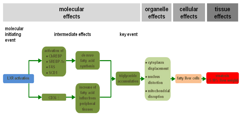
Steatosis is a prototypical type of drug-induced liver injury that refers to the process of abnormal retention of lipids, mainly triglycerides, within hepatocytes. It reflects the impairment of normal synthesis and elimination of triglycerides, and is triggered by a plethora of drugs, such as valproic acid (14). Steatosis can develop further into non-alcoholic steatohepatitis, which is characterized by hepatocellular injury and inflammation (15, 16). Liver steatosis may occur in a microvesicular or in a macrovesicular pattern. In microvesicular steatosis, numerous small lipid droplets are present in the hepatocyte cytoplasm, which do not displace the cell nucleus. By contrast, large droplets that move the hepatocyte nucleus to the periphery are observed in macrovesicular steatosis (14, 17–19). Since interaction of drugs with nuclear receptors is a frequent mechanism observed in liver steatosis, it has been considered as the main MIE in an established liver steatosis AOP (Fig. 2). In particular, activation of the liver X receptor induces an array of effects, such as enhanced transcription of genes encoding mediators of cholesterol and lipid metabolism. This leads to the increased influx of fatty acids from peripheral tissues into the liver and equally drives do novo synthesis of fatty acids. Consequently, triglycerides tend to accumulate in hepatocytes, which is considered as a KE in this AOP. At the organelle level, hepatocellular lipid accumulation may provoke cytoplasm displacement, nucleus distortion, mitochondrial toxicity and endoplasmic reticulum stress. All together, these effects underlie the acquisition of the typical fatty liver cell phenotype, which in turn causes a clinically relevant increase in liver weight (20). This AOP has been generated according to OECD guidelines, including critical consideration of the Bradford-Hill criteria for weight-of-evidence assessment and the OECD key questions for evaluating AOP confidence (9, 20).
Figure 2. AOP for drug-induced liver steatosis.
Activation of the liver X receptor (LXR), which is the MIE (blue), induces a number of transcriptional changes, including activation of the expression of carbohydrate response element binding protein (ChREBP), sterol response element binding protein 1c (SREBP-1c), fatty acid synthase (FAS) and stearoyl-coenzyme A desaturase 1 (SCD1). As a result, de novo synthesis of fatty acids is enhanced in the liver. At the same time, fatty acid translocase (CD36) production is upregulated, which mediates increased hepatic influx of fatty acids from peripheral tissues. All together, these intermediate steps drive accumulation of triglycerides, which is considered a key event (dark green). At the organelle level, this evokes cytoplasm displacement, distortion of the nucleus and mitochondrial disruption. This ultimately burgeons into the appearance of fatty liver cells (orange) and further into the clinical diagnosis of liver steatosis (red) (adapted from (20)).
3.2. Liver fibrosis
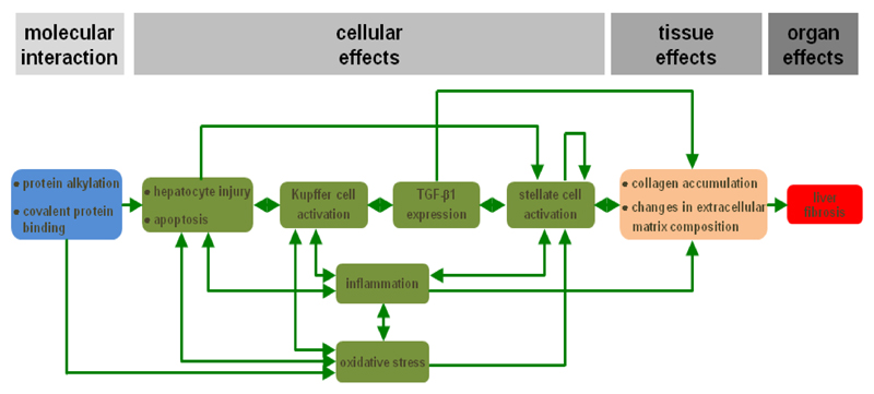
Liver fibrosis is a reversible wound healing response to either acute or chronic cellular injury that reflects a balance between liver repair and scar formation. It can be activated by a number of drugs, such as methotrexate. A central event in liver fibrosis is the activation of hepatic stellate cells, which occurs in two phases, namely the initiation stage and the perpetuation stage (14, 21–23). In the initiation phase, quiescent hepatic stellate cells become responsive to growth factors. This may be triggered by a variety of signals, including reactive oxygen species and apoptotic bodies originating from dying hepatocytes. In the perpetuation phase, the primed hepatic stellate cells undergo several changes related to proliferation, contractility, fibrogenesis, chemotaxis, extracellular matrix degradation and retinoid loss, whereby they adopt a myofibroblast-like phenotype. Hepatic stellate cell activation may be counteracted in a resolution phase through apoptosis, senescence or reversion to the quiescent phenotype (21, 22). Protein alkylation is considered as the MIE in an established AOP on liver fibrosis (Fig. 3), whereas the obvious AO at the organ level is liver fibrosis. Different steps at the cellular and tissue level have been defined, including hepatocyte injury and cell death, activation of Kupffer cells, expression of transforming growth factor beta 1, activation of hepatic stellate cells, oxidative stress and chronic inflammation, collagen accumulation and changes in hepatic extracellular matrix composition. The postulated AOP has been assessed by evaluation of the strength of evidence that supports the MIE, the KEs and the AO (9, 20).
Figure 3. AOP for drug-induced liver fibrosis.
The MIE (blue) is considered protein alkylation and covalent protein binding in the liver. This serves as a trigger to provoke hepatocyte injury, including apoptosis, which in turn activates Kupffer cells. As a result, transforming growth factor beta 1 (TGF-β1) expression is induced, which is a key factor for stellate cell activation. The latter goes hand in hand with the occurrence of inflammation and oxidative stress. The different events at the cellular level (green) are interconnected in several ways. The overall end result is accumulation of collagen and changes in the extracellular matrix composition in the liver (orange), which becomes clinically manifested as the AO, namely liver fibrosis (red) (adapted from (20)).
3.3. Cholestasis
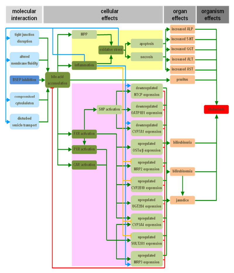
Cholestasis is another manifestation of drug-induced liver injury for which an AOP has been introduced (Fig. 4). Cholestasis can be caused by drugs such as bosentan. The MIE in this AOP is the direct cis-inhibition of the bile salt export pump. As a result of this, toxic bile acids accumulate into hepatocytes or bile canaliculi. These bile salts trigger a direct deteriorative response and an adaptive response (14). At the cellular level, the deteriorative response is accompanied by the formation of the mitochondrial permeability pore, which leads to mitochondrial impairment, inflammation, the production of reactive oxygen species and ultimately to the onset of cell death by both apoptotic and necrotic mechanisms (24, 25). Because of the latter, cytosolic enzymes start to leak from hepatocytes and cholangiocytes and become measurable in the serum (26, 27). A hallmark of cholestasis at the cellular level includes the induction of an adaptive response, which is aimed at counteracting bile accumulation and thus cholestatic liver injury. Accordingly, a complex machinery of transcriptionally coordinated mechanisms involving nuclear receptors is activated by bile acids, which collectively decrease the uptake and increase the export of bile acids and bilirubin into and from hepatocytes, respectively. Simultaneously, detoxification of bile acids is enhanced, while their synthesis becomes downregulated (28–30). The increased effort of cholestastic hepatocytes to remove bilirubin causes bilirubinuria and hyperbilirubinemia. As a result, a yellowish pigmentation of the skin and the conjunctival membranes over the sclera becomes visible, known as jaundice. Furthermore, the elevated presence of bile acids in the serum is thought to account for the typical skin itching in cholestasis patients (26, 27, 30). The development of this AOP was performed according to OECD guidance, including consideration of the Bradford-Hill criteria for weight-of-evidence assessment and the OECD key questions for evaluating AOP confidence. Proposed KEs are the accumulation of bile, the induction of oxidative stress and inflammation, and the activation of nuclear receptors. Furthermore, the AOP distinguishes direct adverse and indirect adaptive effects, and takes a number of alternative MIEs mechanisms into account (9, 31).
Figure 4. AOP for drug-induced cholestasis.
The response matrix between the MIE (dark blue) and AO (red), the inhibition of the bile salt export pump (BSEP) and cholestasis, respectively, spans over the cellular and organ levels. Identified KEs (dark green) include the accumulation of bile, the induction of oxidative stress and inflammation, and the activation of the nuclear receptors pregnane X receptor (PXR), farnesoid X receptor (FXR) and constitutive androstane receptor (CAR). Together with a number of intermediate steps, these KEs drive both a deteriorative cellular response (yellow), which underlies directly caused cholestatic injury, and an adaptive cellular response (purple), which is aimed at counteracting the primary cholestatic insults. Direct inducing and inhibiting effects are indicated with green and red arrows, respectively. Secondary inducing and inhibiting effects of oxidative stress and/or inflammation are indicated with blue and orange arrows, respectively (31) (5′-NT, 5′-nucleotidase; ALP, alkaline phosphatase; ALT, alanine aminotransferase; AST, aspartate aminotransferase; CYP2B10/3A4/7A1, cytochrome P450 2B10/3A4/7A1; GGT, gamma-glutamyl transpeptidase; MPP, mitochondrial permeability pore; MRP2/3, multidrug resistance-associated protein 2/3; NTCP, sodium/taurocholate cotransporter; OATP1B1, organic anion transporter 1B1; OSTα/β organic solute transporter α/β; SHP, small heterodimeric partner; SULT2A1, dehydroepiandrosterone sulfotransferase; UGT2B4, uridine 5′-diphosphate-glucuronosyltransferase 2B4).
4. AOP applications
4.1. Establishment of (quantitative) structure-activity relationships
As the MIE in each AOP involves a rather specific interaction of chemicals with biological systems, it can be used as the basis for generating structure-activity relationships, whether or not quantifiable. In turn, such information can be used for chemical grouping and read-across approaches, thus facilitating predictive and mechanism-based toxicology (1). Using quantitative structure-activity relationship (QSAR) approaches, it has been demonstrated that chemicals with an ester bound to a carbon atom of a heterocyclic group or carbocyclic systems with a least one aromatic ring positively contribute to bile salt export pump inhibition, being the MIE in the AOP on drug-induced cholestasis, while the presence of hydroxyl groups bound to aliphatic carbon atoms has a negative contribution (32, 33). In silico modeling further showed the role of hydroxyl groups in the interaction of chemicals with the bile salt export pump (34). Two-dimensional and three-dimensional QSAR studies have also been performed on ligands of the liver X receptor, which constitutes the MIE in the AOP on drug-induced steatosis. By doing so, a number of chemical features, such as the presence of phenyl rings, chloro groups and methyl moieties, have been identified as determinants of liver X receptor binding and activation (35).
4.2. Elaboration of prioritization strategies
Prioritization of chemicals denotes the process in which less complex, cheaper and faster assays are used to determine which chemicals are subjected first to more complex, expensive and slower testing (36). AOPs have great potential with respect to prioritization strategies. Indeed, they can increase confidence in the integration of information, such as obtained from in vitro assays, for prioritizing chemicals for further assessment. The use of AOPs for the hepatotoxic endpoints described in this chapter in the context of the prioritization has not yet been described in current scientific literature. However, there are some examples for other adverse effects, including developmental toxicity. At present, the most promising alternative vertebrate models for screening of chemicals for developmental toxicity are fish embryos, in particular zebrafish. Using paraoxon, an acetylcholinesterase inhibitor, as a reference chemical, an AOP providing quantitative linkages across levels of biological organization during zebrafish embryogenesis has been proposed. Based on a series of experiments, it was found that normal acetylcholinesterase activity is not required for secondary motor neuron development and that acetylcholinesterase inhibition, the MIE, may not be associated with an increased frequency of spontaneous tail contractions following paraoxon exposure. This AOP may support chemical screening and prioritization strategies with respect to developmental toxicity testing (37).
4.3. Development of in vitro tests
An essential step during AOP development is the labeling of KEs. In turn, this may serve as the basis for the characterization of biomarkers and simultaneously for the establishment of ex vivo, but especially in vitro, toxicity screening assays applicable for regulatory testing purposes. Furthermore, such new non-animal tests might be implemented into integrated testing strategies, thereby contributing to the refinement, reduction and replacement of conventional in vivo testing. Reversely, by linking proposals for the development of in vitro test methods to KEs in an AOP, the relationship to hazard endpoints relevant for regulatory purposes can be established (1, 12).
5. Conclusions and perspectives
Although conceptually not entirely new, AOPs have found their way to the human risk assessment arena in recent years, including the safety evaluation of drugs. The potential use of AOPs in this field is indeed considerably larger than the mode-of-action concept, as, at least ideally, it considers an exposure aspect and because it is not restricted to the tissue and individual level. However, despite the introduction of OECD guidance on AOP development and evaluation (1, 9), this area is still in its infancy and will greatly benefit from fine-tuning in the upcoming years (Table 3). A major criticism on AOPs nowadays is their simplicity and thus their poor reflection of complex toxicological processes. AOPs are presented as stand-alone linear events, yet the reality is likely to be much less straightforward, since parallel cascades and crossing of pathways may be involved. It is important that the overall toxicological scenario does not become lost when using AOPs. Furthermore, AOPs are to be considered as open and flexible structures that should be continuously refined by feeding in old and new data. Such iterative refinement exercises should ideally include the elaboration and quantification of the KERs as well as the specification of toxicokinetic conditions governing the activation of an AOP. Thus, classical kinetic determinants, like absorption, distribution, metabolism and excretion, as well as more specific events, such as hormonal influences and adaptive responses, must be considered in AOP development. Another hurdle to overcome in near future relates to the weight-of-evidence of data that are proposed to substantiate an AOP. Basically, anyone can propose an AOP, but not all AOPs are sufficiently supported by data. In order to develop confidence in the accuracy and utility of AOPs, there needs to be a transparent evaluation process that includes all stakeholders. In addition to hazard identification and the establishment of dose-response relationships, the risk assessment paradigm also includes implementation of exposure data. Thus far, this has gained little attention in the context of AOP development, thereby defining another challenge lying ahead. Several efforts are currently ongoing around the globe to tackle these issues, including at the OECD level (1, 9), the US Hamner Institutes of Health (38), the US Center for Alternatives to Animal Testing (39) and the European research program called Safety Evaluation Ultimately Replacing Animal Testing (40, 41). Such projects are anticipated to yield robust and reliable AOP tools that can be used for a variety of purposes pertinent to toxicology and risk assessment, including the safety evaluation of new drug candidates.
Table 3. Major challenges for future AOP development.
|
Acknowledgements
This work was financially supported by the grants of the University Hospital of the Vrije Universiteit Brussel - Belgium (Willy Gepts Fonds UZ-VUB), the University of São Paulo - Brazil (USP), the São Paulo Research Foundation - Brazil (FAPESP), the Fund for Scientific Research - Flanders (FWO-Vlaanderen), the European Research Council (project CONNECT), the European Union (FP7) and Cosmetics Europe (projects DETECTIVE and HeMiBio).
References
- 1.OECD. Proposal for a template and guidance on developing and assessing the completeness of adverse outcome pathways. Series on Testing and Assessment. 2013;184:1–45. [Google Scholar]
- 2.US EPA. Guidelines for carcinogen risk assessment. Washington D.C: 2005. [Google Scholar]
- 3.Bogdanffy MS, Daston G, Faustman EM, et al. Harmonization of cancer and noncancer risk assessment: proceedings of a consensus-building workshop. Toxicol Sci. 2001;61:18–31. doi: 10.1093/toxsci/61.1.18. [DOI] [PubMed] [Google Scholar]
- 4.Julien E, Boobis AR, Olin SS. The key events dose-response framework: a cross-disciplinary mode-of-action based approach to examining dose-response and thresholds. Crit Rev Food Sci Nutr. 2009;49:682–689. doi: 10.1080/10408390903110692. [DOI] [PMC free article] [PubMed] [Google Scholar]
- 5.Meek ME, Bucher JR, Cohen SM, et al. A framework for human relevance analysis of information on carcinogenic modes of action. Crit Rev Toxicol. 2003;33:591–653. doi: 10.1080/713608373. [DOI] [PubMed] [Google Scholar]
- 6.Seed J, Carney EW, Corley RA, et al. Overview: using mode of action and life stage information to evaluate the human relevance of animal toxicity data. Crit Rev Toxicol. 2005;35:664–672. doi: 10.1080/10408440591007133. [DOI] [PubMed] [Google Scholar]
- 7.NRC. Toxicity testing in the 21st century: a vision and a strategy. The National Academies Press; Washington DC: 2007. [Google Scholar]
- 8.Ankley GT, Bennett RS, Erickson RJ, et al. Adverse outcome pathways: a conceptual framework to support ecotoxicology research and risk assessment. Environ Toxicol Chem. 2010;29:730–741. doi: 10.1002/etc.34. [DOI] [PubMed] [Google Scholar]
- 9. https://aopkb.org/ (consulted February 2015)
- 10.Villeneuve DL, Crump D, Garcia-Reyero N, et al. Adverse outcome pathway (AOP) development I: strategies and principles. Toxicol Sci. 2014;142:312–320. doi: 10.1093/toxsci/kfu199. [DOI] [PMC free article] [PubMed] [Google Scholar]
- 11.Villeneuve DL, Crump D, Garcia-Reyero N, et al. Adverse outcome pathway development II: best practices. Toxicol Sci. 2014;142:321–330. doi: 10.1093/toxsci/kfu200. [DOI] [PMC free article] [PubMed] [Google Scholar]
- 12.Vinken M. The adverse outcome pathway concept: a pragmatic tool in toxicology. Toxicology. 2013;312:158–165. doi: 10.1016/j.tox.2013.08.011. [DOI] [PubMed] [Google Scholar]
- 13.Hill AB. The environment and disease: association or causation? Proc R Soc Med. 1965;58:295–300. doi: 10.1177/003591576505800503. [DOI] [PMC free article] [PubMed] [Google Scholar]
- 14.Vinken M, Maes M, Vanhaecke T, Rogiers V. Drug-induced liver injury: mechanisms, types and biomarkers. Curr Med Chem. 2013;20:3011–3021. doi: 10.2174/0929867311320240006. [DOI] [PubMed] [Google Scholar]
- 15.Begriche K, Massart J, Robin MA, et al. Drug-induced toxicity on mitochondria and lipid metabolism: mechanistic diversity and deleterious consequences for the liver. J Hepatol. 2011;54:773–794. doi: 10.1016/j.jhep.2010.11.006. [DOI] [PubMed] [Google Scholar]
- 16.Cohen JC, Horton JD, Hobbs HH. Human fatty liver disease: old questions and new insights. Science. 2011;332:1519–1523. doi: 10.1126/science.1204265. [DOI] [PMC free article] [PubMed] [Google Scholar]
- 17.Amacher DE. The mechanistic basis for the induction of hepatic steatosis by xenobiotics. Expert Opin Drug Metab Toxicol. 2011;7:949–965. doi: 10.1517/17425255.2011.577740. [DOI] [PubMed] [Google Scholar]
- 18.Ramachandran R, Kakar S. Histological patterns in drug-induced liver disease. J Clin Pathol. 2009;62:481–492. doi: 10.1136/jcp.2008.058248. [DOI] [PubMed] [Google Scholar]
- 19.Zimmerman HJ. Drug-induced liver disease. Clin Liver Dis. 2000;4:73–96. doi: 10.1016/s1089-3261(05)70097-0. [DOI] [PubMed] [Google Scholar]
- 20.Landesmann B, Goumenou M, Munn S, Whelan M. Description of prototype modes-of-action related to repeated dose toxicity. JRC Scientific and Policy Report. 2012:75689. [Google Scholar]
- 21.Friedman SL. Mechanisms of hepatic fibrogenesis. Gastroenterology. 2008;134:1655–1669. doi: 10.1053/j.gastro.2008.03.003. [DOI] [PMC free article] [PubMed] [Google Scholar]
- 22.Friedman SL. Evolving challenges in hepatic fibrosis. Nat Rev Gastroenterol Hepatol. 2010;7:425–436. doi: 10.1038/nrgastro.2010.97. [DOI] [PubMed] [Google Scholar]
- 23.Lee UE, Friedman SL. Mechanisms of hepatic fibrogenesis. Best Pract Res Clin Gastroenterol. 2011;25:195–206. doi: 10.1016/j.bpg.2011.02.005. [DOI] [PMC free article] [PubMed] [Google Scholar]
- 24.Schoemaker MH, Conde de la Rosa L, Buist-Homan M, et al. Tauroursodeoxycholic acid protects rat hepatocytes from bile acid-induced apoptosis via activation of survival pathways. Hepatology. 2004;39:1563–1573. doi: 10.1002/hep.20246. [DOI] [PubMed] [Google Scholar]
- 25.Woolbright BL, Jaeschke H. Novel insight into mechanisms of cholestatic liver injury. World J Gastroenterol. 2012;18:4985–4993. doi: 10.3748/wjg.v18.i36.4985. [DOI] [PMC free article] [PubMed] [Google Scholar]
- 26.Hofmann AF. Bile acids and the enterohepatic circulation. In: Arias IM, Alter HJ, Boyer JL, Cohen DE, Fausto N, Shafritz DA, Wolkoff AW, editors. The liver: biology and pathobiology. Wiley-Blackwell; Oxford: 2009. pp. 289–304. [Google Scholar]
- 27.Padda MS, Sanchez M, Akhtar AJ, Boyer JL. Drug-induced cholestasis. Hepatology. 2011;53:1377–1387. doi: 10.1002/hep.24229. [DOI] [PMC free article] [PubMed] [Google Scholar]
- 28.Zollner G, Trauner M. Molecular mechanisms of cholestasis. Wien Med Wochenschr. 2006;156:380–385. doi: 10.1007/s10354-006-0312-7. [DOI] [PubMed] [Google Scholar]
- 29.Zollner G, Trauner M. Mechanisms of cholestasis. Clin Liver Dis. 2008;12:1–26. doi: 10.1016/j.cld.2007.11.010. [DOI] [PubMed] [Google Scholar]
- 30.Wagner M, Zollner G, Trauner M. New molecular insights into the mechanisms of cholestasis. J Hepatol. 2009;51:565–580. doi: 10.1016/j.jhep.2009.05.012. [DOI] [PubMed] [Google Scholar]
- 31.Vinken M, Landesmann B, Goumenou M, et al. Development of an adverse outcome pathway from drug-mediated bile salt export pump inhibition to cholestatic liver injury. Toxicol Sci. 2013;136:97–106. doi: 10.1093/toxsci/kft177. [DOI] [PubMed] [Google Scholar]
- 32.Hirano H, Kurata A, Onishi Y, et al. High-speed screening and QSAR analysis of human ATP-binding cassette transporter ABCB11 (bile salt export pump) to predict drug-induced intrahepatic cholestasis. Mol Pharm. 2006;2:252–265. doi: 10.1021/mp060004w. [DOI] [PubMed] [Google Scholar]
- 33.Saito H, Osumi M, Hirano H, et al. Technical pitfalls and improvements for high-speed screening and QSAR analysis to predict inhibitors of the human bile salt export pump (ABCB11/BSEP) AAPS J. 2009;11:581–589. doi: 10.1208/s12248-009-9137-9. [DOI] [PMC free article] [PubMed] [Google Scholar]
- 34.Warner DJ, Chen H, Cantin LD, et al. Mitigating the inhibition of human bile salt export pump by drugs: opportunities provided by physicochemical property modulation, in silico modeling, and structural modification. Drug Metab Dispos. 2012;40:2332–2341. doi: 10.1124/dmd.112.047068. [DOI] [PubMed] [Google Scholar]
- 35.Honorio KM, Salum LB, Garratt RC, et al. Two- and three-dimensional quantitative structure–activity relationships studies on a series of liver X receptor ligands. Open Med Chem J. 2008;2:87–96. doi: 10.2174/1874104500802010087. [DOI] [PMC free article] [PubMed] [Google Scholar]
- 36.Judson R, Kavlock R, Martin M, et al. Perspectives on validation of high-throughput assays supporting 21st century toxicity testing. ALTEX. 2013;30:51–56. doi: 10.14573/altex.2013.1.051. [DOI] [PMC free article] [PubMed] [Google Scholar]
- 37.Yozzo KL, McGee SP, Volz DC. Adverse outcome pathways during zebrafish embryogenesis: a case study with paraoxon. Aquat Toxicol. 2013;126:346–354. doi: 10.1016/j.aquatox.2012.09.008. [DOI] [PubMed] [Google Scholar]
- 38.Andersen ME, Clewell R, Bhattacharya S. Developing in vitro tools sufficient by themselves for 21st century risk assessment. In: Gocht T, Schwarz M, editors. Towards the replacement of in vivo repeated dose systemic toxicity testing. Vol. 2. Imprimerie Mouzet; France: 2012. pp. 347–360. [Google Scholar]
- 39. http://caat.jhsph.edu/ (consulted February 2015)
- 40. http://www.seurat-1.eu/ (consulted February 2015)
- 41.Vinken M, Pauwels M, Ates G, et al. Screening of repeated dose toxicity data present in SCC(NF)P/SCCS safety evaluations of cosmetic ingredients. Arch Toxicol. 2012;86:405–412. doi: 10.1007/s00204-011-0769-z. [DOI] [PubMed] [Google Scholar]


