. 2017 May 19;6:e24109. doi: 10.7554/eLife.24109

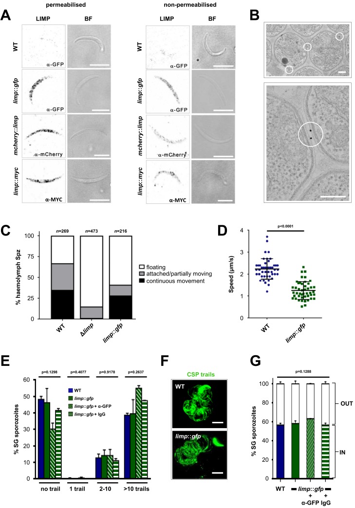
Figure 4. LIMP localises to the parasite plasma membrane but its function cannot be targeted by blocking antibodies.

(A) Immunofluorescence assays of WT, limp::gfp, mcherry::limp and limp::myc salivary gland sporozoites. Sporozoites were stained with anti-GFP (α-GFP), α-mCherry or α-MYC antibodies at days 17–20 p.i. BF=brightfield images. Scale bars = 5 µm. (B) Immuno-electron microscopy of limp::gfp salivary gland sporozoites with anti-GFP antibodies identifies LIMP::GFP (black dots inside white circles) at the parasite plasma membrane. Scale bars = 200 nm. (C) Live motility assay to investigate movement patterns of WT, Δlimp and limp::gfp haemolymph sporozoites (Spz). Most Δlimp sporozoites display a severe attachment phenotype and thus are incapable of moving. The indicated total number of sporozoites derived from three independent experiments in case of WT and six or two independent assays were performed for Δlimp or limp::gfp, respectively. (D) Moving limp::gfp haemolymph sporozoites show reduced gliding speed. Dot plots show means±SEM of 46 analysed sporozoites from three independent experiments per parasite line; p-value for Mann-Whitney test. (E) CSP shedding-based gliding motility assay of WT and limp::gfp salivary gland (SG) sporozoites in the presence and absence of anti-GFP (α-GFP) blocking antibodies or control IgGs. Bars show means±SEM. WT (one experiment; n = 3; 280 sporozoites analysed); limp::gfp (two independent experiments; n = 5; 492 sporozoites analysed); limp::gfp + α-GFP (one experiment; n = 3; 197 sporozoites analysed); limp::gfp + IgG (one experiment; n = 3; 234 sporozoites analysed). (F) Representative images of WT and limp::gfp (no antibody) CSP gliding trails. Scale bars = 10 µm. (G) WT and limp::gfp salivary gland (SG) sporozoite hepatocyte invasion assay in the presence and absence of anti-GFP (α-GFP) blocking antibodies or control IgGs. Bars show means±SEM of invading (IN) and non-invading (OUT) parasites. Data are from one experiment (n = 3) for all parasite lines and conditions. Numbers of sporozoites analysed: WT = 346, limp::gfp = 763, limp::gfp + α-GFP = 807, limp::gfp + IgG = 742. (E and G) p-values for Kruskal-Wallis test.

DOI: http://dx.doi.org/10.7554/eLife.24109.010

Figure 4.

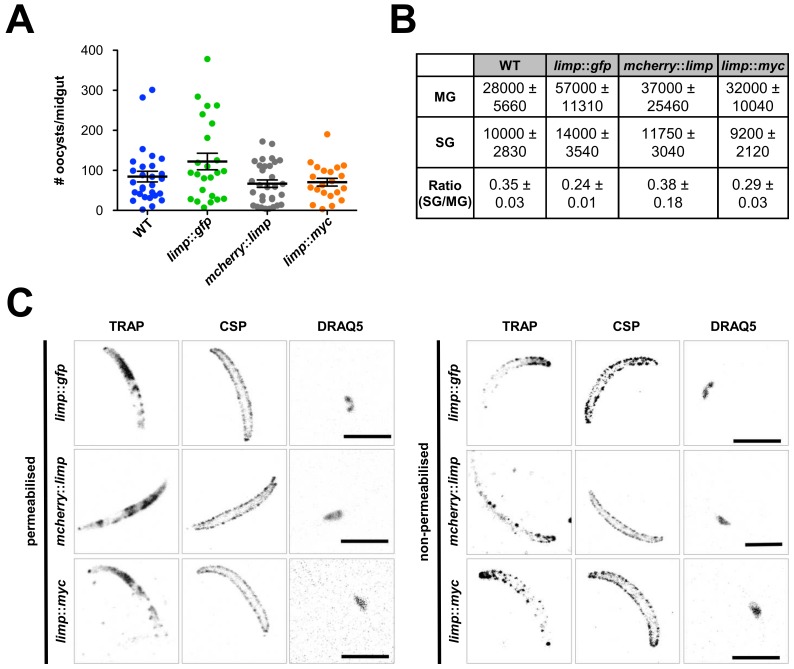
Figure 4—figure supplement 1. Phenotypic characterisation of tagged limp parasite lines.

Figure 4—figure supplement 1.

Oocysts per mosquito midgut. All tagged lines show oocyst numbers similar to WT parasites indicating no defect in midgut colonisation. Lines show means±SEM from two independent experiments. WT (n = 28); limp::gfp (n = 24); mcherry::limp (n = 31); limp::myc (n = 21). (B) Analysis of salivary gland invasion by determining midgut (MG) and salivary gland (SG) sporozoite numbers on day 17 p.i. Values show means±SD from two independent experiments. (C) Immunofluorescence assays of limp::gfp, mcherry::limp and limp::myc salivary gland sporozoites. Sporozoites were stained for the surface marker CSP and the surface/microneme marker TRAP at days 17–20 p.i. DNA stained with DRAQ5. Scale bars = 5 µm.
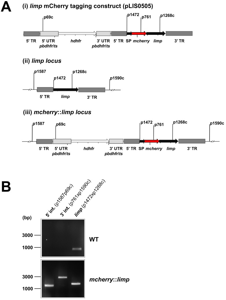
Figure 4—figure supplement 2. Generation and genotyping of the mcherry::limp parasite line.

Figure 4—figure supplement 2.

(A) mCherry limp tagging construct pLIS0505 (i) was obtained by inserting the mcherry gene downstream and in frame of the signal peptide (SP) of limp. To ensure correct cleavage of the N-terminal signal peptide, the sequence after the cleavage site is integrated again downstream of mcherry followed by the limp ORF and endogenous 3' UTR which, together with the endogenous limp promoter, serve as targeting regions (TRs). This construct includes human dhfr selectable marker cassette under the control of P. berghei dhfr/ts 5' and 3' UTRs. The construct was integrated into the limp locus (ii) of cl15cy1 by double homologous recombination, resulting in replacement of endogenous limp by a N-terminally mCherry-tagged version of limp (iii). (B) Correct tagging of limp was shown by diagnostic PCR analyses. PCR analyses in clonal mcherry::limp parasites confirm 5' and 3' integration (int.) of pLIS0505 and electrophoretic mobility shift of limp ORF due to the presence of the mcherry tag.
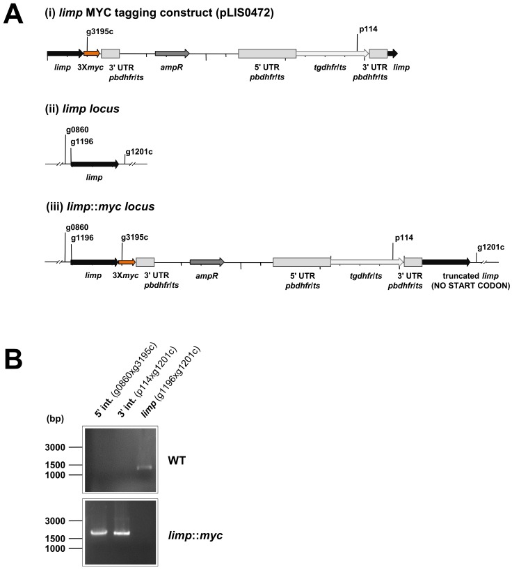
Figure 4—figure supplement 3. Generation and genotyping of the limp::myc parasite line.

Figure 4—figure supplement 3.

(A) limp MYC tagging construct pLIS0472 (i) was obtained analogously to limp GFP tagging construct by cloning the last 1149 bp of the limp ORF excluding the stop codon upstream and in frame with the triple tandem copy myc tag (3Xmyc). This construct includes Toxoplasma gondii dhfr/ts selectable marker cassette under the control of P. berghei dhfr/ts 5' and 3' UTRs. The construct was integrated into the limp locus (ii) of cl15cy1 by single homologous recombination, resulting in the fusion of limp to myc in limp::myc parasites (iii). (B) Correct tagging of limp was shown by diagnostic PCR analyses. PCR analyses in clonal limp::myc parasites confirm 5' and 3' integration (int.) of pLIS0472 and absence of WT limp ORF.