

Abstract
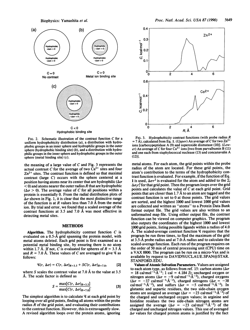
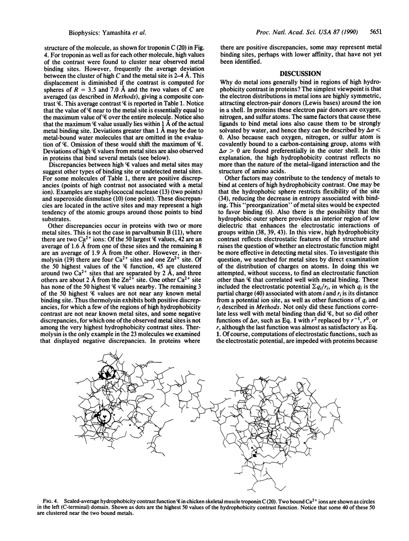
The environments of metal ions (Li+, Na+, K+, Ag+, Cs+, Mg2+, Ca2+, Mn2+, Cu2+, Zn2+) in proteins and other metal-host molecules have been examined. Regardless of the metal and its precise pattern of ligation to the protein, there is a common qualitative feature to the binding site: the metal is ligated by a shell of hydrophilic atomic groups (containing oxygen, nitrogen, or sulfur atoms) and this hydrophilic shell is embedded within a larger shell of hydrophobic atomic groups (containing carbon atoms). That is, metals bind at centers of high hydrophobicity contrast. This qualitative observation can be described analytically by the hydrophobicity contrast function, C, evaluated from the structure. This function is large and positive for a sphere of hydrophilic atomic groups (characterized by atomic solvation parameters, delta sigma, having values less than 0) at the center of a larger sphere of hydrophobic atomic groups (characterized by delta sigma greater than 0). In the 23 metal-binding molecules we have examined, the maximum values of the contrast function lie near to observed metal binding sites. This suggests that the hydrophobicity contrast function may be useful for locating, characterizing, and designing metal binding sites in proteins.
Full text
PDF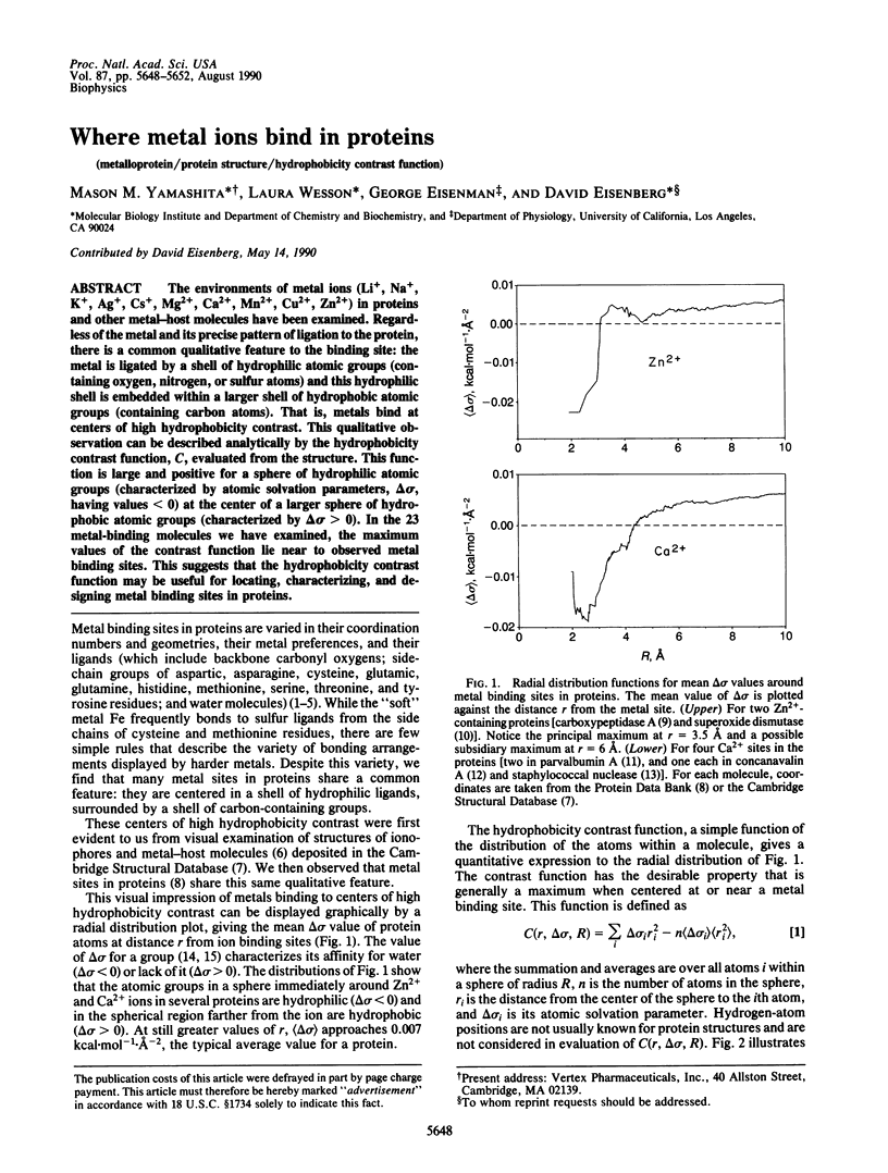

Selected References
These references are in PubMed. This may not be the complete list of references from this article.
- Babu Y. S., Bugg C. E., Cook W. J. Structure of calmodulin refined at 2.2 A resolution. J Mol Biol. 1988 Nov 5;204(1):191–204. doi: 10.1016/0022-2836(88)90608-0. [DOI] [PubMed] [Google Scholar]
- Bernstein F. C., Koetzle T. F., Williams G. J., Meyer E. F., Jr, Brice M. D., Rodgers J. R., Kennard O., Shimanouchi T., Tasumi M. The Protein Data Bank: a computer-based archival file for macromolecular structures. Arch Biochem Biophys. 1978 Jan 30;185(2):584–591. doi: 10.1016/0003-9861(78)90204-7. [DOI] [PubMed] [Google Scholar]
- Cotton F. A., Bier C. J., Day V. W., Hazen E. E., Jr, Larsen S. Some aspects of the structure of staphylococcal nuclease. I. Crystallographic studies. Cold Spring Harb Symp Quant Biol. 1972;36:243–249. doi: 10.1101/sqb.1972.036.01.032. [DOI] [PubMed] [Google Scholar]
- Eisenberg D., McLachlan A. D. Solvation energy in protein folding and binding. Nature. 1986 Jan 16;319(6050):199–203. doi: 10.1038/319199a0. [DOI] [PubMed] [Google Scholar]
- Gilson M. K., Honig B. H. Calculation of electrostatic potentials in an enzyme active site. Nature. 1987 Nov 5;330(6143):84–86. doi: 10.1038/330084a0. [DOI] [PubMed] [Google Scholar]
- Hardman K. D., Ainsworth C. F. Structure of concanavalin A at 2.4-A resolution. Biochemistry. 1972 Dec 19;11(26):4910–4919. doi: 10.1021/bi00776a006. [DOI] [PubMed] [Google Scholar]
- Holmes M. A., Matthews B. W. Structure of thermolysin refined at 1.6 A resolution. J Mol Biol. 1982 Oct 5;160(4):623–639. doi: 10.1016/0022-2836(82)90319-9. [DOI] [PubMed] [Google Scholar]
- Honig B. H., Hubbell W. L., Flewelling R. F. Electrostatic interactions in membranes and proteins. Annu Rev Biophys Biophys Chem. 1986;15:163–193. doi: 10.1146/annurev.bb.15.060186.001115. [DOI] [PubMed] [Google Scholar]
- Ibers J. A., Holm R. H. Modeling coordination sites in metallobiomolecules. Science. 1980 Jul 11;209(4453):223–235. doi: 10.1126/science.7384796. [DOI] [PubMed] [Google Scholar]
- Komine S., Yoshida K., Yamashita H., Masaki Z. Voiding dysfunction in patients with human T-lymphotropic virus type-1-associated myelopathy (HAM). Paraplegia. 1989 Jun;27(3):217–221. doi: 10.1038/sc.1989.32. [DOI] [PubMed] [Google Scholar]
- Kretsinger R. H. Calcium coordination and the calmodulin fold: divergent versus convergent evolution. Cold Spring Harb Symp Quant Biol. 1987;52:499–510. doi: 10.1101/sqb.1987.052.01.057. [DOI] [PubMed] [Google Scholar]
- Linse S., Brodin P., Johansson C., Thulin E., Grundström T., Forsén S. The role of protein surface charges in ion binding. Nature. 1988 Oct 13;335(6191):651–652. doi: 10.1038/335651a0. [DOI] [PubMed] [Google Scholar]
- Moews P. C., Kretsinger R. H. Refinement of the structure of carp muscle calcium-binding parvalbumin by model building and difference Fourier analysis. J Mol Biol. 1975 Jan 15;91(2):201–225. doi: 10.1016/0022-2836(75)90160-6. [DOI] [PubMed] [Google Scholar]
- Pantoliano M. W., Whitlow M., Wood J. F., Rollence M. L., Finzel B. C., Gilliland G. L., Poulos T. L., Bryan P. N. The engineering of binding affinity at metal ion binding sites for the stabilization of proteins: subtilisin as a test case. Biochemistry. 1988 Nov 1;27(22):8311–8317. doi: 10.1021/bi00422a004. [DOI] [PubMed] [Google Scholar]
- Rees D. C., Lewis M., Lipscomb W. N. Refined crystal structure of carboxypeptidase A at 1.54 A resolution. J Mol Biol. 1983 Aug 5;168(2):367–387. doi: 10.1016/s0022-2836(83)80024-2. [DOI] [PubMed] [Google Scholar]
- Satyshur K. A., Rao S. T., Pyzalska D., Drendel W., Greaser M., Sundaralingam M. Refined structure of chicken skeletal muscle troponin C in the two-calcium state at 2-A resolution. J Biol Chem. 1988 Feb 5;263(4):1628–1647. [PubMed] [Google Scholar]
- Serpersu E. H., Hibler D. W., Gerlt J. A., Mildvan A. S. Kinetic and magnetic resonance studies of the glutamate-43 to serine mutant of staphylococcal nuclease. Biochemistry. 1989 Feb 21;28(4):1539–1548. doi: 10.1021/bi00430a018. [DOI] [PubMed] [Google Scholar]
- Serpersu E. H., McCracken J., Peisach J., Mildvan A. S. Electron spin echo modulation and nuclear relaxation studies of staphylococcal nuclease and its metal-coordinating mutants. Biochemistry. 1988 Oct 18;27(21):8034–8044. doi: 10.1021/bi00421a010. [DOI] [PubMed] [Google Scholar]
- Serpersu E. H., Shortle D., Mildvan A. S. Kinetic and magnetic resonance studies of active-site mutants of staphylococcal nuclease: factors contributing to catalysis. Biochemistry. 1987 Mar 10;26(5):1289–1300. doi: 10.1021/bi00379a014. [DOI] [PubMed] [Google Scholar]
- Serpersu E. H., Shortle D., Mildvan A. S. Kinetic and magnetic resonance studies of effects of genetic substitution of a Ca2+-liganding amino acid in staphylococcal nuclease. Biochemistry. 1986 Jan 14;25(1):68–77. doi: 10.1021/bi00349a011. [DOI] [PubMed] [Google Scholar]
- Stallings W. C., Pattridge K. A., Strong R. K., Ludwig M. L. The structure of manganese superoxide dismutase from Thermus thermophilus HB8 at 2.4-A resolution. J Biol Chem. 1985 Dec 25;260(30):16424–16432. [PubMed] [Google Scholar]
- Tainer J. A., Getzoff E. D., Beem K. M., Richardson J. S., Richardson D. C. Determination and analysis of the 2 A-structure of copper, zinc superoxide dismutase. J Mol Biol. 1982 Sep 15;160(2):181–217. doi: 10.1016/0022-2836(82)90174-7. [DOI] [PubMed] [Google Scholar]
- Warshel A., Russell S. T. Calculations of electrostatic interactions in biological systems and in solutions. Q Rev Biophys. 1984 Aug;17(3):283–422. doi: 10.1017/s0033583500005333. [DOI] [PubMed] [Google Scholar]