Abstract
Capsule endoscopy was conceived by inventive minds of good people. In the beginning there was a will to do something for medicine. The idea fomented after a discourse between the talented engineer with his physician friend. It took years to develop the concept. Then excellent engineers created de novo the necessary components to turn the capsule into a viable reality. The story is a tribute to human ingenuity.
Keywords: History, capsule endoscopy, complementary metal oxide semiconductor imager (CMOS imager)
The history of time for capsule endoscopy
Capsule endoscopy entered the domain of clinical gastroenterology in the year 2001 with the clearance by the Food and Drug Administration (FDA) and obtaining CE Mark certification. The journey from conception to implementation started 20 years earlier. In 1981, Gavriel Iddan took a sabbatical from his regular position as a senior engineer at the electro optical design section of Rafael, the research and development (R&D) branch of the Israeli Ministry of Defense. Iddan had taken an interest in medical imaging and spent his leave to study X-ray and ultrasound imaging for Elscint (a company dedicated to medical imaging) in Boston. His neighbor, Eitan Scapa, an Israeli gastroenterologist spent his sabbatical in a medical institution in Boston as well. The two befriended each other and Iddan learned about the use of fiber optics in the gastrointestinal tract. Scapa explained to Iddan that the technology of fiber optics left the small bowel out of reach for medical inspection. That is when Iddan became aware of the medical challenge and started thinking about solutions. In the meanwhile, small charged coupled device (CCD) imaging chips reached the market. Ten years later, on a second sabbatical, Iddan considered using the CCD miniature camera connected to an electrical umbilical cord. The length of the small bowel precluded this option. That is when he suggested cutting the umbilical cord, in its stead attaching a mini transmitter to the CCD camera and letting the device travel on its own. However, the CCD elements consume a lot of energy and miniature batteries would permit at best 10 min transmission time. More problems accumulated. How would the capsule guarantee adequate visibility, debris could obscure the surface of the camera? A capsule study of the small intestine is likely to take many hours and occupy patient and physician at the screen for a large amount of time. How would it be possible to free patient and physician from a lengthy exam and limit the time necessary at the screen? In 1993, Iddan came up with the brilliant idea to split the system into three parts. Part one: the camera and its transmitter. Part two: a recorder attached to a sensor array placed on the surface of the patient’s abdomen. Part three: a software package that processes the stored information on the recorder to generate a study that can be reviewed by the physician at his leisure in a reasonable amount of time. The problem of the energy guzzling CCD camera was solved nearly by coincidence. While reading a journal on optical engineering (1), Iddan came across an article by Eric Fossum. He described the use of a Silicon chip (CMOS—complementary metal oxide semiconductor) and in near clairvoyance predicted that CMOS would replace CCD. This would happen for multiple reasons not the least that they consume just one percent the amount of energy of CCDs. Fossum would later assist the Given Imaging team of engineers (2). Now the pieces were beginning to fall into place. Further miniaturization of the components helped along.
The translation of concept to reality would require an organization with funds but first and foremost the expertise of very innovative engineers to create the elements for a functioning capsule camera. In 1994, Gavriel Iddan met Gavriel Meron who precisely did that, namely raised the necessary funds and recruited the talented physicists and engineers (Here is a list of some of the people involved: Dov Avni, Avner Badichi, A. Reichert, B. Ehrlich, H. Kislev, Y. Avron, D. Adler, M. Frish, A. Glukhovsky, S. Friedman and other talented people. Many of these were students of Prof. Y. Kidron from the Technion in Haifa). The original patent of Gavriel Iddan (awarded in 1997) envisioned a CCD chip, which was too bulky and consumed too much energy. The light generated by a heated filament consumed too much energy, was too weak and did not turn on/off fast enough. The optical design was not suitable. Meron understood that he could not buy the miniature camera elements off the shelf. His team would have to create them. Dov Avni’s specialty was analog video. He too had spent many years at Rafael, the same R&D branch of the Israeli Ministry of Defense that Gavriel Iddan was working at. Avni was charged to create de novo an imager, a light source and miniaturize all the components. Once he informed Meron that he had solved the technical challenges of the CMOS chip imager, Meron was ready to move. Meron’s team consulted with the Sarnoff Corporation at Princeton, an institute dedicated to the research in vision, video and semiconductor technology. The leading research expert of the Sarnoff Corporation predicted that it would be impossible to generate sharp images with CMOS technology because at body temperature the signal to noise ratio was too low due to the presence of random photons. Avni though, knew that engineers at Tower Semiconductor (Migdal Haemek, Israel) had solved this problem. They confirmed Avni’s design. Together they had developed a process to manufacture CMOS imagers that overcame this particular problem. Then Meron founded Given Imaging in 1998. He defined the company’s mission “to develop, produce… swallowable disposable electronic capsule”. Avni and his team mate Oded Koren were the first to use Nakamura’s invention of LED as a light source for an optical device. This allowed for energy saving and management at the microsecond level. The optical dome received an ellipsoid shape to prevent light reflection.
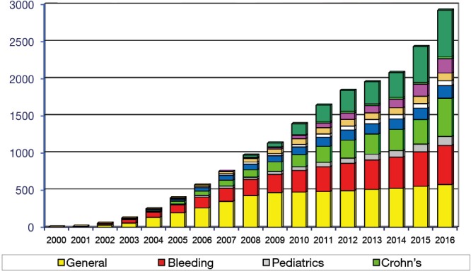
While this activity was in full progress in Israel, Paul Swain, independently in the United Kingdom experimented with wireless transmission from mini cameras and succeeded to send live images from a pig’s stomach to a video screen (3). Meron met Swain in 1997 at the United European Gastroenterology Week (UEGW) in Birmingham. They decided to join forces. Swain was keenly aware of the colossal achievements made in Israel. He reports “… successfully overcoming the enormous obstacles of size, transmission strength, battery power, image resolution, among many others working prototypes were produced in January 1999 by the Given Imaging R&D group headed by Dr. Arkady Glukhovsky” (4). Paul Swain received the honor to be the first human on planet earth to swallow a capsule endoscope. This took place October 1999 in Dr. Scapa’s office in Ramat Hasharon near Tel Aviv. Eitan Scapa, Paul Swain and nearly the entire Given Imaging team (8 to 10 people) gathered in his basement, their eyes glued to the screen. A minor drama took place. The capsule did not leave Paul Swain’s stomach for over three hours. Scapa passed an endoscope without sedation into Swain and with a snare moved the capsule into the duodenum. Three years later, in April 2002, the first international capsule endoscopy conference took place in Rome, Italy. I was among the over 90 physicians who had convened from 12 different countries to share their first experience on some 850 studies. I remember the excitement that was palpable in the hall during the three-day conference when every presenter produced new images and videos of small intestinal pathology that had remained hidden to the human eye until then. All of us were aware that we had just set foot on terra incognita and that the capsule had moved the border of knowledge in gastroenterology another mile forward. A month later, at Digestive Disease Week (DDW) in San Francisco, physicians presented more than 30 abstracts on capsule endoscopy. Since then the number of peer-reviewed publications on capsule endoscopy has skyrocketed (see Figure 1). At DDW I ran into Gavriel Iddan, the dreamer of capsule endoscopy. Beaming with happiness, he said to me, do you know what a physician just told me? Because of your capsule, I was able to save a patient’s life! Iddan had realized his dream. He had wanted to help patients*.
Figure 1.

More than 2,500 peer reviewed publications on capsule endoscopy as of Dec. 01, 2016.
Acknowledgements
None.
Gavriel Iddan ends his article on “A Short History of the Gastrointestinal Capsule” published in Atlas of Video Capsule Endoscopy, by Keuchel M, Hagenmueller F, Fleischer DE, Springer Verlag 2006, 1-3, with the remarkable sentence: “Letters of gratitude from doctors and patients who were helped by the capsule are often received at the company›s headquarters.”
Conflicts of Interest: The author has no conflicts of interest to declare.
References
- 1.Fossum ER. Active image sensors: are CCDs Dinosaurs? Proc SPIE 1993;1900:2-14. 10.1117/12.148585 [DOI] [Google Scholar]
- 2.Personal communication Dov Avni and Gavriel Meron , 2017
- 3.Swain CP, Gong F, Mills TN. Wireless transmission of a colour television moving image from the stomach using a miniature CCD camera, light source and microwave transmitter. Gut 1997;45:AB40. [Google Scholar]
- 4.Iddan GJ, Swain CP. History and development of capsule endoscopy. Gastrointest Endosc Clin N Am 2004;14:1-9. 10.1016/j.giec.2003.10.022 [DOI] [PubMed] [Google Scholar]


