Abstract
The entry of infectious agents in rodent colonies occurs despite robust sentinel monitoring programs, strict quarantine measures, and stringent biosecurity practices. In light of several outbreaks with Aspiculuris tetraptera in our facilities, we investigated the presence of anthelmintic resistance and the use of exhaust air dust (EAD) PCR for early detection of A. tetraptera infection. To determine anthelmintic resistance, C57BL/6, DBA/2, and NCr nude mice were experimentally inoculated with embryonated A. tetraptera ova harvested from enzootically infected mice, followed by treatment with 150 ppm fenbendazole in feed, 150 ppm fenbendazole plus 5 ppm piperazine in feed, or 2.1 mg/mL piperazine in water for 4 or 8 wk. Regardless of the mouse strain or treatment, no A. tetraptera were recovered at necropsy, indicating the lack of resistance in the worms to anthelmintic treatment. In addition, 10 of 12 DBA/2 positive-control mice cleared the A. tetraptera infection without treatment. To evaluate the feasibility of EAD PCR for A. tetraptera, 69 cages of breeder mice enzootically infected with A. tetraptera were housed on a Tecniplast IVC rack as a field study. On day 0, 56% to 58% of the cages on this rack tested positive for A. tetraptera by PCR and fecal centrifugation flotation (FCF). PCR from EAD swabs became positive for A. tetraptera DNA within 1 wk of placing the above cages on the rack. When these mice were treated with 150 ppm fenbendazole in feed, EAD PCR reverted to pinworm-negative after 1 mo of treatment and remained negative for an additional 8 wk. The ability of EAD PCR to detect few A. tetraptera positive mice was investigated by housing only 6 infected mice on another IVC rack as a field study. The EAD PCR from this rack was positive for A. tetraptera DNA within 1 wk of placing the positive mice on it. These findings demonstrate that fenbendazole is still an effective anthelmintic and that EAD PCR is a rapid, noninvasive assay that may be a useful diagnostic tool for antemortem detection of A. tetraptera infection, in conjunction with fecal PCR and FCF.
Abbreviations: AHU, air-handling unit; CRL, Charles River Laboratories; EAD, exhaust air dust; FCF, fecal centrifugation flotation; VHP, vaporized hydrogen peroxide
The prime objective of a sentinel program is early detection of pathogens before research data is compromised due to confounding variables such as infection caused by viruses, bacteria or parasites. Despite a stringent sentinel program, meticulous husbandry practices, high quality equipment and biosecurity, outbreaks of pathogens still occur in rodent colonies. Two major oxyurids found in mice are Syphacia obvelata and Aspiculuris tetraptera. Their immunomodulatory effect is well documented, and thus they act as a confounding factor especially in immunologic studies.3,5,9,33,46,63 These murine pinworms are the most prevalent among all the mouse parasites in rodent colonies in various parts of the world.51,55 Due to their biology, including intermittent shedding of ova and environmentally resistant ova, these agents can go undetected in animal facilities and thus become chronic and persistent. Current methods of detection of murine pinworms are either too invasive or not fully reliable, thus resulting in many false-negative results. Many facilities treat the incoming rodents with fenbendazole prophylactically during the quarantine period regardless of their health status. These mice may be shipped to other institutions as part of collaborative effort. The question arises of whether these practices could create mouse pinworms that are resistant to anthelmintics as well as perpetuate resistance. During the last decade (2006 through 2015), 24 outbreaks of A. tetraptera infection have been detected in rodent facilities on the campus of University of North Carolina at Chapel Hill. Therefore the current studies were aimed to investigate anthelmintic resistance as well as better methods of antemortem detection of pinworms in our mouse colonies enzootically infected with A. tetraptera.
Scant information is available regarding anthelmintic resistance in laboratory bred rodent colonies. However, during the last 2 decades, anthelmintic resistance has surged worldwide, especially in the control of gastrointestinal nematodes in various species of livestock such as sheep, goats, cattle, and horses.37,49,61,62,70 Resistance against antiparasitic drugs for protozoa such as Plasmodium, Giardia, and Eimeria in people and chickens has also been reported.62 The term ‘global worming’ has been used to describe the indiscriminate use of broad-spectrum anthelmintic drugs that has contributed to the development of resistance.37 Repeated anthelmintic treatments provide a positive selective advantage for the survival of the worms that carry the mutation for resistance, and these resistance genes are inherited by their progeny.27 Repeated use of the antiparasitic drugs can lead to selection pressure and even changes in the biology of the worms.57 The in vitro tests used to diagnose anthelmintic resistance in animals include the egg-hatch test and microagar larval development test, neither of which were used in the current study.14 The in vivo tests to detect resistance include fecal egg count reduction testing as well as treatment and necropsy assays.14,70 The latter in vivo test involves isolating potentially resistant parasites, inoculating animals and conducting sensitivity assays by performing necropsy of treated and untreated animals, which was done in the current study. Once anthelmintic resistance is identified in a parasitic population, the various molecular tests used to detect molecular markers of resistance include pyrosequencing assays designed to measure resistance-associated allelic frequencies, as well as PCR-based assays, such as allele-specific PCR, restriction fragment-length polymorphism analysis, and tandem competitive PCR.37,62,70 Molecular monitoring of parasite populations to evaluate anthelmintic susceptibility has become part of the parasite control programs in nonrodent species.62
Drugs that have been used to treat pinworm infection in mice include fenbendazole, thiabendazole, ivermectin, piperazine, moxidectin, doramectin, levamisole, mebendazole, and netobimin.15,56,67 In the current study, we used the in vivo assay mentioned above to test the anthelminthic resistance of A. tetraptera, which was the only murine oxyurid detected in our mice colonies during the past decade. This large scale in vivo assay to test for resistance can be done in mice because of the availability of large numbers of mice for testing, low cost, ease of testing procedure and ease of necropsy. We tested the resistance of A. tetraptera against fenbendazole and piperazine. Fenbendazole is a methylcarbamate benzimidazole broad-spectrum anthelmintic with ovicidal, larvicidal, and adulticidal activity as well as wide margin of safety. Fenbendazole acts by binding and damaging tubulin in helminths, thereby inhibiting tubulin polymerization, microtubule formation, and the intracellular microtubular transport system.54 Piperazine causes flaccid paralysis of the worms by blocking acetylcholine at the neuromuscular junction.54 Piperazine also binds to GABA-gated chloride channels located on somatic muscle cells of the parasite. The resulting increased permeability of chloride into the cell causes relaxation and paralysis of the musculature.39 We hypothesized that the reason for the high number of outbreaks of A. tetraptera at our facility was that the worms had acquired resistance to anthelmintics. To test this hypothesis, we evaluated the resistance of our endogenous populations of A. tetraptera against fenbendazole and piperazine by evaluating 3 strains of mice and 2 methods of worm inoculation.
Another component of the prevention of pinworm outbreaks is better methods for detecting infection. Pinworm ova persist in the environment for a long time, thus presenting a challenge to completely eradicating this parasite.45 Open-top cages have the highest risk of disease transmission through aerosols, fomites and potential contact between cages. In the last 2 decades, many animal facilities have transitioned to using IVC, which provide both biocontainment and bioexclusion. A low prevalence of pinworms is hard to detect in dirty-bedding sentinels housed in IVC because ova are diluted in the bedding, thereby decreasing the chances that sentinel mice will ingest them and get infected. Susceptibility to pinworm infection is dependent on age, sex, strain and immune status of the host.34,43,44,68 The effectiveness of soiled-bedding sentinels to detect pinworm infection is influenced by factors such as amount of bedding transferred, quantity of viable ova in the bedding, frequency of bedding transfer, diagnostic test used, and time elapsed between first exposure of sentinels to dirty bedding and diagnostic testing.24,25 Current methods to detect A. tetraptera include fecal centrifugation flotation (FCF), PCR of fecal pellets, and gross examination of cecum and colon.22,24,26,45,50 Real-time PCR was found to be 4 times more sensitive than FCF in detecting pinworm DNA in fecal samples, and results correlated well with gut checks.22 Histologic examination of sections of colon may help to detect very low worm burden.
Accurate detection of pinworm infection ante mortem is challenging because pinworm ova are shed intermittently leading to a high probability of false negatives.13 Pinworm eggs persist in the environment, such as in dust, equipment, and ventilation intake ducts.31 Pinworm eggs have been detected in the dust of the ventilation system, dirty cages, and even on the hands of technicians working in a rat breeding facility.42 The idea of direct detection of infectious agents by swabbing surfaces such as cages and racks originated a decade ago.16,17 Exhaust air from the rack has been monitored for infectious agents in the past by housing sentinel mice in customized cages that received a portion of the exhaust air from IVC rack prior to HEPA filtration and by testing gauze filters on the inner surface of the exhaust prefilter of the IVC rack.17 Exhaust air sentinels and gauze filters were very effective in detecting mouse hepatitis virus, Sendai virus, and Helicobacter spp. but less effective in detecting mouse parvovirus.17 A recent publication was the first report of successful detection of fur mite DNA using swabs from horizontal exhaust manifolds, with 94% probability of detection within a month of placing the cage with infected mice on the IVC rack.35 However environmental sampling carries a high risk of getting false-positive results if PCR primers are nonspecific. A recent report identified a preponderance of false-positive PCR results from exhaust air dust (EAD) swabs for mouse pinworms because of nonspecific PCR primers.41
The mouse populations in 2 long-standing rodent facilities on our campus were enzootically infected with A. tetraptera. One of these vivaria was fully renovated and repopulated with pinworm- free rodent colonies. Last year, the plans for renovating the second enzootically A. tetraptera infected vivarium with conventional open top cages, were formulated. The renovations of this vivarium provided the opportunity to conduct the field studies described in the second half of this manuscript. These field studies were single experimental manipulations followed by observations. In these studies, we investigated if we could detect A. tetraptera DNA in components of the IVC rack as well as air handling unit using real time PCR and the ability of this method to detect very few A. tetraptera-positive mice on the IVC rack. Environmental decontamination is highly recommended after treatment is initiated in pinworm-infested mice to avoid the risk of reinfection.19,31 We also evaluated our decontamination methods for the room and the equipment by testing the EAD samples with real-time PCR for pinworm DNA. These studies were conducted with the primary objective of improving the institutional rodent health surveillance program.
Materials and Methods
Animals.
For study 1, male (age, 3 to 4 wk) DBA/2NTac (DBA/2), C57BL/6NTac (C57BL/6), and CrTac:NCr-Foxn1nu (NCr) nude mice, were obtained from Taconic Biosciences (New York, NY). These vendor mice were negative for minute virus of mice, Theiler murine encephalomyelitis virus, mouse hepatitis virus, mouse norovirus, mouse parvovirus, enzootic diarrhea of infant mice virus, pneumonia virus of mice, ectromelia virus, mouse adenovirus, lymphocytic choriomeningitis virus, mouse cytomegalovirus, polyoma virus, lactate dehydrogenase elevating virus, pinworms, and fur mites. These mice were housed in groups of 3 or 4 in static filter microisolation cages (Allentown Caging, Allentown, PA) with irradiated corncob bedding (The Andersons Lab Bedding, Maumee, OH) and a 12:12-h light:dark cycle. They were fed an irradiated diet (no. 5058, Purina LabDiet, St Louis, MO) ad libitum and had ad libitum access to hypochlorinated reverse-osmosis–purified water from bottles.
For field study 2, a total of 69 breeder cages from a colony of transgenic mice on C57BL/6J background were enrolled in the study. Each cage housed 2 or 3 mice (age, 2 to 8 mo) for pair or trio mating. The sentinel cage for these breeders containing 2 Crl:CD1(ICR) female mice (age, 3 mo), was placed on the same IVC rack as the breeders. The mice for field study 3 were progeny of these breeder mice before their treatment to clear A. tetraptera infection. All the mice in this vivarium were initially housed in static open-top cages with weekly cage changes. The sentinels from this vivarium were consistently positive for A. tetraptera and mouse hepatitis virus. These mice had never been treated for pinworm infestation. The sentinels were negative by serology for ectromelia virus, enzootic diarrhea of infant mice virus, lymphocytic choriomeningitis virus, Mycoplasma, murine parvovirus, minute virus of mice, polyoma virus, pneumonia virus of mice, reovirus 3, Theiler murine encephalomyelitis virus, Sendai virus, mouse adenovirus types 1 and 2, and mouse cytomegalovirus. For field studies 2 and 3, the mice were transferred to autoclaved IVC cages on a Green line rack with a Smartflow air handling unit (AHU; Tecniplast USA, West Chester, PA). Mice were fed irradiated RMH 3000 diet (Purina LabDiet) ad libitum, were given autoclaved hypochlorinated reverse-osmosis–purified water in bottles, and were housed on autoclaved irradiated corncob bedding (The Andersons Lab Bedding). IVC were changed once every 2 wk in a cage-changing station (Cs5 Evo Changing Station, Tecniplast USA), and water bottles were changed once a week. The sentinel mice were exposed to a teaspoonful of dirty bedding from each cage at the time of cage change. The mice were maintained on a 12:12-h light:dark cycle, ventilation of 75 air changes per hour, temperature of 21 to 23 °C (70 to 74 °F), and 30% to 70% humidity. All animal procedures were reviewed and approved by the IACUC of the University of North Carolina at Chapel Hill (UNC). The animal care program of the UNC has full AAALAC accreditation.
Harvest and amplification of Aspiculuris tetraptera worms for study 1.
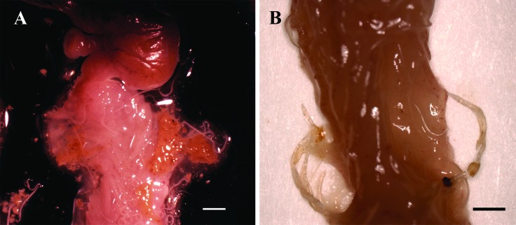
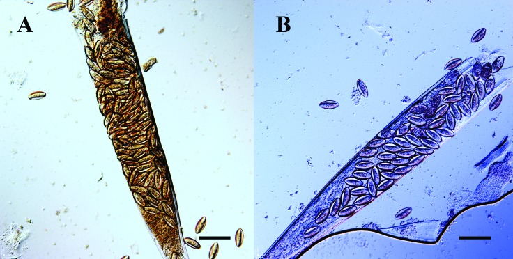
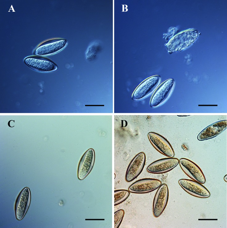
A. tetraptera worms were harvested from mice with a known history of A. tetraptera infection. The mucosa as well as fecal contents of entire cecum and colon were examined to locate the worms (Figure 1). The worms were placed in a 100 × 15-mm Petri dish containing 20 mL distilled water. The opened colons were kept in 100 × 15 mm Petri dishes with a small amount of distilled water overnight for reexamination the next day. A square was drawn in the center of a 75 × 25 mm glass slide by using a wax pencil, and 6 to 8 drops of distilled water were placed within the square. Ten gravid female worms were placed in the water and macerated partially with wooden sticks to release some eggs (Figure 2). To prevent the slides from drying out, each slide with worms was placed on 2 wooden sticks in a Petri dish with a folded water-saturated lab wipe (Kimtech Science Kimwipes Delicate Task Wipers, Kimberly–Clark Professional, Roswell, GA) at the bottom (Figure 3). The slides were gently aerated by using a plastic pipette once daily and water was added to the lab wipes once daily until the eggs were harvested. Each gravid female worm had approximately 200 eggs. Eggs started to embryonate beginning on day 3 at room temperature (20 °C [68 °F]; in the drops of distilled water (Figure 4). Movement was seen in these eggs under light microscope. In order to amplify and sustain A. tetraptera infection, 3-d-old embryonated eggs were used to infect male NCr nude mice via oral gavage. At 4 wk after gavage, fecal pellets from nude mice were checked for pinworm ova by using FCF. Nude mice were euthanized, and gravid A. tetraptera worms were harvested 3 to 4 d prior to each inoculation date.
Figure 1.
Anatomic location of Aspiculuris tetraptera worms. (A) A. tetraptera worms in the proximal colon. Bar, 2 mm. (B) A. tetraptera worms embedded in the crypts of colon. Bar, 1 mm. Magnification, 6.25×.
Figure 2.
Parts of macerated female Aspiculuris tetraptera worms filled with embryonated ova on day 3 of culture in distilled water at room temperature. Bar, 150 µm. Magnification, 40×.
Figure 3.
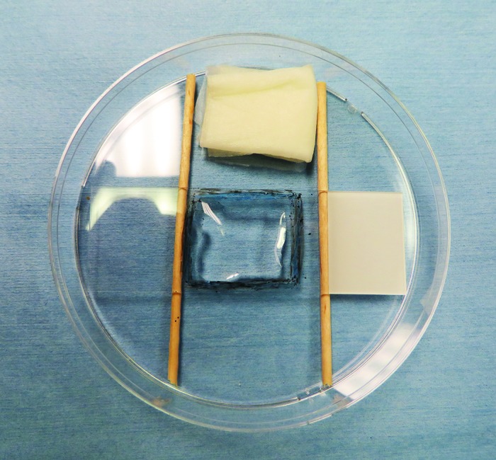
Slide set-up for culturing Aspiculuris tetraptera worms at room temperature. A square was drawn in the center of a 75 × 25 mm glass slide by using a wax pencil, and 6 to 8 drops of distilled water were placed within the square. The gravid female worms were placed in the distilled water and macerated to release eggs. Each slide with worms was placed on 2 wooden sticks in a Petri dish with folded Kimwipe saturated with water in the bottom.
Figure 4.
Embryonated Aspiculuris tetraptera ova on day 3 of culture in distilled water at room temperature. (A) 2 live ova. (B) 2 live and 1 dead ova. Embryonated Aspiculuris tetraptera ova on day 5 of culture in distilled water at room temperature. (C) 2 live ova. (D) Multiple live ova with one dead ovum. Bar, 50 µm. The unembryonated ova prior to day 3 of culture have a round nucleus inside an ellipsoidal outer shell. The embryonated ova have an elongated embryo which can be seen moving inside the outer shell. The embryonated ova look very similar to each other from day 3 to day 5 of the culture. Magnification, 40×.
Study 1: Assessment of anthelmintic resistance of A. tetraptera against fenbendazole and piperazine in various strains of mice.
Study 1A: Evaluation of anthelmintic resistance in A. tetraptera in DBA/2, C57BL/6, and NCr mice after oral inoculation of A. tetraptera ova.
Newly arrived male DBA/2, C57BL/6, and NCr nude mice (Taconic Biosciences) were individually tested for A. tetraptera and S. obvelata by FCF and tape test. After 3 to 4 wk of acclimation, mice from each strain were randomly assigned to 5 groups for each strain. The numbers of mice per group are listed in Table 1. (Table 1). Four groups of mice were inoculated with 0.1 to 0.3 mL of distilled water containing 200 to 400 embryonated A. tetraptera eggs via oral gavage. The fifth group was gavaged with distilled water only as the negative control. Mice were tested by FCF at 4 wk after inoculation to verify infection and shedding. Upon confirmation of presence of infection in inoculated mice and absence of infection in the negative control group, 3 groups of A. tetraptera-positive C57BL/6 and DBA/2 mice, were treated with 3 different anthelmintics for 8 wk. The fourth group in each strain served as the untreated positive control. The 3 groups of A. tetraptera-positive NCr nude mice were treated with these anthelmintics for 4 wk only. The anthelmintics were: 150 ppm fenbendazole in feed; 150 ppm fenbendazole plus 5 ppm piperazine in feed; and 2.1 mg/mL piperazine in drinking water. Mice were tested for pinworm infection by using FCF every week for a total of 4 wk after starting the treatment. C57BL/6 and DBA/2 mice were necropsied after 8 wk of treatment, and NCr nude mice were necropsied after 4 wk of treatment. The mucosa and contents of cecum and colon were examined for pinworms at necropsy.
Table 1.
Average number of A. tetraptera ova obtained by fecal centrifugation flotation per cage and average number of A. tetraptera worms per mouse at necropsy in 3 strains of mice treated with different anthelmintic drugs for 8 wk (DBA/2, C57BL/6) and for 4 wk (NCr nude)
| Strain of mice | Treatment group | n | Average no. of ova after treatment |
Average no. (range) of worms at necropsy | |||
| Week 1 | Week 2 | Week 3 | Week 4 | ||||
| DBA/2 | NEG | 10 | 0 | 0 | 0 | 0 | 0 (0) |
| POS | 12 | 19 | 15 | 5 | 21 | 1 (0–6) | |
| FEN | 12 | 0 | 0 | 0 | 0 | 0 (0) | |
| FEN + PIP | 11 | 0 | 0 | 0 | 0 | 0 (0) | |
| PIPW | 12 | 0 | 0 | 0 | 0 | 0 (0) | |
| C57BL/6 | NEG | 10 | 0 | 0 | 0 | 0 | 0 (0) |
| POS | 10 | 25 | 30 | 118 | 208 | 155 (112–221) | |
| FEN | 12 | 0 | 0 | 0 | 0 | 0 (0) | |
| FEN + PIP | 12 | 0 | 0 | 0 | 0 | 0 (0) | |
| PIPW | 13 | 0 | 0 | 1 nonviable ovum | 0 | 0 (0) | |
| NCr | NEG | 7 | 0 | 0 | 0 | 0 | 0 (0) |
| POS | 7 | 5 | 1 | 6 | 4 | 5 (5–10) | |
| FEN | 6 | 0 | 0 | 0 | 0 | 0 (0) | |
| FEN + PIP | 6 | 0 | 0 | 0 | 0 | 0 (0) | |
| PIPW | 8 | 0 | 0 | 0 | 0 | 0 (0) | |
FEN, 150 ppm fenbendazole in feed; FEN + PIP, 150 ppm fenbendazole and 5 ppm piperazine in feed; NEG, negative-control mice that were orally gavaged with distilled water only; PIPW, 2.1 mg/mL piperazine in drinking water; POS, positive-control mice that were orally gavaged with A. tetraptera ova but were not treated.
Study 1B: Evaluation of anthelmintic resistance in A. tetraptera in NCr mice after oral and topical inoculation of A. tetraptera ova.
In a separate experiment, CrTac:NCr-Foxn1nu (NCr) nude mice were divided into 9 groups. The numbers of mice per group are listed in Table 2. (Table 2). Four groups of mice were infected with greater than 100 A. tetraptera embryonated ova in distilled water via oral gavage. Another 4 groups of mice were infected topically with A. tetraptera by dripping distilled water containing more than 100 embryonated eggs on the mouse's head, shoulders, as well as on the bedding. The ninth group was gavaged with distilled water only, as the negative control. All mice were tested by FCF at 4 wk after inoculation to verify infection and shedding. Upon confirmation of presence of infection in inoculated mice and absence of infection in negative control group, 6 groups of A. tetraptera-positive mice were treated with 3 different anthelmintics for 8 wk. The anthelmintic combinations were: 150 ppm fenbendazole in feed; 150 ppm fenbendazole plus 5 ppm piperazine in feed; and 2.1 mg/mL piperazine in drinking water. Mice were tested for pinworm infection by using FCF every week for the first 4 wk and then during the seventh and eighth weeks, after starting the treatment. Mice were euthanized at the end of the treatment period. The mucosa and contents of cecum and colon were examined for pinworms at necropsy.
Table 2.
Average number of A. tetrapteraova by fecal centrifugation flotation per cage and average number of A. tetrapteraworms per mouse at necropsy in CrTac:NCr-Foxn1nunude mice inoculated with A. tetrapteraova via oral gavage or topically and then treated with different anthelmintic drugs for 8 wk
| Method of inoculation of ova | Treatment Group | n | Average no. of ova after treatment |
Average no. (range) of worms at necropsy | |||
| Week 1 | Week 2 | Week 3 | Week 4 | ||||
| Oral gavage | NEG | 3 | 0 | 0 | 0 | 0 | 0 (0) |
| POS | 6 | 3 | 8 | 9 | 29 | 58 (5–110) | |
| FEN | 11 | 0 | 0 | 0 | 0 | 0 (0) | |
| FEN + PIP | 11 | 0 | 0 | 0 | 0 | 0 (0) | |
| PIPW | 7 | 0 | 0 | 0 | 0 | 0 (0) | |
| Topical | POS | 8 | 26 | 99 | 46 | 5 | 107 (2–144) |
| FEN | 8 | 0 | 0 | 0 | 0 | 0 (0) | |
| FEN + PIP | 12 | 0 | 0 | 0 | 0 | 0 (0) | |
| PIPW | 7 | 0 | 0 | 0 | 0 | 0 (0) | |
FEN, 150 ppm fenbendazole in feed; FEN + PIP, 150 ppm fenbendazole and 5 ppm piperazine in feed; NEG, negative-control mice that were inoculated with distilled water only; PIPW, 2.1 mg/mL piperazine in drinking water; POS, positive-control mice that were inoculated with A. tetraptera ova but were not treated.
Study 2: Field study to test enzootically infected mouse colony for A. tetraptera by PCR from EAD swabs.
A brand-new 140-cage capacity Green line IVC rack (Tecniplast USA) with new autoclavable hoses and new Smartflow AHU (Tecniplast USA) were swabbed, and samples were sent for pinworm PCR to 2 commercial diagnostic laboratories (IDEXX BioResearch, Columbia, MO, and Charles River Laboratories [CRL], Wilmington, MA). Pinworm PCR was negative from both laboratories. New exhaust and supply prefilters were placed on the AHU. A total of 69 breeder cages from the colony enzootically infected with A. tetraptera and 1 sentinel cage were transferred to the above new IVC rack and AHU. A 4 × 18 cm strip of 3M Filtrete 1900 filter paper (Filtrete, Maplewood, MN) marked with 2 × 2-cm squares, was affixed with tape on the underside of the exhaust prefilter (Figure 5). The contaminated air from the IVC rack first comes in contact with underside of the exhaust prefilter before it is filtered. Fecal pellets at different stages of desiccation were collected from the bedding of each cage for FCF to detect pinworm ova. Fresh fecal samples and fur swabs were collected from mice in the cages that were negative for pinworm ova by FCF. Fecal samples were submitted to IDEXX BioResearch, whereas fecal samples and fur swabs were submitted to CRL for pinworm PCR. This evaluation was done to establish the prevalence of pinworm infestation in the mice at the onset of the study.
Figure 5.

View of the underside of the exhaust prefilter in the air handling unit. A 4 × 18 cm strip of 3M Filtrete 1900 filter paper, with 2 × 2-cm marked squares, was affixed with tape on the underside of the exhaust prefilter.
EAD swabbing of the components of the IVC rack and AHU was performed weekly, starting from 1 wk after the mice were placed on the IVC rack. These breeder mice were fed irradiated RMH 3000 diet (Purina LabDiet, St. Louis, MO). After EAD swabs tested positive for A. tetraptera by PCR, mice were switched to irradiated 2920X diet (Harlan Laboratories, Madison, WI) for 1 wk to get them acclimated. After 1 wk of the new diet, all of the mice including sentinels, were given irradiated diet containing 150 ppm fenbendazole (TD.130910, Harlan Laboratories), fed ad libitum. After 4 wk of treatment by feeding the fenbendazole-medicated diet, all the mice were transferred to another decontaminated IVC rack and AHU. The second IVC rack, hoses, and AHU were confirmed negative for pinworm PCR before transferring the study mice to them. The study room was sanitized and decontaminated as described later. EAD swabbing was commenced again 1 wk after cleaning the room and transferring mice to a new IVC rack. The swabs were sent to both IDEXX BioResearch and CRL for pinworm PCR. After disinfection of the housing room and 4 wk of treatment, the supply prefilter on AHU was also swabbed every week for 4 wk until the end of fenbendazole treatment in order to detect pinworm DNA in the room air. Fenbendazole treatment was done for a total of 8 wk. The EAD swabbing was continued every week for 1 mo after finishing the fenbendazole treatment. The filter paper in the sentinel cage top was tested by PCR for pinworm DNA every 2 wk by CRL as described below, from the beginning of study until the end of fenbendazole treatment of mice.
At the end of this 2-mo period, the breeder mice that needed to be culled from the colony were necropsied, and cecum and colon were examined for the presence of pinworms. A total of 73 mice were necropsied from 33 culled cages. Then fecal pellets at different stages of desiccation were collected from the bedding of the remaining cages to test for pinworm ova by FCF. This was done to confirm that the mice were no longer shedding pinworms. Fresh fecal samples and fur swabs were collected from mice in cages that were negative for pinworm ova by FCF. These samples were sent to the 2 diagnostic laboratories for pinworm PCR. Sentinel mice on the study rack were necropsied, and the cecum and colon were examined for pinworms. Any mice that were euthanized or found dead throughout the study period were necropsied, and the cecum and colon were examined for pinworms.
Study 3: Field studies to determine the ability of EAD PCR to detect A. tetraptera DNA with few A. tetraptera-positive mice on the IVC rack.
Study 3A: Field study to determine the ability of EAD PCR to detect A. tetraptera DNA with 2 A. tetraptera-positive mice on the IVC rack.
A 140-cage capacity Green line IVC rack, hoses, and Smartflow AHU (Tecniplast USA) were cleaned and decontaminated. Various components of this equipment were swabbed and confirmed negative by pinworm PCR. A 18 × 4 cm strip of 3M Filtrete 1900 filter paper was attached on the underside of the exhaust prefilter in the AHU and filter paper was sampled as described in the EAD swabbing procedure. Only 3 cages were placed on this IVC rack and the remaining 137 spaces were left empty. We confirmed with the manufacturer that the pressure inside the cages, the travel of air in the rack and air changes per hour are not adversely affected by having only 3 cages on the Techniplast Green line IVC rack. One cage contained 2 mice (age, 8 to 12 wk) that were negative for A. tetraptera ova by FCF. Another cage contained 2 mice (age, 8 to 12 wk), one of which was negative and one was positive for A. tetraptera ova by FCF. These 4 mice were weaned progeny from the untreated breeder colony that was enzootically infected with A. tetraptera. The mice negative for A. tetraptera by FCF were added to the study cages to generate dust. The third cage contained 2 sentinel Crl:CD1(ICR) mice (age, 3 to 4 wk), which were exposed to dirty bedding from the 2 cages at the beginning of the study and every 2 wk at the time of cage change. Weekly EAD swabbing was performed on the components of IVC rack and AHU, beginning 1 wk after the mice were placed on the IVC rack. The swabs were sent to both IDEXX BioResearch and CRL to test by pinworm PCR. Fecal samples from bedding of the 2 cages with study mice were periodically tested for pinworm ova by FCF. Fresh fecal samples from each of the 4 study mice were periodically tested by PCR for pinworms. After 6 wk of EAD swabbing, both the study mice and sentinel mice were necropsied. Their cecum and colon were examined for pinworms at necropsy.
Study 3B: Field study to determine the ability of EAD PCR to detect A. tetraptera DNA with 6 A. tetraptera-positive mice on the IVC rack.
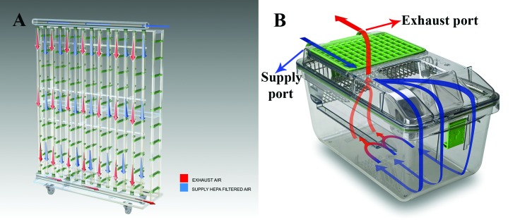
The same room, IVC rack, hoses, and AHU as in study 3A were used for study 3B. The exhaust and supply prefilters in AHU were replaced with new prefilters. A new 18 × 4 cm strip of 3M Filtrete 1900 filter paper was attached on the underside of the exhaust prefilter in the AHU, and filter paper was sampled as described in the EAD swabbing procedure. Sixty-seven spaces on the rack were filled with cages containing bedding and feed in the feed hopper (but not mice) in order to simulate the normal air flow pattern on this 140-cage capacity IVC rack (Figure 6). Again, only 3 cages of mice were placed on this IVC rack. The first cage contained 4 mice (age, 12 to 16 wk) that were negative for A. tetraptera ova by FCF, and the second cage contained 4 mice (age, 12 to 16 wk) that were positive for A. tetraptera ova by FCF. These 8 mice were weaned progeny from the untreated breeder colony that was enzootically infected with A. tetraptera. The third cage contained 2 sentinel Crl:CD1(ICR) mice (age, 3 to 4 wk). Weekly EAD swabbing was performed on the components of IVC rack and AHU, beginning from 1 wk after the mice were placed on the IVC rack. The swabs were sent to both IDEXX BioResearch and CRL to test by pinworm PCR. Fecal samples from the bedding of the 2 cages with study mice were periodically tested for A. tetraptera ova by FCF. Fresh fecal samples of each of the 8 study mice were periodically tested by PCR for pinworms. The 8 study mice in 2 cages were necropsied after we obtained result of positive PCR from EAD swabs collected 1 wk after the start of the study. Their cecum and colon were examined for A. tetraptera worms. The sentinel mice were exposed to dirty bedding from pinworm-positive and -negative mice for 1 wk only. After exposure, the sentinel mice were transferred to another clean sterile cage and kept on the IVC rack for another 5 wk to cover the prepatent period of A. tetraptera. The sentinel mice were necropsied at the end of that period. Their cecum and colon were examined for pinworms at necropsy.
Figure 6.
Schematic diagram of pattern of flow of exhaust air and HEPA-filtered supply air through (A) Tecniplast Green line IVC rack and (B) Tecniplast Green line IVC cage. Blue arrows indicate supply airflow, and red arrows indicate exhaust airflow. Image courtesy of Tecniplast USA (West Chester, PA).
Fecal centrifugation and flotation.
Fecal samples were collected in 1.5-mL Eppendorf microfuge tubes. The tube was filled with zinc sulfate solution (specific gravity, 1.180) to the 1-mL mark. If the fecal pellets were dry, they were left for about 1 h to soak. When the sample became soft after soaking, the solution was mixed well and centrifuged for 5 min at 6000 rpm (2000 × g; Spectrafuge Mini Centrifuge, Labnet International, Edison, NJ). Then zinc sulfate solution was added to form a small meniscus at the top of the vial. A cover slip was placed on top of the meniscus for 15 min and then placed on a glass slide. The slide was examined at 40× magnification for pinworm ova.
Exhaust air dust swabbing.
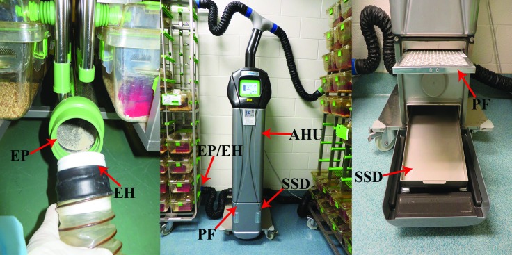
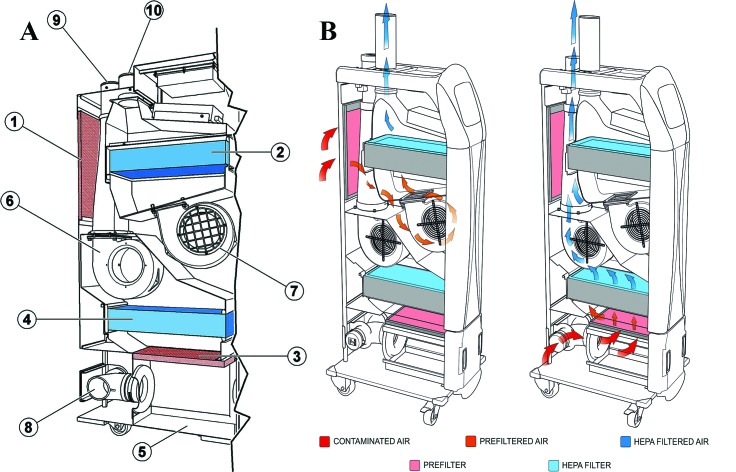
EAD swabs were collected from 4 different components of the Tecniplast Green line IVC rack and Smartflow AHU, namely, the stainless steel drawer below the exhaust prefilter in AHU, the underside of the exhaust prefilter, the inside of exhaust plenum at the bottom of the IVC rack and inside of the hose connected to the exhaust plenum of the IVC rack leading to AHU (Figure 7). The exhaust air from the entire IVC rack comes in contact with underside of the exhaust prefilter first, prior to filtration in the prefilter and HEPA filtration (Figure 8). Diagnostic sampling was done by gently rotating the swab all over the surface in a systematic fashion. Effort was made to collect as much of visible dust on the surface as possible. Diagnostic specimens were collected at the aforementioned four locations using 4 different sticky swabs provided by CRL, and the swabs were pooled as one sample in a 5-mL microfuge tube provided by CRL, for sending to CRL for pinworm PCR. The above 4 locations were also swabbed using four different polyester-tipped cotton swabs (263000 BD CultureSwab, Becton Dickinson, Franklin Lakes, NJ). These swabs were pooled as one sample in a 15-mL sterile conical tube for sending to IDEXX BioResearch for pinworm PCR. In addition, a strip of 4 × 18 cm 3M Filtrete 1900 HVAC filter was attached to the underside of exhaust prefilter in the AHU using tape, upon recommendations from IDEXX BioResearch. Several 2 × 2cm squares were marked on the filter paper (Figure 5). One of these squares was also cut using sterile scissors and forceps at each swabbing and placed with the BD polyester tipped swabs in the 15-mL tube, for inclusion as a single sample for sending to IDEXX BioResearch for pinworm PCR.
Figure 7.
The 4 sites for exhaust air dust (EAD) swabbing on Tecniplast Smartflow air-handling unit (AHU) and Green line IVC rack are shown as: EH, the inside of the exhaust hose connected to the exhaust plenum of the IVC rack leading to AHU; EP, the inside of exhaust plenum at the bottom of the IVC rack; PF, the underside of the exhaust prefilter; and SSD, the stainless steel drawer below the exhaust prefilter.
Figure 8.
Schematic diagram showing the features of Tecniplast Smartflow air handling unit.(A) Components inside the air-handling unit: 1, environment air inlet prefilter; 2, supply air HEPA filter; 3, exhaust air prefilter; 4, exhaust air HEPA filter; 5, dust collection tray; 6, exhaust air blower motor; 7, supply air blower motor; 8, exhaust air inlet; 9, exhaust air outlet to the environment; and 10, supply air outlet to rack. (B) Supply and exhaust air flow through the air handling unit. Image courtesy of Tecniplast USA (West Chester, PA).
Decontamination methods.
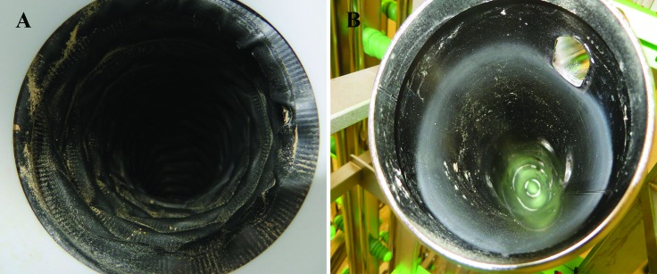
To clean and decontaminate equipment for studies 2 and 3 at the outset of the study, the Green line IVC rack and autoclavable hoses (part no. ACSCVF75M11RGM, Tecniplast USA) were washed for 30 min at 82 °C (180 °F) and autoclaved. The Smartflow AHU and cage-changing station were cleaned using vaporized hydrogen peroxide (VHP; Clarus C Hydrogen Peroxide Vapor Generator, Bioquell, Horsham, PA). After the cage change every 2 wk, the dirty cages with feed and bedding were autoclaved prior to disassembling them for cleaning. After 4 wk of treatment with fenbendazole, the mice were transferred to another IVC rack and AHU that were decontaminated as described earlier. Brand-new supply and exhaust prefilters were placed in this decontaminated AHU. The floor, ceiling, and walls of the room were mopped with diluted Vimoba 128 (1 oz. per 1 gal. water; Quip Labs, Wilmington, DE). Vimoba 128 is a cationic detergent containing quaternary ammonium chloride and has bactericidal as well as viricidal properties. Mechanical scrubbing of the surfaces with detergent to remove A. tetraptera ova has been recommended as a method of environmental decontamination.13 All disposable materials were removed from the mouse room. All carts were cleaned and wiped with diluted Vimoba. The dirty IVC rack and hoses were autoclaved followed by a hot-water wash and then were autoclaved again. Some dust, firmly adhered in the hoses and plenum, remained after this procedure (Figure 9). The dirty AHU and cage-changing station were cleaned using VHP. Swabs from the plenum, hoses, and stainless steel drawer of the AHU from this cleaned equipment, were sent for pinworm PCR to both diagnostic laboratories.
Figure 9.
After 4 wk of treatment with fenbendazole-medicated feed in study 2, the dirty IVC rack was autoclaved followed by washing at 82 °C (180 °F) for 30 min and a second autoclave cycle. The image shows the adhered dust that remained (A) inside the exhaust hose connected to the exhaust plenum of the IVC rack; and (B) inside the exhaust plenum on the IVC rack. PCR analyses from swabs of this adhered dust were negative for Aspiculuris tetraptera DNA.
Testing of sentinel cage-top filters.
The IVC cages for the mouse colony enzootically infected with A. tetraptera were changed every 2 wk. The sentinel mice were exposed to dirty bedding from each cage at the time of cage change. After each cage change, the cage-top filter of the dirty sentinel cage was removed from the lid of the cage. This filter was tested based on recommendations of CRL using the protocol provided by them. A 2.5- to 3-in. square piece of the filter paper was cut using sterile instruments. This piece was rolled and placed in a 50-mL sterile conical tube such that the dirty side, exposed directly to the cage, was on the inside. The filter paper was analyzed by pinworm PCR by CRL every 2 wk, from the beginning of field study 2 until the end of the 8 wk of fenbendazole treatment of mice.
Statistical analysis
Analysis of the data relied on descriptive tabular statistical methods. Interpretation of the results was appropriately focused on statistical estimates (for example, outcome rates) representing the magnitudes of the effects of interest. For study 1, descriptive tabulations were used to characterize the occurrences of infections and their sensitivity to effective anthelmintics. Finding a single worm at necropsy after completion of the treatment of mice for A. tetraptera infection in studies 1A and 1B was considered a significant result. For the field studies (2, 3A, 3B), descriptive tabulations were used to summarize results about the capability of the EAD PCR approach for detecting A. tetraptera infection.
Results
Evaluation of anthelmintic resistance in A. tetraptera in DBA/2, C57BL/6 and NCr mice after oral inoculation of A. tetraptera ova (study 1A).
Newly arrived DBA/2, C57BL/6, and NCr nude mice were negative for A. tetraptera and S. obvelata as determined by FCF and tape test. All the mice gavaged with embryonated A. tetraptera eggs were shedding ova in fecal pellets, as evident by positive FCF for A. tetraptera ova, at 4 wk after inoculation. In comparison, the mice gavaged with distilled water as negative controls remained pinworm-negative 4 wk after the gavage. The numbers of A. tetraptera ova for 4 wk after treatment and A. tetraptera worms at necropsy are shown in Table 1 for various treatment groups in these 3 strains of mice. None of the treated mice had A. tetraptera ova based on testing by FCF beginning 1 wk after treatment with different anthelmintics. All 3 anthelmintic treatments were effective in eliminating A. tetraptera infections after 4 wk of treatment in NCr mice and after 8 wk of treatment in DBA/2 and C57BL/6 mice. Ten of a total of 12 positive-control DBA/2 mice cleared the A. tetraptera infection without treatment, as determined by the absence of worms in the cecum and colon at necropsy. In the remaining 2 DBA/2 mice, one mouse had one worm, and the second mouse had 6 worms in the colon. In contrast, A. tetraptera worms thrived and multiplied well in C57BL/6 mice, as evident by the large numbers of worms found in the cecum and colon of the positive-control C57BL/6 mice at necropsy.
Evaluation of anthelmintic resistance in A. tetraptera in NCr nude mice after oral and topical inoculation of A. tetraptera ova (study 1B).
All of the 8 groups of NCr nude mice inoculated with embryonated A. tetraptera ova via oral gavage or topically were shedding A. tetraptera ova, as determined by positive FCF, at 4 wk after inoculation. The negative-control mice, which were gavaged with distilled water only, remained negative for A. tetraptera ova at 4 wk after inoculation. The numbers of A. tetraptera ova present in the feces for 4 wk after treatment and A. tetraptera worms at necropsy, are shown in Table 2. None of the treated mice had any A. tetraptera ova upon testing by FCF beginning 1 wk after treatment with each of the tested anthelmintics. All 3 anthelmintics were effective in eliminating A. tetraptera infestations after 8 wk of treatment. Large numbers of A. tetraptera worms were present in the colon and cecum of positive control NCr nude mice at necropsy.
Evaluation of PCR from EAD swabs to detect A. tetraptera DNA using mouse colony enzootically infected with A. tetraptera as field study (study 2).
The components of the IVC rack and AHU were tested by pinworm PCR prior to housing the A. tetraptera-infected study mice on them. Pinworm PCR was negative for both A. tetraptera and S. obvelata, as determined by both IDEXX BioResearch and CRL. A total of 20 of 69 (29%) of the breeder mice cages were positive for A. tetraptera ova by FCF. Fresh fecal samples and fur swabs from mice in cages negative by FCF, were sent for pinworm PCR. Among those 49 cages, 19 were positive for A. tetraptera PCR from CRL, whereas 20 were positive for A. tetraptera PCR from IDEXX BioResearch. This difference in the results was possibly because sample collection was done on 2 different days. Thus 56% to 58% of breeder cages were positive for A. tetraptera at the outset of the study. The EAD swabs from the IVC rack and AHU collected 1 wk after starting the study were positive for A. tetraptera, as confirmed by PCR at both diagnostic laboratories. Then mice were provided pelleted diet containing fenbendazole for 8 wk to treat A. tetraptera infection. After 4 wk of treatment, the room was sanitized, and the mice were transferred to another decontaminated IVC rack and AHU. The EAD swabs of the second IVC rack and AHU were confirmed negative for pinworm PCR before the mice were transferred onto them. The EAD swabs from the first IVC rack and AHU were still positive for A. tetraptera by PCR after 4 wk of fenbendazole treatment. This dirty IVC rack was autoclaved, then washed for 30 min at 82 oC (180 oF), followed by autoclaving again. The exhaust prefilter on the AHU was thrown away, and the AHU was decontaminated using vaporized hydrogen peroxide. Although some dust remained adhered inside the plenum and hoses of first IVC rack (Figure 9), the EAD swabs from these cleaned IVC rack and AHU were negative for pinworm DNA by PCR.
EAD swabs of the second IVC rack and AHU were collected every week for 8 wk, comprising the final 4 wk of fenbendazole treatment and an additional 4 wk afterwards. All swabs tested negative for A. tetraptera PCR as confirmed by both diagnostic laboratories. EAD swabs of the supply prefilter in the AHU were collected every week for 4 wk after decontaminating the room and placing mice on the second cleaned IVC rack and AHU. These swabs were negative for pinworm DNA by PCR. Sentinel cage-top filters, collected every 2 wk from start of the study through the end of the fenbendazole treatment, were negative for pinworm DNA by PCR as tested by CRL. No A. tetraptera worms were found on examination of cecum and colon of the 73 mice from 33 cages culled from this breeding colony, after treatment with fenbendazole. The fecal pellets collected from the bedding of the breeder cages remaining at the end of the study, were negative for A. tetraptera ova by FCF. Fresh fecal samples and fur swabs were collected from breeder cages that were negative for A. tetraptera ova by FCF. All of these samples were negative for pinworm DNA by PCR as determined by both diagnostic laboratories. A total of 11 mice died during the course of the study due to unrelated reasons, such as dystocia. The presence or absence of pinworms in the cecum and colon of these 11 mice paralleled the results of FCF and pinworm PCR. The mice in this breeding colony have remained free of pinworms a year after the end of the study, as determined by the quarterly sentinel monitoring program.
Evaluation of ability of PCR from EAD swabs to detect A. tetraptera DNA with 2 infected mice on IVC rack as field study (study 3A).
The components of decontaminated IVC rack and AHU were confirmed as negative for pinworm PCR prior to beginning this study. Three cages of mice were placed on IVC rack. The first cage contained 2 sentinel mice, the second cage contained 2 mice negative for A. tetraptera ova by FCF, and the third cage contained one mouse positive for A. tetraptera ova and one negative for A. tetraptera ova by FCF. The EAD swabs, taken every week for 6 wk, were negative by pinworm PCR as tested by both diagnostic laboratories. Fecal pellets from the bedding of 2 study cages were tested periodically by FCF for A. tetraptera ova. The third cage with the A. tetraptera-positive mouse was inconsistently positive by FCF during the 6-wk period. Pinworm PCR from fresh fecal samples from individual mice revealed that one of the mice in the second cage that was negative for pinworm ova by FCF, was actually positive for A. tetraptera by PCR. However, the positive PCR results were inconsistent for both A. tetraptera-infected mice during the 6-wk period. The negative cagemates in both of the study cages stayed negative for A. tetraptera as tested by PCR. The results of examination of fecal pellets by FCF and by A. tetraptera PCR prior to necropsy as well as from examination of cecum and colon from the 4 study mice and 2 sentinel mice, at the end of the study, are shown in Table 3.
Table 3.
Results of examination of fecal pellets by fecal centrifugation flotation (FCF) and by A.tetrapteraPCR prior to necropsy as well as examination of cecum and colon at necropsy of the mice in field studies 3A and 3B to evaluate ability of exhaust air dust (EAD) PCR for detecting low levels of A. tetrapterainfection
| Study | Cage no. | Animal ID | Presence of ova on FCF | Pinworm PCR | Aspiculuris worms at necropsy | Description of the worms |
| 3A | 1 | 1-1 | Negative | Negative | Negative | |
| 1 | 1-2 | Positive | Negative | Positive | 1 female and 1 male worms | |
| 2 | 2-1 | Negative | Positive | Positive | 1 female worm | |
| 2 | 2-2 | Negative | Negative | Negative | ||
| Sentinel | S-1 | Negative | Negative | Negative | ||
| Sentinel | S-2 | Negative | Negative | Negative | ||
| 3B | 1 | 1-1 | Positive | Nd | Positive | Many adult worms |
| 1 | 1-2 | Positive | Nd | Positive | Few adult worms | |
| 1 | 1-3 | Positive | Nd | Positive | Numerous adult worms | |
| 1 | 1-4 | Positive | Nd | Positive | Many adult worms | |
| 2 | 2-1 | Negative | Negative | Positive | Few juvenile worms | |
| 2 | 2-2 | Negative | Negative | Negative | ||
| 2 | 2-3 | Negative | Negative | Negative | ||
| 2 | 2-4 | Negative | Negative | Positive | Many adult worms | |
| Sentinel | S-1 | Nd | Nd | Positive | One juvenile female worm | |
| Sentinel | S-2 | Nd | Nd | Negative |
Nd, not done
Evaluation of ability of PCR from EAD swabs to detect A. tetraptera DNA with 6 infected mice on IVC rack as field study (study 3B).
Three cages of mice were placed on IVC rack. The first cage contained 4 mice that were negative for A. tetraptera ova by FCF, the second cage contained 4 mice positive for A. tetraptera ova by FCF, and the third cage contained 2 sentinel mice. Sixty-seven spaces on the rack were filled with cages that contained feed and bedding only. EAD swabs from the components of IVC rack and AHU, collected after 1 wk of starting the study, were positive for A. tetraptera DNA by PCR as confirmed by both CRL and IDEXX BioResearch. The results of examination of fecal pellets by FCF and by A. tetraptera PCR prior to necropsy as well as from examination of cecum and colon from the 8 study mice are shown in Table 3. Although the fecal pellets of 4 mice in one cage on the IVC rack were negative for A. tetraptera ova by FCF and by A. tetraptera PCR, 2 of these mice had A. tetraptera worms in the colon at necropsy. Therefore the PCR-positive EAD swab after 1 wk represented the accumulation of A. tetraptera DNA from 6 positive mice on the rack. To compare the use of sentinel mice with EAD PCR to detect A. tetraptera infection, sentinel mice in third cage were exposed to dirty bedding from the 2 study cages for 1 wk only. The sentinel mice were euthanized after another 5 wk. One of the sentinel mice was positive for 1 juvenile female A. tetraptera worm at necropsy (Table 3).
Discussion
We investigated the presence of anthelmintic resistance in our enzootic A. tetraptera worm populations against fenbendazole and piperazine by performing in vivo sensitivity assay in DBA/2, C57BL/6, and NCr nude mice. Our 2 major conclusions from these studies (1A and 1B) were: 1) absence of anthelmintic resistance against fenbendazole and piperazine in our enzootic A. tetraptera worm populations when inoculated in DBA/2, C57BL/6, and NCr nude strains of mice; and 2) the majority of positive-control DBA/2 mice cleared the A. tetraptera infection without treatment. We also conducted field studies, which were single experimental manipulations followed by observations. These field studies involved utilization of real-time PCR technology to evaluate dust collected from components of an IVC rack and AHU as a diagnostic tool to detect A. tetraptera DNA. We also evaluated the ability of EAD PCR to detect A. tetraptera DNA from IVC racks that contained very low numbers of mice enzootically infected with A. tetraptera. Furthermore, we assessed whether our current decontamination methods for the environment comprising the mouse room, IVC rack, AHU, and cage-changing station were effective in eliminating A. tetraptera ova. Our 3 major observations from the field studies (2, 3A, and 3B) were: 1) A. tetraptera PCR from EAD swabs became positive within 1 wk of housing very high as well as very low numbers of mice enzootically infected with A. tetraptera on the IVC racks; 2) one sentinel mouse was positive for a single A. tetraptera worm by direct colon examination at 6 wk after exposure to dirty bedding for 1 wk from 6 A. tetraptera infected mice; and 3) the combination of decontaminating AHU and cage-changing station with VHP as well as washing at 82 oC (180 oF) followed by autoclaving the IVC rack were effective decontamination methods for the elimination of A. tetraptera ova (including DNA) from the equipment.
We hypothesized that treatment failure might be a contributing cause of the A. tetraptera outbreaks in our facilities during the last decade, and we therefore investigated the presence of anthelmintic resistance in the enzootic A. tetraptera worm populations infecting our mouse colonies. Resistance can arise due to novel mutations, preexisting alleles for resistance, recurrent mutations, linkage disequilibrium, meiotic recombination, and migration of alleles.27 Other factors influencing development of anthelmintic resistance include husbandry practices, host species, climate, size of herds or flocks, and the duration, frequency, dose, and type of anthelmintics used. The resistant worm populations can be genetically divergent depending on the origin of resistance.27 The comparison of different susceptible and resistant field isolates can help to identify various genes associated with the resistance phenotype in these worm populations.27 The in vivo sensitivity assay in this study was performed by orally gavaging 3 different strains of mice with embryonated A. tetraptera ova. These ova were harvested from A. tetraptera female worms found in enzootically infected mice populations on campus because we wanted to evaluate resistance in our enzootic pinworm isolate. Although adult female worms can release eggs after incubation in saline for 2 to 3 h at 37 °C,1 we followed the procedure of releasing the ova by macerating female worms in culture conditions.43 Previous studies on hatching characteristics of A. tetraptera ova revealed that the highest hatching rate is achieved at 30 oC to 40 oC and that ova reached the infective stage after 9 to 10 d in culture.1 In contrast, we were able to obtain embryonated eggs beginning on day 3 of culture. Temperature and humidity are critical factors in the development of the eggs outside the host. Our results are similar to those of earlier studies in which ova released from macerated adult A. tetraptera female worms developed first-stage larva by the third day of incubation in aerated distilled water at room temperature.2,53 We used the embryonated eggs collected on the third day of culture to inoculate mice because mice fed eggs containing immature embryos after 24 h of culture did not develop pinworm infection.53
A group of NCr nude mice was inoculated with embryonated A. tetraptera ova topically by suspending the ova in distilled water and dripping the suspension on the head and shoulders of the mice as well as on their bedding. Other researchers have used only oral gavage as the route of inoculation of pinworm ova for producing experimental infection in mice.1,43,44 Our rationale for using topical inoculation of A. tetraptera ova was based on the observation that mice are social animals that interact closely with each other and huddle together in the cage. Thus cagemates can possibly ingest pinworm ova stuck to the fur as well as on the bedding. The study mice were successfully inoculated with A. tetraptera ova by topical application, as indicated by the presence of ova by FCF. In addition, large worm burden was found in the positive-control NCr nude mice at necropsy, indicating that topical inoculation was equivalent to oral gavage in achieving a high worm burden. This novel method of experimental inoculation of A. tetraptera ova by topical application offers a refinement of the experimental technique, by reducing the stress of handling and gavaging the mice. Male mice were used in studies 1A and 1B because they are more susceptible to A. tetraptera infection due to testosterone effects that lower their immune response to parasitic infection.44 Athymic nude mice were used to amplify and sustain A. tetraptera because of their high susceptibility to pinworm infection.34 Parasite burden differs among different strains of mice after acute infection.20 Athymic nude mice, AKR, DBA, C57BL/6, and C3H mice are more susceptible to pinworm infection as compared with other inbred strains.23,34,38,68Therefore we chose male mice of DBA/2, C57BL/6, and NCr athymic nude strains to investigate anthelmintic resistance so that we could achieve a high worm burden in the host to test the efficacy of treatment.
Both methods of anthelmintics delivery, namely, in feed and drinking water, were effective in eliminating A. tetraptera worms. The dosages of anthelmintics used in this study (150 ppm fenbendazole in feed, 5 ppm piperazine in feed, and 2.1 mg/mL piperazine in drinking water) are the same as published dosages for these drugs for treating A. tetraptera infection.56 In one study, treatment with 2.1 mg/mL piperazine in drinking water for 2 wk followed by 2 wk of ivermectin, followed by a second 2-wk treatment with piperazine, successfully eliminated S. muris, Syphacia obvelata and Aspiculuris tetraptera.72 Several published studies report that feed containing 150 ppm fenbendazole fed continuously or during alternate weeks for 5 to 8 wk eliminated A. tetraptera.7,30 We observed that feeding fenbendazole-medicated feed continuously for 8 wk was not only an effective treatment, but it also reduced feed wastage and ensured compliance from the husbandry staff. The delivery of ivermectin via the automated drinking water system was found to be a less-expensive option for treating A. tetraptera when compared with fenbendazole-medicated feed. However the delivery of the drug required modification of the watering equipment, which could be expensive and time consuming.29 In addition, ivermectin cannot be administered to breeders because of its toxic effects in mouse pups.58,59,65 In contrast, fenbendazole-medicated feed can be administered at the time of cage change and requires minimal additional labor, and no additional precautions are needed for personnel handling the medicated feed.
No A. tetraptera ova were noted by FCF in any of the treatment groups at one wk after starting the treatment with anthelmintics. However, the mice were not tested by PCR of fecal pellets for A. tetraptera DNA or by direct visualization of colonic contents at this time point. Thus we are not concluding that 1 wk of treatment was sufficient to clear A. tetraptera infection. A 1-wk treatment may have arrested the ova production in the adult A. tetraptera worms without killing them. However, FCF and direct examination of the cecum were done at 4 wk in NCr mice (study 1A) that showed no A. tetraptera ova or worms in the mice. Although these NCr mice cleared the infection after 4 wk of treatment, we gave the anthelmintics to C57BL/6 and DBA/2 mice for the entire 8-wk treatment period to ensure that no resistant worms developed and that all the mice had the opportunity to consume an adequate therapeutic dosage. A concentration of 150 ppm fenbendazole achieves a dose of 8 to 12 mg/kg daily.69 Although reduced litter size was reported in rats fed fenbendazole feed for 7 wk,36 other researchers did not observe an adverse effect of fenbendazole at therapeutic doses on traits such as reproduction, behavior, and carcinogenesis.69 No A. tetraptera worms were found in the mice in any of the treatment groups at necropsy, indicating that both piperazine and fenbendazole were effective against A. tetraptera infection. Thus our findings did not support the existence of resistance against fenbendazole and piperazine in the A. tetraptera populations evaluated.
Ten of the 12 DBA/2 mice in the positive control group did not have any worms in the colon at necropsy. Although all 12 mice were positive for A. tetraptera ova by FCF at the outset, 10 of those mice reverted to negative status without anthelmintic treatment. The remaining 2 mice had 1 and 6 worms, respectively. DBA/2 mice predominantly have a Th2 cytokine response to antigens, characterized by the production of interleukins such as IL4, IL5, IL13, and IL10.48,60,64 These cytokines evoke antibody responses, including IgE, and activate eosinophils for clearance of extracellular bacteria and parasites such as helminths. The DBA/2 mice in this study likely mounted a protective Th2-biased immune response that caused expulsion of the worms. In contrast, C57BL/6 mice have a Th1-polarized response to antigens, which is characterized by the production of IFNγ, IL2, and TNFβ.4,48,60,64 These cytokines cause the production of complement-fixing antibodies by B cells and the activation of macrophages to kill intracellular bacteria and viruses. These immunological characteristics likely caused the C57BL/6 positive control mice in our study to not expel A. tetraptera worms and thus they had a high worm burden at necropsy. One publication reported that A. tetraptera worms were expelled due to innate immunity upon primary exposure in CFLP mice, which is an outbred laboratory stock in the United Kingdom.6 The CFLP mice in this study subsequently developed acquired immunity and were able to eliminate worms rapidly upon reinfection.6 In another study, infection with S. obvelata in Balb/c mice induced protective Th2 immune responses , and IL13 was found to be the dominant cytokine responsible for worm expulsion in the host.46 Swiss Webster mice exposed to bedding infected with S. obvelata for 8 wk cleared the infection by wk 14 of the study without treatment.11 Further studies revealed that MHC II genes regulate susceptibility to S. obvelata infection, as indicated by significantly higher worm prevalence in MHC II−/− mice compared with MHC II+/+ mice.66 Athymic nude mice are highly susceptible to pinworm infection because they lack the T-cell mediated immune response for pinworm expulsion.34 The positive control NCr mice in the current study also had large number of A. tetraptera worms in the cecum and colon at necropsy. These results support the well-documented role of T cell-mediated immunity in parasite clearance from the host.
In order to test environmental sampling as a diagnostic tool for detecting A. tetraptera DNA, we used real-time PCR to test swabs of dust collected from the exhaust plenum and exhaust hoses of the IVC rack as well as the exhaust prefilter and the stainless steel drawer of AHU. Based on the results of FCF and PCR, 56% to 58% of the 69 breeder cages on the IVC rack had mice infected with A. tetraptera at the outset of the field study. The pooled fecal pellets collected from the bedding at different stages of desiccation, representing different times of defecation, were tested using FCF. This method increased the chances of detecting A. tetraptera ova because female A. tetraptera worms release ova intermittently in the fecal pellets.52 Real-time PCR is an effective method for ante-mortem detection of various infectious agents from feces, fur swabs, and oropharyngeal swabs.28 Real-time PCR for the detection of A. tetraptera was found to be 10 times more sensitive than fecal flotation and 4 times more sensitive than FCF.22 The components of IVC rack and AHU were confirmed negative by pinworm PCR before experimental mice were housed on them. This was done to ensure that any pinworm DNA detected in EAD swabs came from mice housed on the IVC rack rather than the components of the rack and AHU. Interestingly, the EAD swab collected after 1 wk of housing above breeder mice on the IVC rack, was positive for A. tetraptera DNA by PCR. This result was obtained by both CRL and IDEXX BioResearch, which attests to the accuracy of our results.
Clearly, the A. tetraptera DNA present in the fecal pellets, bedding, and even fur of the mice infected with A. tetraptera became airborne, was captured in the components of the IVC rack and AHU, and was sufficient to be detected by real-time PCR. Environmental sampling has previously been explored as a strategy to detect pathogenic agents that are transmitted by aerosolization. One group of researchers placed sentinel mice in custom-designed Bioscreen cages that received a portion of the exhaust air from the entire IVC rack prior to HEPA filtration. These sentinels were used to detect pathogens by exposure to exhaust air from the rack.8,17 Mouse hepatitis virus and Sendai virus were detected in the exhaust-air sentinels placed in Bioscreen cages for 12 wk.17 This creative environmental sampling is effective for detecting pathogens such as pinworms as well, that are transmitted by the fecal–oral route. Mice run through bedding mixed with fecal pellets all the time and some of the contaminated fecal particles can become airborne. Another group of investigators reliably detected fur mite DNA by using PCR from swabs of the horizontal manifolds adjacent to each row on an Allentown IVC rack. They obtained positive fur mite PCR results in 59%, 88%, 88%, and 94% of the infested racks within 1, 2, 3, and 4 wk, respectively, of placing the cages containing live fur mites on the rack.35 Recently, researchers have reported successful detection of murine norovirus and Helicobacter hepaticus in gauze pieces affixed to the exhaust prefilter in the AHU when the infected mice were housed on the IVC racks.47,73 Thus PCR of environmental samples as a routine procedure in health monitoring protocols can be used to augment the information gathered from dirty-bedding sentinels for early detection of pathogens.10 We hypothesized that the microisolation cage filter on the sentinel cage might acquire some A. tetraptera DNA by aerosolization of particles from the dirty bedding from A. tetraptera-infected mice. However, the sentinel cage-top filter never became PCR-positive despite being tested every 2 wk from the start of the study till the end of fenbendazole treatment. The majority of exhaust air in the IVC cages exits through the exhaust port on the back of the cage. Only a small amount of exhaust air exits through the filter in the lid of cage, due to the pressure barrier in the cage. As a consequence, testing of the sentinel cage-top filter was not effective for the detection of A. tetraptera infestation within the rack.
After obtaining PCR-positive EAD swab for A. tetraptera DNA, the next steps included treating the mice with fenbendazole-medicated feed and environmental decontamination so that we could determine if the EAD swab reverted to negative status. All of these mice were treated by feeding fenbendazole-medicated feed continuously for 8 wk. At the end of the study, none of the culled mice from the breeder colony had A. tetraptera worms in the colon at necropsy. In addition, no A. tetraptera infection was found by FCF or PCR of fecal pellets from the remaining cages. Once again, 150 ppm fenbendazole in the feed proved to be an effective treatment for eradicating A. tetraptera from our mouse colonies. After 4 wk of treatment of the mice with fenbendazole-medicated feed, a comprehensive environmental decontamination was performed to test if the mice were still releasing A. tetraptera ova. After decontamination, weekly EAD PCR was negative for A. tetraptera DNA for 2 mo, which marked the end of the study. During the course of our studies, it became apparent that a positive EAD swab for A. tetraptera DNA would have never become negative on its own, even if the mice were treated with fenbendazole-medicated feed, because the dust containing A. tetraptera DNA accumulated and adhered in the components of IVC rack and AHU. To test the efficacy of anthelmintic treatment, removal of this contaminated dust by the processes of autoclaving, washing and decontamination was essential. The exhaust prefilter needed to be replaced after 4 wk of treatment because the dust can never be fully removed from the porous filter in the prefilter. At the same time, a minimum of 4 wk of anthelmintic treatment was needed to cause cessation of ova release from adult A. tetraptera worms.
Environmental decontamination is crucial for removing the residual ova to prevent reinfection and for removing residual DNA to avoid false-positive PCR results. S. muris was eradicated from rats and voles for several months, by feeding fenbendazole-medicated diet on alternating weeks for 9 wk without any environmental decontamination.32 However, the knowledge that pinworm ova survive and remain infective in the environment behooves facility veterinarians to invest time, money, and effort into thorough environmental decontamination. The decontamination of a room is paramount after treating pinworm-infected mice in production-oriented mouse facilities with conventional mouse caging and no cage-changing station. During a study published in 1961, mice housed in conventional caging got reinfected with pinworms when they were not moved to a clean room after treatment.31 These researchers found pinworm ova in the dust, equipment, and air vents.31 The decontamination of microisolation cages, IVC racks, AHU, cage-changing station, and surfaces in the room are critical in preventing reinfection of mice colonies with pinworms after anthelmintic treatment. The PCR of swab from supply prefilter was negative every week after 4 wk of fenbendazole treatment and environmental decontamination, indicating that the A. tetraptera DNA had been eliminated from room air. Formaldehyde gas, chloride dioxide, 100 °C dry heat for 30 min, and ethylene oxide have successfully made 94% to 100% of S. muris eggs nonviable.19,21,45 Physical methods such as steam cleaning, scrubbing with detergent, and painting also provide effective environmental decontamination for pinworms.13 Although no published report on the efficacy of VHP in killing pinworm ova is available, VHP has been well documented as an effective chemical for biodecontamination.40 VHP is also known to be efficacious against a variety of bacteria, viruses, fungi, bacterial endospores, and Caenorhabditis elegans. VHP is not listed as carcinogen by OSHA, IARC, and NTP and does not leave a residue after decontamination.18 Further research is needed to investigate the length of time for which pinworm eggs remain infective in the environment, which will help to identify the most suitable disinfectant.
Because the pinworm infection on a 140 cage capacity rack may be limited to 1 or 2 cages in nonexperimental conditions, the ability of EAD PCR to detect low levels of infection was evaluated in 2 short field studies. When only 2 A. tetraptera-positive mice were housed on a 140-cage–capacity IVC rack, the weekly PCR from EAD swab never became positive throughout 6 wk of this study. These 2 positive mice were inconsistently positive for A. tetraptera by PCR and FCF of fecal pellets during the study period. The examination of colons at necropsy revealed a single female worm in one positive mouse and two worms in the second positive mouse. These results indicated that both mice had very low worm burden, and they were shedding ova intermittently in undetectably low numbers. Various reasons for negative pinworm PCR results but positive pinworm gut checks include the presence of male worms only, immature female worms, low levels of worm infestation, low copy numbers of pinworm DNA in the feces, and PCR inhibitors in the feces.46 Hence, caution is needed in the interpretation of negative pinworm PCR results from EAD swabs because mice with very low pinworm burden may still be present on the rack. The ability of PCR from EAD swabs to detect A. tetraptera DNA was investigated again by housing 6 A. tetraptera positive mice on the rack. In addition to the 3 cages of study mice, 67 spaces on one side of the IVC rack were filled with fully assembled cages without the mice in order to simulate the air flow in at least half-filled IVC rack. The air flow in the rack needs to be uniform in order to detect infectious agents in the exhaust air.17 We wanted to increase the amount of dust in the exhaust air because bedding and feed generate some dust, which is essentially the sample collected during EAD swabbing. Under these conditions, the EAD swab became positive for A. tetraptera DNA by PCR within 1 wk of placing the study mice on the rack. At necropsy, all 6 mice had large number of A. tetraptera worms in the colon. The results of this study suggested that PCR from exhaust air sampling has potential to be a sensitive and rapid method for the detection of pinworm infection in mice on the IVC racks even when as few as 6 A. tetraptera positive mice housed in 2 cages are present on the rack. However, it is likely that the mice in those cages would need a large worm burden and shed large numbers of ova consistently for the positive PCR result to occur. We were not comparing studies 3A and 3B but rather providing different experimental conditions in these 2 short field studies to investigate capability of EAD PCR to detect A. tetraptera infection when very few positive mice were housed on IVC rack. The 3 cages containing study mice were placed in the same positions in IVC rack in both studies. Further research may be done to investigate the effect of location of the cage containing pinworm positive mice on the rack on the ability to detect pinworm DNA by EAD PCR.
The field studies 3A and 3B provided some insight into the use of dirty-bedding sentinels to detect A. tetraptera infection in comparison with PCR from EAD swabs. During these studies, the sentinel mice did not become infected with A. tetraptera when the 2 positive mice on the rack had very low worm burden. The dirty bedding placed in the sentinel cage every 2 wk most likely did not have an adequate infective dose of A. tetraptera ova. In the subsequent study with 6 A. tetraptera-positive mice with heavy worm burden, 1 juvenile worm was found at necropsy in 1 of the sentinel mice. These sentinel mice were exposed to dirty bedding for 1 wk, and the sentinel cage was held for 6 wk after this exposure to allow any ingested ova to complete their life cycle. These findings suggested that dirty-bedding sentinels may not constitute a fully reliable and efficient detection tool for A. tetraptera. Similar results regarding the variable reliability of NOD.CB17-Prkdcscid/NCrHsd (NOD SCID), RjOr1:Swiss (Swiss), and Hsd:ICR (CD1) dirty-bedding sentinels to detect pinworm infection in mice housed in IVC caging, were reported in another study.25 In contrast, PCR for A. tetraptera DNA from EAD swabs may turn out to be a much faster, noninvasive, and simpler method of detecting the infection. In theory, A. tetraptera eggs take 3 to 8 d to embryonate and become infective in the environment, which provides sufficient time for the sentinel mice to ingest contaminated fecal pellets during a 2-wk IVC cage changing schedule. However, the effectiveness of soiled-bedding sentinels to detect pinworm infection is influenced by factors such as the amount of bedding transferred, quantity of viable ova in the bedding, frequency of bedding transfer, diagnostic test used, and time elapsed between first exposure of sentinels to dirty bedding and diagnostic testing.24,25
PCR from environmental sampling has the inherent disadvantage that false-negative or false-positive results can be obtained due to environmental contaminants. However, such a caveat can be minimized by using very specific PCR primers for the rodent pathogen of interest.41 In a recent study, a large number of false-positive pinworm EAD PCR results were obtained because of nonspecific primers that amplified DNA from rhabditid nematodes which were contaminants in the nonirradiated and nonautoclaved corncob bedding.41 Furthermore they obtained true-negative EAD PCR results for pinworm DNA, using the same nonspecific primers, from racks that housed mice in autoclaved cages, indicating that heat generated during autoclaving denatured the DNA in the contaminated corncob bedding.41 We eliminated these environmental contaminants by autoclaving the irradiated corncob bedding with the cage prior to housing mice in the cage, as confirmed by negative EAD PCR for pinworm DNA for 8 consecutive weeks in study 2. In addition, the previous researchers stated that pinworm PCR from swabs collected from horizontal exhaust manifolds of Allentown IVC racks was not reliable in detecting pinworm infection in a naturally infected mouse colony.41 However, they did not indicate the extent of pinworm infection in their mouse colony. In contrast, our field studies were conducted with mice enzootically infected with A. tetraptera with high worm burden, housed on Tecniplast IVC racks. To our knowledge, ours is the first report in a peer-reviewed journal regarding the detection of A. tetraptera by real-time PCR of exhaust air dust collected from components of an IVC rack and AHU.
The scope of our field studies regarding PCR from exhaust air dust was limited by available resources and thus did not include a comprehensive and fully replicated definitive study design that would establish the levels of sensitivity and specificity of the EAD PCR diagnostic test. Nonetheless, our findings suggest a new hypothesis that the EAD PCR diagnostic test is promising. Our results were obtained with careful attention to accuracy, reliability, and validity of measurements. For example, EAD PCR testing for A. tetraptera was supplemented with ante-mortem as well as post-mortem testing of the mice. We verified the presence and absence of A. tetraptera infection by FCF, PCR using fecal pellets and fur swabs, and examination of the cecum and colon at necropsy. The results of positive EAD swabs were confirmed 3 times, and results of negative EAD swabs were confirmed 8 wk in a row. The results of weekly EAD swabs were independently confirmed by 2 different diagnostic laboratories, IDEXX BioResearch and CRL. Basically the sampling sites in these studies were the terminal sections, where the exhaust air from the entire rack passed through prior to HEPA filtration. The sampling at these 4 locations enabled us to survey the entire IVC rack. The potential usefulness of PCR using EAD swabs lies in the fact that a single assay may help to quickly diagnose the presence of pinworm infection in the entire rack within 1 wk, rather than requiring sentinel mice to begin shedding pinworm ova after nearly 23 to 25 d of prepatent period or finding pinworms in sentinel mice at necropsy during quarterly testing. This PCR-based methodology embodies the principles of 3 Rs for health monitoring programs. We hope that the findings from our field studies will serve as an impetus to conduct further scientific experiments that will improve our understanding of the strengths and limitations of EAD PCR for health monitoring for pinworms in rodent colonies.
The balance among prophylactic or curative use of anthelmintics, development of anthelmintic resistance, and evidence based parasite control is delicate. Treatment failures occur if the anthelmintic does not have larvicidal, ovicidal, and adulticidal modes of action. A drug that merely paralyzes and causes expulsion of the worms (for example, piperazine) is not effective in eradication because reinfection can occur through retrofection and autoingestion. There is no report to date of anthelmintic resistance in rodent colonies, and our results do not prove otherwise. We recommend judicious and strategic use of anthelmintics to maintain susceptibility in the worm populations and to prevent the development of resistance.
PCR from EAD swabs holds promise as an effective, less labor-intensive, sensitive, rapid and noninvasive method for detecting A. tetraptera infection in the mice on an IVC rack. No reports are available regarding detection of S. obvelata by PCR from EAD swabs. A. tetraptera ova are less likely to become airborne as they are found in mucus covering the fecal pellets. In contrast, S. obvelata ova are laid on the skin and hair of perianal region and they can easily aerosolize.71 So it should also be possible to detect S. obvelata DNA using PCR from EAD swabs. We envision using this diagnostic method as an adjunct to our routine quarterly dirty bedding sentinel testing, for isolating the IVC rack with infection in the case of an outbreak, and for confirming the elimination of infection at the end of a treatment protocol. Few researchers have recommended the use of EAD PCR as the stand-alone method for rodent health monitoring programs, with the elimination of sentinel animals altogether.12 In our opinion, the concept of eliminating sentinels is still in its infancy, and further research is needed to fully characterize the diagnostic sensitivity and specificity of environmental sampling for the detection of multiple rodent pathogens. False-negative PCR results from EAD swabs do occur as was observed in this study, when 2 A. tetraptera worms were found in the colon of a mouse with negative pinworm PCR from fecal pellets and negative pinworm EAD PCR from the IVC rack. Therefore, PCR from environmental samples should never be the sole assay to detect pinworms in mice and can be used in conjunction with other diagnostic approaches such as PCR from fecal pellets and direct visualization of worms at necropsy.
References
- 1.Anya AO. 1966. Experimental studies on the physiology of hatching of eggs of Aspiculuris tetraptera Schulz (Oxyuridea: Nematoda). Parasitology 56:733–744. [DOI] [PubMed] [Google Scholar]
- 2.Anya AO. 1966. Studies on the biology of some oxyurid nematodes. I. Factors in the development of eggs of Aspiculuris tetraptera Schulz. J Helminthol 40:253–260. [DOI] [PubMed] [Google Scholar]
- 3.Baird SM, Beattie GM, Lannom RA, Lipsick JS, Jensen FC, Kaplan NO. 1982. Induction of lymphoma in antigenically stimulated athymic mice. Cancer Res 42:198–206. [PubMed] [Google Scholar]
- 4.Barthold SW, Griffey SM, Percy DH. 2016. Mouse, p 1–117. In: Barthold SW, Griffey SM, Percy DH. Pathology of laboratory rodents and rabbits, 4th ed Ames (IA): Wiley Blackwell. [Google Scholar]
- 5.Beattie GM, Baird SM, Lipsick JS, Lannom RA, Kaplan NO. 1981. Induction of T and B lymphocyte responses in antigenically stimulated athymic mice. Cancer Res 41:2322–2327. [PubMed] [Google Scholar]
- 6.Behnke JM. 1975. Immune expulsion of the nematode Aspiculuris tetraptera from mice given primary and challenge infections. Int J Parasitol 5:511–515. [DOI] [PubMed] [Google Scholar]
- 7.Boivin GP, Ormsby I, Hall JE. 1996. Eradication of Aspiculuris tetraptera using fenbendazole-medicated food. Contemp Top Lab Anim Sci 35:69–70. [PubMed] [Google Scholar]
- 8.Brielmeier M, Mahabir E, Needham JR, Lengger C, Wilhelm P, Schmidt J. 2006. Microbiological monitoring of laboratory mice and biocontainment in individually ventilated cages: a field study. Lab Anim 40:247–260. [DOI] [PubMed] [Google Scholar]
- 9.Bugarski D, Jovcić G, Katić-Radivojević S, Petakov M, Krstić A, Stojanović N, Milenković P. 2006. Hematopoietic changes and altered reactivity to IL17 in Syphacia obvelata infected mice. Parasitol Int 55:91–97. [DOI] [PubMed] [Google Scholar]
- 10.Carty AJ. 2008. Opportunistic infections of mice and rats: Jacoby and Lindsey revisited. ILAR J 49:272–276. [DOI] [PubMed] [Google Scholar]
- 11.Clarke CL, Perdue KA. 2004. Detection and clearance of Syphacia obvelata infection in Swiss Webster and athymic nude mice. Contemp Top Lab Anim Sci 43:9–13. [PubMed] [Google Scholar]
- 12.Clifford CB, Henderson KS, Chungu C. 2014. A guide to modern strategies for infection surveillance of rodent populations: Beyond sentinels. Charles River Guidebook Series. Wilmington (MA): Charles River Laboratories Publishing. [Google Scholar]
- 13.Clifford CB, Watson J. 2008. Old enemies, still with us after all these years. ILAR J 49:291–302. [DOI] [PMC free article] [PubMed] [Google Scholar]
- 14.Coles GC, Jackson F, Pomroy WE, Prichard RK, von Samson-Himmelstjerna G, Silvestre A, Taylor MA, Vercruysse J. 2006. The detection of anthelmintic resistance in nematodes of veterinary importance. Vet Parasitol 136:167–185. [DOI] [PubMed] [Google Scholar]
- 15.Comley JC. 1980. The expulsion of Aspiculuris tetraptera and Syphacia spp. from mice after anthelmintic treatment. Int J Parasitol 10:205–211. [DOI] [PubMed] [Google Scholar]
- 16.Compton SR, Homberger FR, MacArthur Clark J. 2004. Microbiological monitoring in individually ventilated cage systems. Lab Anim (NY) 33:36–41. [DOI] [PMC free article] [PubMed] [Google Scholar]
- 17.Compton SR, Homberger FR, Paturzo FX, Clark JM. 2004. Efficacy of three microbiological monitoring methods in a ventilated cage rack. Comp Med 54:382–392. [PubMed] [Google Scholar]
- 18.Czarneski MA, Lorcheim K. 2011. A discussion of biologic safety cabinet decontamination methods: formaldehyde, chlorine dioxide, and vapor-phase hydrogen peroxide. Appl Biosaf 16:26–33. [Google Scholar]
- 19.Czarra JA, Adams JK, Carter CL, Hill WA, Coan PN. 2014. Exposure to chlorine dioxide gas for 4 h renders Syphacia ova nonviable. J Am Assoc Lab Anim Sci 53:364–367. [PMC free article] [PubMed] [Google Scholar]
- 20.Derothe JM, Loubès C, Orth A, Renaud F, Moulia C. 1997. Comparison between patterns of pinworm infection (Aspiculuris tetraptera) in wild and laboratory strains of mice, Mus musculus. Int J Parasitol 27:645–651. [DOI] [PubMed] [Google Scholar]
- 21.Dix J, Astill J, Whelan G. 2004. Assessment of methods of destruction of Syphacia muris eggs. Lab Anim 38:11–16. [DOI] [PubMed] [Google Scholar]
- 22.Dole VS, Zaias J, Kyricopoulos-Cleasby DM, Banu LA, Waterman LL, Sanders K, Henderson KS. 2011. Comparison of traditional and PCR methods during screening for and confirmation of Aspiculuris tetraptera in a mouse facility. J Am Assoc Lab Anim Sci 50:904–909. [PMC free article] [PubMed] [Google Scholar]
- 23.Eaton GJ. 1972. Intestinal helminths in inbred strains of mice. Lab Anim Sci 22:850–853. [PubMed] [Google Scholar]
- 24.Effler JC, Hickman-Davis JM, Erwin JG, Cartner SC, Schoeb TR. 2008. Comparison of methods for detection of pinworms in mice and rats. Lab Anim (NY) 37:210–215. [DOI] [PubMed] [Google Scholar]
- 25.Eguíluz C, Rossi M, Viguera E. 2015. Pinworm detection in mice with immunodeficient (NOD SCID) and immunocompetent (CD1 and Swiss) soiled bedding sentinels in individually ventilated cage systems. Lab Anim 49:302–310. [DOI] [PubMed] [Google Scholar]
- 26.Feldman SH, Bowman SG. 2007. Molecular phylogeny of the pinworms of mice, rats, and rabbits and its use to develop molecular beacon assays for the detection of pinworms in mice. Lab Anim (NY) 36:43–50. [DOI] [PubMed] [Google Scholar]
- 27.Gilleard JS, Beech RN. 2007. Population genetics of anthelmintic resistance in parasitic nematodes. Parasitology 134:1133–1147. [DOI] [PubMed] [Google Scholar]
- 28.Henderson KS, Perkins CL, Havens RB, Kelly MJ, Francis BC, Dole VS, Shek WR. 2013. Efficacy of direct detection of pathogens in naturally infected mice by using a high-density PCR array. J Am Assoc Lab Anim Sci 52:763–772. [PMC free article] [PubMed] [Google Scholar]
- 29.Hickman D, Swan M, Hartman GP. 2008. A cost-effective and efficacious method of pinworm treatment for large colonies of mice. Lab Anim (NY) 37:308–312. [DOI] [PMC free article] [PubMed] [Google Scholar]
- 30.Hill WA, Randolph MM, Lokey SJ, Hayes E, Boyd KL, Mandrell TD. 2006. Efficacy and safety of topical selamectin to eradicate pinworm (Syphacia spp.) infections in rats (Rattus norvegicus) and mice (Mus musculus). J Am Assoc Lab Anim Sci 45:23–26. [PubMed] [Google Scholar]
- 31.Hoag WG. 1961. Oxyuriasis in laboratory mouse colonies. Am J Vet Res 22:150–153. [PubMed] [Google Scholar]
- 32.Huerkamp MJ, Benjamin KA, Zitzow LA, Pullium JK, Lloyd JA, Thompson WD, Webb SK, Lehner ND. 2000. Fenbendazole treatment without environmental decontamination eradicates Syphacia muris from all rats in a large, complex research institution. Contemp Top Lab Anim Sci 39:9–12. [PubMed] [Google Scholar]
- 33.Ilić V, Krstić A, Katić-Radivojević S, Jovcić G, Milenković P, Bugarski D. 2010. Syphacia obvelata modifies mitogen-activated protein kinases and nitric oxide synthases expression in murine bone marrow cells. Parasitol Int 59:82–88. [DOI] [PubMed] [Google Scholar]
- 34.Jacobson RH, Reed ND. 1974. The thymus dependency of resistance to pinworm infection in mice. J Parasitol 60:976–979. [PubMed] [Google Scholar]
- 35.Jensen ES, Allen KP, Henderson KS, Szabo A, Thulin JD. 2013. PCR testing of a ventilated caging system to detect murine fur mites. J Am Assoc Lab Anim Sci 52:28–33. [PMC free article] [PubMed] [Google Scholar]
- 36.Johnston NA, Bieszczak JR, Verhulst S, Disney KE, Montgomery KE, Toth LA. 2006. Fenbendazole treatment and litter size in rats. J Am Assoc Lab Anim Sci 45:35–39. [PubMed] [Google Scholar]
- 37.Kaplan RM, Vidyashankar AN. 2012. An inconvenient truth: global worming and anthelmintic resistance. Vet Parasitol 186:70–78. [DOI] [PubMed] [Google Scholar]
- 38.King VM, Cosgrove GE. 1963. Intestinal helminths in various strains of laboratory mice. Lab Anim Care 13:46–48. [PubMed] [Google Scholar]
- 39.Köhler P. 2001. The biochemical basis of anthelmintic action and resistance. Int J Parasitol 31:336–345. [DOI] [PubMed] [Google Scholar]
- 40.Krause J, McDonnell G, Riedesel H. 2001. Biodecontamination of animal rooms and heat-sensitive equipment with vaporized hydrogen peroxide. Contemp Top Lab Anim Sci 40:18–21. [PubMed] [Google Scholar]
- 41.Leblanc M, Berry K, Graciano S, Becker B, Reuter JD. 2014. False-positive results after environmental pinworm PCR testing due to rhabditid nematodes in corncob bedding. J Am Assoc Lab Anim Sci 53:717–724. [PMC free article] [PubMed] [Google Scholar]
- 42.Lytvynets A, Langrova I, Lachout J, Vadlejch J. 2013. Detection of pinworm eggs in the dust of laboratory animal breeding facility, in the cages, and on the hands of the technicians. Lab Anim 47:71–73. [DOI] [PubMed] [Google Scholar]
- 43.Mathies AW. 1959. Certain aspects of the host–parasite relationship of Aspiculuris tetraptera, a mouse pinworm. I. Host specificity and age resistance. Exp Parasitol 8:31–38. [DOI] [PubMed] [Google Scholar]
- 44.Mathies AW. 1959. Certain aspects of the host–parasite relationship of Aspiculuris tetrapetra, a mouse pinworm. II. Sex resistance. Exp Parasitol 8:39–45. [DOI] [PubMed] [Google Scholar]
- 45.Meade TM, Watson J. 2014. Characterization of rat pinworm (Syphacia muris) epidemiology as a means to increase detection and elimination. J Am Assoc Lab Anim Sci 53:661–667. [PMC free article] [PubMed] [Google Scholar]
- 46.Michels C, Goyal P, Nieuwenhuizen N, Brombacher F. 2006. Infection with Syphacia obvelata (pinworm) induces protective Th2 immune responses and influences ovalbumin-induced allergic reactions. Infect Immun 74:5926–5932. [DOI] [PMC free article] [PubMed] [Google Scholar]
- 47.Miller M, Ritter B, Zorn J, Brielmeier M. 2016. Exhaust air particle PCR detects Helicobacter hepaticus infections at low prevalence. J Vet Sci Technol 7:1000343. [Google Scholar]
- 48.Mills CD, Kincaid K, Alt JM, Heilman MJ, Hill AM. 2000. M-1/M-2 macrophages and the Th1/Th2 paradigm. J Immunol 164:6166–6173.10843666 [Google Scholar]
- 49.Papadopoulos E, Gallidis E, Ptochos S. 2012. Anthelmintic resistance in sheep in Europe: a selected review. Vet Parasitol 189:85–88. [DOI] [PubMed] [Google Scholar]
- 50.Parel JD, Galula JU, Ooi HK. 2008. Characterization of rDNA sequences from Syphacia obvelata, Syphacia muris, and Aspiculuris tetraptera and development of a PCR-based method for identification. Vet Parasitol 153:379–383. [DOI] [PubMed] [Google Scholar]
- 51.Perec-Matysiak A, Okulewicz A, Hildebrand J, Zaleśny G. 2006. Helminth parasites of laboratory mice and rats. Wiad Parazytol 52:99–102. [PubMed] [Google Scholar]
- 52.Phillipson RF. 1974. Intermittent egg release by Aspiculuris tetraptera in mice. Parasitology 69:207–213. [DOI] [PubMed] [Google Scholar]
- 53.Philpot F. 1924. Notes on the eggs and early development of some species of Oxyuridae. J Helminthol 2:239–252. [Google Scholar]
- 54.Plumb DC. 2011. Plumb's veterinary drug handbook. 7th ed Stockholm (WI): Wiley Blackwell. [Google Scholar]
- 55.Pritchett-Corning K, Cosentino J, Clifford CB. 2009. Contemporary prevalence of infectious agents in laboratory mice and rats. Lab Anim 43:165–173. [DOI] [PubMed] [Google Scholar]
- 56.Pritchett KR, Johnston NA. 2002. A review of treatments for the eradication of pinworm infections from laboratory rodent colonies. Contemp Top Lab Anim Sci 41:36–46. [PubMed] [Google Scholar]
- 57.Reinemeyer CR. 2012. Anthelmintic resistance in nonstrongylid parasites of horses. Vet Parasitol 185:9–15. [DOI] [PubMed] [Google Scholar]
- 58.Ricart Arbona RJ, Lipman NS, Riedel ER, Wolf FR. 2010. Treatment and eradication of murine fur mites. I. Toxicologic evaluation of ivermectin-compounded feed. J Am Assoc Lab Anim Sci 49:564–570. [PMC free article] [PubMed] [Google Scholar]
- 59.Ricart Arbona RJ, Lipman NS, Wolf FR. 2010. Treatment and eradication of murine fur mites. III. Treatment of a large mouse colony with ivermectin-compounded feed. J Am Assoc Lab Anim Sci 49:633–637. [PMC free article] [PubMed] [Google Scholar]
- 60.Romagnani S. 2000. T-cell subsets (Th1 versus Th2). Ann Allergy Asthma Immunol 85:9–18. [DOI] [PubMed] [Google Scholar]
- 61.Rose H, Rinaldi L, Bosco A, Mavrot F, de Waal T, Skuce P, Charlier J, Torgerson PR, Hertzberg H, Hendrickx G, Vercruysse J, Morgan ER. 2015. Widespread anthelmintic resistance in European farmed ruminants: a systematic review. Vet Rec 176:546–547. [DOI] [PubMed] [Google Scholar]
- 62.Sangster N, Batterham P, Chapman HD, Duraisingh M, Le Jambre L, Shirley M, Upcroft J, Upcroft P. 2002. Resistance to antiparasitic drugs: the role of molecular diagnosis. Int J Parasitol 32:637–653. [DOI] [PubMed] [Google Scholar]
- 63.Sato Y, Ooi HK, Nonaka N, Oku Y, Kamiya M. 1995. Antibody production in Syphacia obvelata-infected mice. J Parasitol 81:559–562. [PubMed] [Google Scholar]
- 64.Sellers RS, Clifford CB, Treuting PM, Brayton C. 2012. Immunological variation between inbred laboratory mouse strains: points to consider in phenotyping genetically immunomodified mice. Vet Pathol 49:32–43. [DOI] [PubMed] [Google Scholar]
- 65.Skopets B, Wilson RP, Griffith JW, Lang CM. 1996. Ivermectin toxicity in young mice. Lab Anim Sci 46:111–112. [PubMed] [Google Scholar]
- 66.Stewart PW, Chapes SK. 2003. Role of major histocompatibility complex class II in resistance of mice to naturally acquired infection with Syphacia obvelata. Comp Med 53:70–74. [PubMed] [Google Scholar]
- 67.Sueta T, Miyoshi I, Okamura T, Kasai N. 2002. Experimental eradication of pinworms (Syphacia obvelata and Aspiculuris tetraptera) from mice colonies using ivermectin. Exp Anim 51:367–373. [DOI] [PubMed] [Google Scholar]
- 68.Taffs LF. 1976. Pinworm infections in laboratory rodents: a review. Lab Anim 10:1–13. [DOI] [PubMed] [Google Scholar]
- 69.Villar D, Cray C, Zaias J, Altman NH. 2007. Biologic effects of fenbendazole in rats and mice: a review. J Am Assoc Lab Anim Sci 46:8–15. [PubMed] [Google Scholar]
- 70.von Samson-Himmelstjerna GV. 2006. Molecular diagnosis of anthelmintic resistance. Vet Parasitol 136:99–107. [DOI] [PubMed] [Google Scholar]
- 71.Whary MT, Baumgarth N, Fox JG, Barthold SW. 2015. Biology and diseases of mice, p 43–149. In: Fox JG, Anderson LC, Otto GM, Pritchett-Corning KR, Whary MT. Laboratory animal medicine, 3rd edition San Diego (CA): Academic Press. [Google Scholar]
- 72.Zenner L. 1998. Effective eradication of pinworms (Syphacia muris, Syphacia obvelata, and Aspiculuris tetraptera) from a rodent breeding colony by oral anthelmintic therapy. Lab Anim 32:337–342. [DOI] [PubMed] [Google Scholar]
- 73.Zorn J, Ritter B, Miller M, Kraus M, Northrup E, Brielmeier M. 2016. Murine norovirus detection in the exhaust air of IVCs is more sensitive than serological analysis of soiled-bedding sentinels. Lab Anim [Epub ahead of print] [DOI] [PubMed] [Google Scholar]


