Abstract
Primary cardiac tumors are rare with angiosarcoma being the most common among malignant cardiac tumor. We present a case of 30-year-old female patient in whom F-18-fluorodeoxyglucose positron emission tomography/computed tomography demonstrated a necrotic mass in right atrium with multiple fluorodeoxyglucose avid lesions in both upper and lower alveolus, liver, multiple bones, and bilateral lungs. Patient underwent biopsy from gum swelling which revealed metastatic angiosarcoma.
Keywords: Angiosarcoma, F-18-fluorodeoxyglucose positron emission tomography/computed tomography, gingival metastasis, primary cardiac tumor
Case Report
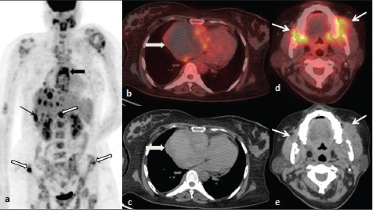
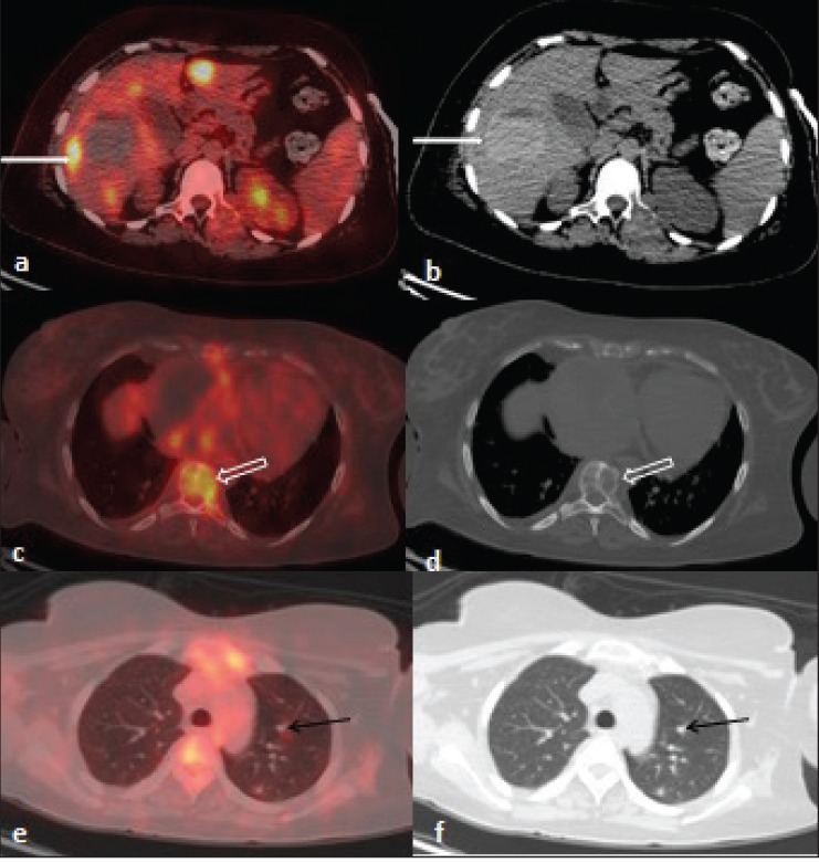
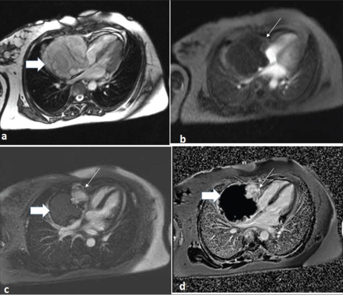
A 30-year-old female patient presented with a history of weakness and breathlessness. Along with these complaints she also had pain in gums on both sides. Patient underwent F-18-fluorodeoxyglucose (FDG) positron emission tomography/computed tomography (CT) for initial evaluation. It demonstrated a necrotic mass in right atrium with multiple FDG avid lesions in both upper and lower alveolus, liver, multiple bones, and bilateral lungs [Figures 1 and 2]. Further, patient underwent cardiac magnetic resonance, which showed a lobulated heterogeneous mass lesion on T1/T2 arising from right atrium with few hyperintense foci within the lesion suggesting hemorrhage / necrosis [Figure 3]. Histopathological examination from the biopsy of gum swelling revealed metastatic angiosarcoma.
Figure 1.

Whole body F-18 FDG PET/CT: (a) Maximum intensity projection demonstrated FDG uptake in the region of heart (thick arrow) and multiple foci of increased tracer uptake involving liver (thin arrow) and skeletal sites (outlined arrow). (b,c) Transaxial fused and CT images at the level of thorax showed large necrotic mass lesion in the region of right atrium with increased radiotracer uptake in the periphery (thick arrow). (d,e) Head and neck transaxial fused and CT images showed irregular soft tissue density lesion with increased FDG uptake involving bilateral alveolar region with subtle necrosis.
Figure 2.

F-18 FDG PET/CT transaxial fused and CT images: (a, b) Transaxial images at the level of liver showed multiple foci of increased tracer uptake and large hyperdense mass lesion in segment V/VI of right lobe with mildly increased tracer uptake in the periphery (thin white arrow). (c-f) Transaxial images showed lytic lesions in multiple vertebrae (thick arrow) with increased radiotracer uptake and multiple nodules in bilateral lungs (thin black arrow) suggestive of metastases.
Figure 3.

Cardiac magnetic resonance images: (a) Large lobulated intracavitary right atrial (RA) mass lesion with RA free wall appearing heterogenous on T2WI- 4CH TRUFI (four chamber true fast imaging with steady-state free precession). (b) perfusion images showed nodular uptake of contrast predominantly in the periphery (thin arrow) with no significant enhancement in rest of the mass (thick arrow). (c,d) Post GAD/PSIR images at 5 min demonstrated patchy nodular intense enhancement (thin arrow).
Primary cardiac tumors are rare and comprise only 0.001-0.0028% of autopsy report. Among these, approximately 20-25% are malignant of which angiosarcoma is the most common.[1,2,3] Angiosarcoma is an aggressive tumor and has high tendency for metastasis. Surgical removal is the definitive treatment for localized angiosarcoma and so preoperative evaluation to accurately localize the primary tumour and detect metastasis is important. CT and magnetic resonance imaging are routinely used for evaluation for cardiac tumor. Although F18 FDG PET/CT has been used in staging of various tumor, its use in staging of cardiac tumor is not well defined. Limited data are available in use of F18 FDG PET/CT in primary cardiac tumor.[4,5,6,7] In present case, there was unusual presentation of gum swelling and along with diagnostic dilemma, the extent of disease was also not clear. F-18 FDG PET/CT in this patient not only localized probable primary site but also helped in accurately staging the disease by detecting metastatic sites.
Financial support and sponsorship
Nil
Conflicts of interest
There are no conflicts of interest.
References
- 1.Burke A, Virmani S. Atlas of tumor pathology. Washington, DC: Washington Armed Force Institute of Tumor Pathology; 1996. [Google Scholar]
- 2.Reynen K. Cardiac myxomas. N Engl J Med. 1995;333:1610–17. doi: 10.1056/NEJM199512143332407. [DOI] [PubMed] [Google Scholar]
- 3.Bussani R, De-Giorgio F, Abbate A, Silvestri F. Cardiac metastases. J Clin Pathol. 2007;60:27–34. doi: 10.1136/jcp.2005.035105. [DOI] [PMC free article] [PubMed] [Google Scholar]
- 4.Kambiz R, Harald S, Michael S, Lars S, Andreas H, Tilmann S, et al. Differentiation of malignant and benign cardiac tumors using 18F-FDG PET/CT. J Nucl Med. 2012;53:856–63. doi: 10.2967/jnumed.111.095364. [DOI] [PubMed] [Google Scholar]
- 5.Bilski M, Kaminski G, Dziuk M. Metabolic activity assessment of cardiac angiosarcoma by 18FDG PET-CT. Nucl Med Rev Cent East Eur. 2012;15:83–4. doi: 10.5603/nmr-18736. [DOI] [PubMed] [Google Scholar]
- 6.Hou CH, Shen DH, Lin LF, Gao HW, Hsu YC, Cheng CY. Aggressive right atrial tumor with extensive FDG-avid metastases in a case of cardiac angiosarcoma. Ann Nucl Med Mol Imaging. 2012;25:201–5. [Google Scholar]
- 7.Tan H, Jiang L, Gao Y, Zen Z, Shi H. 18F-FDG PET/CT imaging in primary cardiac angiosarcoma: Diagnosis and follow up. Clin Nucl Med. 2013;38:1002–5. doi: 10.1097/RLU.0000000000000254. [DOI] [PubMed] [Google Scholar]


