

During development, nerve cells in the body extend and reach forward, spreading axonal tendrils and establishing sensitive connections that ultimately wire the system into a whole. The signals that these nerve cells send and receive to determine the location and specificity of their synapses have captivated the interest of Joshua R. Sanes, recently appointed director of the new interdisciplinary Center for Brain Neuroscience at Harvard University (Cambridge, MA). Sanes has spent his research career studying the nature of synapse formation from embryological and molecular perspectives, and his work led to his election to the National Academy of Sciences in 2002.
In his Inaugural Article in this issue of PNAS, Sanes and colleagues examined a cluster of genes that helps determine how neuronal connections are formed in the spinal cord (1). The protocadherin (Pcdh) genes play roles in specifying synaptic connectivity, and previous research has shown that mice lacking a certain Pcdh gene cluster have decreased spinal cord synapses and are nearly immobile. However, these results were complicated by the fact that the Pcdh ablation also led to apoptosis of spinal interneurons. In his Inaugural Article study, Sanes and his team circumvented this apoptosis problem by preserving spinal interneurons and demonstrated that Pcdh-mutant mice still have decreased numbers of spinal cord synapses and neurological deficiencies. Their findings thus support the direct role of protocadherins in synaptic development.
Mental Fascination
Sanes was born in 1949 in Buffalo, NY, and “wanted to be a scientist when I was pretty young,” he says. His father, who owned an automobile parts supply store, was an avid reader, and there were many books around the house for Sanes to read. Sanes often picked up the popularized science books on psychoanalysis prevalent in the 1950s. Sanes attributes his fascination with mental illness to reading these books, especially while in junior high school. By the time he went to high school, Sanes was already working in the laboratory of a prominent microbiologist, Robert Guthrie, at Children's Hospital (Buffalo, NY). Guthrie, the father of a mentally disabled child, had begun to use bacteria to study mental illness, eventually developing the test for phenylketonuria, a metabolic disorder that can lead to severe retardation if left untreated. Sanes was one of several local students who worked in Guthrie's laboratories on Saturdays and during the summer. The work was meaningful for Sanes. He says, “High school students could do repetitive tests and still make real contributions to the work.”
In college at Yale University (New Haven, CT), Sanes “went in and out of science.” He focused on psychology but was briefly a philosophy major. He says his studies “didn't seem directed, but then I found out about neurobiology. There was a field emerging that was exactly what I was looking for.” Neurobiology seemed to fit his interest in looking at the biological basis for mental illness. Sanes wrote his senior thesis at Yale in the Department of Pharmacology with a “terrific mentor,” Paul Greengard. In Greengard's laboratory, Sanes had the opportunity to contribute to research on protein kinases (2).
After graduating with degrees in biochemistry and psychology in 1970, Sanes attended graduate school, entering the neurobiology program at Harvard. He remained interested in the biological basis of mental illness and structured his thesis to fit his interests, but he found there were limited techniques. “It was hard to be a hardcore biologist and do mental illness research,” he says. “That wouldn't be true now” because of the explosion of molecular biology and its associated techniques, such as gene knockout methods, amplification technologies, or biochip microarray analyses. After a year and a half, Sanes joined John Hildebrand's laboratory at Harvard and studied the development of neurons in the moth Manduca sexta (3–6). One project centered on the role of acetylcholine in the development of antennae (5). Says Sanes, “It was what people did before molecular biology. People worked on big insects because it was easier to study their biochemistry.”
Dr. Sanes Goes to Washington
When Sanes finished his doctorate in 1976, he took a year off to work in the U.S. Congress, joining the Office of Technology Assessment. “I did my thesis during the Watergate years, and I became an unbelievable Watergate junkie,” he admits. Sanes thought it would be interesting to be in the same city as those who had played a part in the political drama. “It was just as cool as I'd hoped it would be,” he says. Sanes worked closely with state senators but found he was more interested in neurobiology than health policy. While in Washington he met his future wife, Susan Corcoran, who was also working in the Office of Technology Assessment after recently returning from the Peace Corps. At the end of the year, Sanes returned as planned to Harvard to the laboratory of U. J. “Jack” McMahan, who had been Sanes' “surrogate mentor” during graduate school. “We cooked up a project that I could come back to,” he says. “As a postdoc, I moved toward the formation of synapses because I thought that was most relevant to my interests.”
The formation of the synapse at the neuromuscular junction, where nerve cells and muscle cells meet, depends on information that each cell type communicates to the other. The junction consists of the presynaptic nerve terminal on one side, the postsynaptic membrane of the muscle fiber on the other, and the basal lamina in between. When Sanes started his research in this area, almost no molecular biology tools existed. He took a molecular biology course in graduate school and explains, “If it was a 16-week course, 15 weeks were on E. coli and lambda phage. There were no genes, no sequences, no introns, no exons, and no PCR.” He remembers how difficult it was to study synapses with biochemical methods because the proteins were in low abundance and membrane-bound.
At the time, in the late 1970s, scientists in the field knew what synapses looked like and how they worked as well as some of the steps in their development, but very little live imaging existed. Says Sanes, “Everything about their development was deduced from static images, and that had the potential to be misleading.” Under McMahan, Sanes studied the anatomy and embryology of synapse formation in the skeletal muscles of frogs (7). “We showed that some of the messages the nerve and muscle send to each other to organize the synapse are contained in the extracellular matrix,” he explains. “That told you where the information was.” According to Sanes, this gave him a good starting platform, and he then spent his second postdoctoral position studying the biochemical angle of synapse formation in rats. Sanes then joined the laboratory of Zach Hall at the University of California, San Francisco, where Hall had moved from Harvard. Under Hall, Sanes studied the biochemical composition of basal lamina for the signaling molecules (8).
A Different Washington
In 1980, Sanes moved on to a faculty position in the Department of Physiology at the Washington University School of Medicine (St. Louis, MO), becoming part of “one of the most preeminent neuroscience groups in the country.” Sanes set about looking for some of the molecules that help direct formation of the neuromuscular junction (9–11). In one study, he used antibodies to purify proteins from the basal lamina and discovered the protein laminin β2 made by muscle cells. “It was the antigen recognized by the very first antibody I made, and I made it, believe it or not,” he says.
A few years later, in 1983, Sanes began collaborating with John Merlie on gene expression at the neuromuscular junction (12). Merlie had joined the Washington University faculty around the same time as Sanes, and they worked closely together until Merlie died unexpectedly in 1995. Sanes calls Merlie “one of the early molecular neurobiologists and certainly one of my best friends.” He continues, “He taught me a huge amount about molecular biology and transgenic and knockout mice. I wouldn't say we were pioneers, but we used things early on.” Sanes and Merlie cloned the gene for laminin β2 (13) and generated knockout mice to study the role of the protein. Mice unable to produce laminin β2 still formed synapses, but the junctions were abnormal (14). They continued to try to determine the components necessary for synapse formation by generating a series of transgenic mice deficient in candidate genes (15, 16). Sanes and Merlie also initiated studies using mouse genetics to determine the components necessary for synapse formation (17), which Sanes later continued on his own. A knockout, however, brought them back to an old problem; it gave only a static picture. In the late 1990s, Sanes found an answer to the limitation of static pictures through work with Jeff Lichtman, also at Washington University. Sanes calls Lichtman “the world's greatest expert in live imaging of synapses as they form.” Together, they fused Sanes' knowledge of molecular genetics with Lichtman's skills in imaging to determine how synapses form and how genotypes affect phenotypes in real time (18–20).
“What is it that makes a cell form and stabilize a particular synapse?”
More recently, Sanes began extending work from the neuromuscular junction studies to investigating the synapses formed between neurons (21, 22). “We also hoped to study the brain,” says Sanes, “but the technical hurdles were too great for us until recently.” In his PNAS Inaugural Article, Sanes examines genes necessary for specificity at the synapse. The protocadherin gene family has almost 60 members clustered in three tandem arrays. Previous work in mice lacking one of these clusters had produced a nearly immobile animal with reduced spinal cord synapses (23). However, the phenotype also produced apoptosis of the same neurons, complicating the interpretation. Sanes and his colleagues avoided this problem by developing a knockout that also lacked the proapoptotic protein. Despite the preservation of spinal cord neurons, the mice still had fewer synapses, suggesting that protocadherins are active in specificity of neuronal connections (1).
Future Connections
“A linear trajectory peppered with wonderful sabbaticals” is how Sanes describes his work on synapse formation. His sabbaticals, which were in France and taken while Sanes was at Washington University, have been good experiences to enjoy with his family, who have traveled with him each time. His son Jesse was born in Paris while Sanes was working at the Pasteur Institute in 1986. Both Jesse and Sanes' daughter Milla attended local schools in France years later while Sanes worked at the Institute of Developmental Biology at the University of Marseille in 1999. Although Sanes does not speak French and says his accent has not improved since high school, he does “know a lot more words.”
Sanes' laboratory will continue to study the rules of synapse formation at the neuromuscular junction and in the brain. “Although we've used molecular methods, we've tried to do that in a biological context,” he says. With live imaging, they can “really see what happens.” Sanes hopes to use live imaging, with the help of Lichtman, to understand how neuronal “experience in the form of electrochemical activity” over time modifies the synapse and its implications for processes such as learning. He also hopes to continue the work described in his Inaugural Article, trying to determine how synapses form where they do. Asks Sanes, “What is it that makes a cell form and stabilize a particular synapse?”
Sanes says he is fortunate to still collaborate actively with Lichtman and be able to relocate to the new Center for Brain Neuroscience at Harvard University. Both men recently moved to the new center, where Sanes is the director. As Sanes describes it, the center is part of a “systems neuroscience effort that uses molecular biology, engineering, and psychology to look at the circuit level questions.” Sanes sees his laboratory as part of this emerging field, and he looks forward to the advancements that this interdisciplinary approach will bring to neuroscience. As he says, “This will be as exciting as development has been in the last 20 years.”
Figure 1.
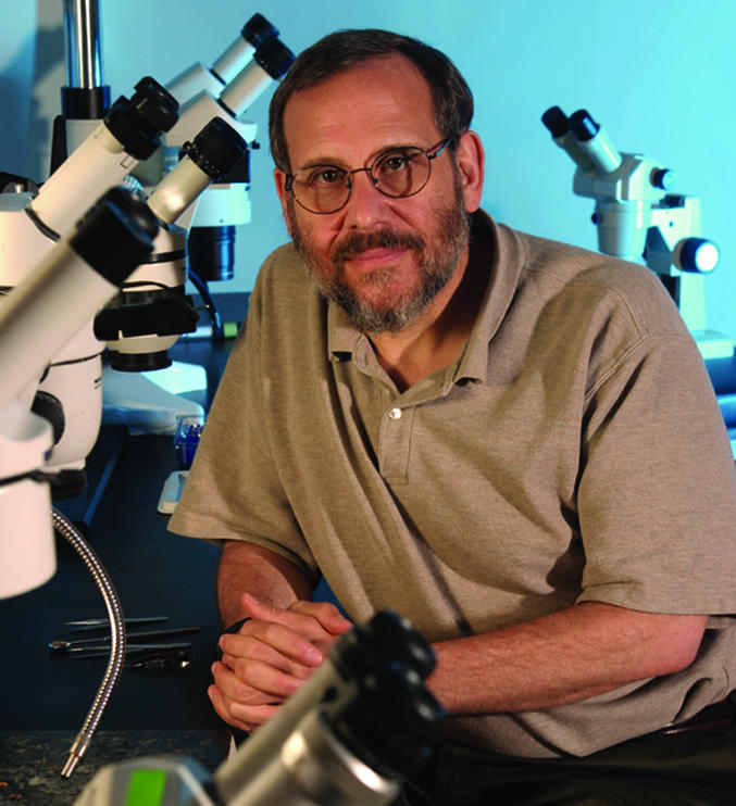
Joshua R. Sanes
This is a Biography of a recently elected member of the National Academy of Sciences to accompany the member's Inaugural Article on page 8.
References
- 1.Weiner, J. A., Wang, X., Tapia, J. C. & Sanes, J. R. (2005) Proc. Natl. Acad. Sci. USA 102, 8-14. [DOI] [PMC free article] [PubMed] [Google Scholar]
- 2.Kuo, J. F., Krueger, B. K., Sanes, J. R. & Greengard, P. (1970) Biochim. Biophys. Acta 212, 79-91. [DOI] [PubMed] [Google Scholar]
- 3.Sanes, J. R. & Hildebrand, J. G. (1976) Dev. Biol. 51, 282-299. [PubMed] [Google Scholar]
- 4.Sanes, J. R. & Hildebrand, J. G. (1976) Dev. Biol. 51, 300-319. [DOI] [PubMed] [Google Scholar]
- 5.Sanes, J. R. & Hildebrand, J. G. (1976) Dev. Biol. 52, 105-120. [DOI] [PubMed] [Google Scholar]
- 6.Sanes, J. R., Hildebrand, J. G. & Prescott, D. J. (1976) Dev. Biol. 52, 121-127. [DOI] [PubMed] [Google Scholar]
- 7.Sanes J. R., Marshall, L. M. & McMahan, U. J. (1978) J. Cell Biol. 78, 176-198. [DOI] [PMC free article] [PubMed] [Google Scholar]
- 8.Sanes, J. R. & Hall, Z. W. (1979) J. Cell Biol. 83, 357-370. [DOI] [PMC free article] [PubMed] [Google Scholar]
- 9.Wigston, D. J. & Sanes, J. R. (1982) Nature 299, 464-467. [DOI] [PubMed] [Google Scholar]
- 10.Sanes, J. R. & Cheney, J. M. (1982) Nature 300, 646-647. [DOI] [PubMed] [Google Scholar]
- 11.Covault, J. & Sanes, J. R. (1985) Proc. Natl. Acad. Sci. USA 82, 4544-4548. [DOI] [PMC free article] [PubMed] [Google Scholar]
- 12.Merlie, J. P. & Sanes, J. R. (1985) Nature 317, 66-68. [DOI] [PubMed] [Google Scholar]
- 13.Hunter, D. D., Porter, B. E., Bulock, J. W., Adams, S. P., Merlie, J. P. & Sanes, J. R. (1989) Cell 59, 905-913. [DOI] [PubMed] [Google Scholar]
- 14.Noakes, P. G., Gautam, M., Mudd, J., Sanes, J. R. & Merlie, J. P. (1995) Nature 374, 258-262. [DOI] [PubMed] [Google Scholar]
- 15.Gautam, M., Noakes, P. G., Mudd, J., Nichol, M., Chu, G. C., Sanes, J. R. & Merlie, J. P (1995) Nature 377, 232-236. [DOI] [PubMed] [Google Scholar]
- 16.Gautam, M., Noakes, P. G., Moscoso, L., Rupp, F., Scheller, R. H., Merlie, J. P. & Sanes, J. R. (1996) Cell 85, 525-535. [DOI] [PubMed] [Google Scholar]
- 17.Grady, R. M., Teng, H., Nichol, M. C., Cunningham, J. C., Wilkinson, R. S. & Sanes, J. R. (1997) Cell 90, 729-738. [DOI] [PubMed] [Google Scholar]
- 18.Nguyen, Q. T., Sanes, J. R. & Lichtman, J. W. (2002) Nat. Neurosci. 5, 861-867. [DOI] [PubMed] [Google Scholar]
- 19.Gan, W. B., Kwon, E., Feng, G., Sanes, J. R. & Lichtman, J. W. (2003) Nat. Neurosci. 6, 956-960. [DOI] [PubMed] [Google Scholar]
- 20.Buffelli, M., Burgess, R. W., Feng, G., Lobe, C., Lichtman, J. W. & Sanes, J. R. (2003) Nature 424, 430-434. [DOI] [PubMed] [Google Scholar]
- 21.Feng, G., Tintrup, H., Kirsch, J., Nichol, M. C., Kuhse, J., Betz, H. & Sanes, J. R. (1998) Science 282, 1321-1324. [DOI] [PubMed] [Google Scholar]
- 22.Yamagata, M., Weiner, J. A. & Sanes, J. R. (2002) Cell 110, 649-660. [DOI] [PubMed] [Google Scholar]
- 23.Wang, X., Weiner, J. A., Levi, S., Tovar, K. R., Craig, A. M., Bradley, A. & Sanes, J. R. (2002) Neuron 36, 843-854. [DOI] [PubMed] [Google Scholar]