Abstract
1,3-β-Glucan synthase is responsible for the synthesis of β-glucan, an essential cell wall structural component in most fungi. We sought to determine whether Coccidioides posadasii possesses genes homologous to known fungal FKS genes that encode the catalytic subunit of 1,3-β-glucan synthase. A single gene, designated FKS1, was identified, and examination of its predicted protein product showed a high degree of conservation with Fks proteins from other filamentous fungi. FKS1 is expressed at similar levels in mycelia and early spherulating cultures, and expression decreases as the spherules mature. We used Agrobacterium-mediated transformation to create strains that harbor ΔFKS1::hygB, a null allele of FKS1, and hypothesize that Fks1p function is essential, due to our inability to purify this allele away from a complementing wild-type FKS1 allele in a heterokaryotic strain. The heterokaryon appears normal with respect to growth rate and arthroconidium production; however, microscopic examination of strains with ΔFKS1::hygB alleles revealed abnormal swelling of hyphal elements.
Coccidioides species (C. immitis and C. posadasii) are primary fungal pathogens that cause disease by the respiratory route in humans and many other species of mammals. The genus has recently been reorganized from one species (C. immitis) into two species, with C. posadasii being localized to Texas, Arizona, Mexico, and South America whereas C. immitis is found only in the area of the San Joaquin Valley of California (13). Although the species are genetically distinct, differences in morphology or pathogenicity have not yet been delineated.
Coccidioides spp. are dimorphic and produce characteristic structures for the saprobic and parasitic growth phases (for a review of the life cycle, see reference 6). The soil-dwelling saprobic phase is mycelial, with asexual spores produced through septation of hyphae and subsequent differentiation of alternate cellular compartments into arthroconidia, which contain on average two nuclei but sometimes contain more (6). Mature conidia 3 to 6 by 2 to 4 μm in size are liberated by physical disruption and disperse readily in the wind. Upon inhalation into the host lung, the arthroconidium undergoes a phase transition to form a large (60 to >100 μm) spherule that undergoes internal septation to produce hundreds of endospores. Most infections are relatively mild and resolve without specific treatment, although about 30% of infections produce flu-like symptoms (39). However, some infections are severe and require extensive antifungal therapy (40).
The fungal cell wall is conceptually attractive as a target for antifungal therapies. It is critical for maintaining cell shape and integrity and contains the polysaccharides 1,3-β-glucan and chitin (4, 38), which are not constituents of mammalian cells. Antifungal agents that act by inhibiting their synthesis could be highly selective.
1,3-β-glucan is the predominant polysaccharide in most fungal cell walls, including those of Saccharomyces cerevisiae, Candida albicans, and Neurospora crassa (14, 38), and is a significant component in the walls of C. posadasii in all growth phases (21, 42). The glucan synthase complex has been best studied in S. cerevisiae and consists of a catalytic subunit encoded by either of two genes (FKS1 or FKS2) and a regulatory subunit (RHO1). The FKS genes encode large polypeptides (∼200 kDa) with 16 membrane-spanning domains that synthesize 1,3-β-glucan from UDP-glucose subunits (4, 11, 33). Rho1p is a small GTPase that in its prenylated form interacts with Fksp catalytic subunits to activate 1,3-β-glucan synthase (22, 23, 32, 37). Glucan synthase activity is essential in yeast. An FKS1 FKS2 double deletion is lethal, although single deletions are viable, suggesting overlapping functions of the two catalytic polypeptides (24, 33). FKS2 is required for sporulation, and a third gene in yeast, FKS3, is nonessential for growth or sporulation (41). FKS1 homologs have been reported in the pathogenic fungi C. albicans, Cryptococcus neoformans, Paracoccidioides brasiliensis, and Pneumocystis carinii and the filamentous fungi Aspergillus nidulans and Aspergillus fumigatus (3, 10, 28, 35, 41). The FKS1 genes have been demonstrated to be essential single-copy genes in C. albicans, C. neoformans, and A. fumigatus (3, 10, 41). The echinocandins and pneumocandins are a class of antifungal drugs that inhibit 1,3-β-glucan synthase (9). Use of one echinocandin, caspofungin (Merck Pharmaceuticals), is successful in the treatment of infections due to Candida spp. and Aspergillus spp. and has been approved by the Food and Drug Administration for such treatment. However, it has not been effective against C. neoformans (2, 25, 29, 36). Caspofungin has been shown to be effective in treating mice infected with Coccidioides spp. (19), but relatively high doses were required, and in earlier studies a different echinocandin, cilofungin, was found not to be useful (17). Although these results suggested that 1,3-β-glucan synthase might not be a therapeutically important antifungal target in Coccidioides spp., other pharmacologic factors could be responsible for these marginal effects. To investigate 1,3-β-glucan synthase as an antifungal target in Coccidioides spp., we sought to identify the gene and disrupt the function of the FKS homolog(s) in C. posadasii. We have identified a single gene, FKS1, with similarity to other fungal FKS genes, and disruption experiments suggest that FKS1 is essential for growth of C. posadasii.
MATERIALS AND METHODS
Strains, media, and growth conditions.
C. posadasii strain Silveira (ATCC 28868), originally isolated from a patient (15), was maintained in the saprobic phase on 2× GYE agar (2% glucose, 1% yeast extract, 1.6% agar) at 28°C or ambient room temperature. To isolate arthroconidia, strains were grown for 3 to 4 weeks on 2× GYE agar and arthroconidia were harvested in sterile water by use of a mini-stir bar as described previously (16, 18). Spherules were cultivated at 39°C and 9% CO2 in modified Converse medium (7). All manipulations of viable preparations were performed using biosafety level 3 conditions and practices in laboratories registered with the Centers for Disease Control for possession of this select agent.
Isolation and sequencing of FKS1.
A C. posadasii genomic library constructed in lambda GEM11 (34) was screened with a 405-bp probe generated by reverse transcriptase PCR (RT-PCR) from mycelial RNA by use of degenerate primers based on the A. nidulans FKSAp (26), asp4U (5′GCIAAYCARGAYAAYTA) and asp5L (5′YTCRCARTGYTTIATIC). The probe was labeled with digoxigenin-UTP (DIG-UTP) according to the vendor's protocol by use of a Roche “Genius” kit (Boehringer Mannheim, Indianapolis, Ind.). The hybridizing λ clone GS5 was digested with SacI, and two subfragments of 3.5 and 6.5 kb were cloned into pBluescript SK− (Stratagene, La Jolla, Calif.) to create pES518 and pES527, respectively. The insert of pES518 was sequenced in its entirety and found to contain sequence with similarity to fungal FKS gene sequences encoding 1,3-β-glucan synthase catalytic subunits. The predicted start and end of the FKS open reading frame were not present in this subclone. The carboxy-terminal sequence of the predicted glucan synthase gene as well as downstream sequence were obtained from pES527. The sequence of a 500-bp SacI fragment predicted to lie between the SacI fragments of pES518 and pES527 was obtained from direct sequencing of the λ clone GS5. A partial sequence was deposited in GenBank (GenBank AF159533).
To obtain further upstream sequence, including the predicted start of the open reading frame, additional λ clones were screened. A 508-bp PCR fragment was generated from the 5′ proximal sequences of pES518 by use of primers ESGS12 (5′TATAGACCCCCTTCTTCC) and ESGS9 (5′TTATCGCCTTCCATAGAC). This PCR product was labeled with [α-32P]dCTP and used to probe Southern blots containing EcoRI digests of four additional λ clones isolated during the original screening. A 3-kb hybridizing fragment of one clone, GS1, was cloned into pBluescript SK+ (Stratagene), generating pAM1203. The insert of pAM1203 was sequenced on both strands and found to contain the predicted N-terminal regions of the C. posadasii 1,3-β-glucan synthase homolog designated FKS1.
Construction of pAM1253 with a FKS1 deletion-replacement cassette.
The plasmid pAM1253 that contained the FKS1 gene replacement cassette was created in multiple steps. A 1-kb PCR fragment was generated from 5′ FKS1-proximal sequence in pAM1203 by use of primers OAM 472 (5′CCCGAATTCCCCATTTAGCGCCCAGGAGTC3′) and OAM473 (5′CCCGGTACCACATGATGAATCAGGAGAAG3′). OAM472 and OAM473 were designed to add 5′ EcoRI and 3′ KpnI restriction sites to the PCR product. The PCR fragment was first cloned into pGEM-T Easy (Promega) to create pAM1245. A 1-kb EcoRI-KpnI fragment was isolated from pAM1245 and cloned into the EcoRI and KpnI sites of pAM1121, creating pAM1248 and placing the FKS1 upstream sequence 5′ to the hphB gene cassette derived from pCB1004 (5). A 2-kb PCR fragment was generated from sequence 3′ of the FKS1 gene by use of primers OAM482 (5′GGATCCAATTTTGCTGTGTCTCCCTTG3′) and OAM483 (5′AGATCTATCGTTGTGCTCATACCACCG3′) and genomic DNA as a template. The primers were designed to add 5′ BamHI and 3′ BglII restriction sites to the PCR fragment. The PCR fragment was cloned into pGEM-T Easy to create pAM1247. To complete construction of the FKS1 gene replacement vector, fragments containing the 3′ flanking sequence, the 5′ flanking sequence fused to hphB, and the binary vector pAM1145 were ligated together. The 3′ flanking sequence were released from pAM1247 as a 2-kb BamHI-BglII fragment; the 5′ flanking sequence and hphB were released from pAM1248 as a 2.6-kb EcoRI-BamHI fragment, and these were cloned into pAM1145 digested with EcoRI and BglII to create pAM1253. The binary vector pAM1145 contains a broad-host-range origin of replication, the right and left borders of the T-DNA from the octopine Ti-plasmid pTiA6, and a mutated form of virG (virGN54D), which obviates the need to add compounds such as acetosyringone to induce T-DNA transfer (20). pAM1145 was previously constructed by insertion of a 5.6-kb KpnI-SalI fragment from plasmid pAD1378 (8) into pAD1310 (27). pAM1253 was introduced into A. tumefaciens strain AD965 by electroporation to create strain A1253.
Transformation of C. posadasii.
C. posadasii was transformed using A. tumefaciens strain A1253 as described previously (1), with some modifications as follows. Arthroconidial germlings and A. tumefaciens were mixed and dispersed with a sterile spatula onto sterile 80-mm-diameter nitrocellulose filters placed onto 100-mm-diameter petri plates (seven plates) containing AB induction medium. Plates were incubated at 28°C, and after 48 h the nitrocellulose filters were transferred to plates containing 2× GYE agar with 50 μg of hygromycin (Hyg)/ml to select for transformed C. posadasii and 100 μg of kanamycin/ml as a counterselection for A. tumefaciens. Transformants were cut from the nitrocellulose filters and transferred to fresh plates of the selection medium. After 2 weeks of incubation at 28°C, arthroconidia from these transformants were streaked onto selective media to isolate monoconidial colonies. These colonies were cut from the agar, transferred to fresh plates of selection medium, and allowed to conidiate. This conidial passage was repeated two more times.
Southern blot analysis of transformants.
C. posadasii genomic DNA was isolated by scraping mycelium (about 0.1 g wet weight) from a young colony, transferring it to a 2-ml screw-cap tube containing about 600 μl of 0.5-mm-diameter acid-treated glass beads and 1 ml of lysis buffer (50 mM Tris-HCl [pH 7.5], 100 mM EDTA [pH 8.0], 100 mM NaCl, 0.5% sodium dodecyl sulfate, 100 mM dithiothreitol), and subjecting it to mechanical disruption. The tubes were shaken either by bead beating (Mini Beadbeater 8; Biospec Products, Bartlesville, Okla.) for three periods of 30 s with 20-s rest intervals in between or by vortexing on a flat 12-tube holder at 3,700 rpm for 10 min. Samples were incubated at 65°C for 30 min and centrifuged in a microcentrifuge at 9,000 × g rpm for 2 min. The supernatant was extracted with phenol-chloroform-isoamyl alcohol (25:24:1), and nucleic acids were precipitated from the aqueous layer with 0.6 volumes of isopropyl alcohol. Pellets were resuspended in 300 μl of Tris-EDTA (pH 7.6) containing 100 μg of RNase A/ml and incubated at 37°C for 30 min. A total of 50 μl of 5 M NaCl and 40 μl of cetyltrimethylammonium bromide-NaCl (10% cetyltrimethylammonium bromide in 0.7 M NaCl) was added to each sample, and the samples were incubated at 68°C for 10 min. The samples were extracted twice with equal volumes of chloroform-isoamyl alcohol, once with phenol-chloroform-isoamyl alcohol, and once again with chloroform-isoamyl alcohol. The DNA was precipitated with isopropyl alcohol and resuspended in 100 μl of water. Genomic DNA (10 μg) was subjected to restriction digestion, separated through 0.8% agarose gels, transferred to nylon membrane, and hybridized with [α-32P]-dCTP radiolabeled probes. The probes for the FKS1 locus and hphB gene were a 1.3-kb EcoRI-PstI fragment isolated from pAM1203 and a 1.4-kb HpaI fragment isolated from pCB1004, respectively.
RNA isolation and real-time RT-PCR analysis.
RNA was prepared from mycelia (48 h of growth in liquid 2× GYE agar at 37°C) and spherules (grown in modified Converse liquid medium at 39°C and 8% CO2). Mycelia were harvested by filtration onto sterile Whatman Type 1 filter paper and washed with ice-cold RNase-free phosphate-buffered saline (PBS). Spherules were collected by centrifugation at 3,700 × g for 40 min at 4°C and washed with ice-cold PBS. Pellets or mycelial mats were divided into 2-ml screw-cap Microfuge tubes, and 0.7 ml of ice-cold breaking buffer (200 mM Tris [pH 7.6], 0.5 M NaCl, 10 mM EDTA [pH 8.0]), 0.6 ml of acid phenol, and 0.5 ml of 0.5 mm zirconium beads (Biospec Products) were added. The tubes were placed on a vortexing apparatus at 3,000 rpm for three 2-min intervals. The tubes were then centrifuged for 10 min at 20,000 × g in a microcentrifuge at room temperature, and the supernatants were removed to new RNase-free tubes. The samples were extracted with phenol, and the RNA was precipitated with isopropanol. The RNA was resuspended in 300 μl of water, precipitated with LiCl, washed, and resuspended in water.
Poly(A)+ RNA was isolated from 300 μg of total RNA from each developmental time point by use of a PolyATract mRNA isolation system III (Promega, Madison, Wis.) following the manufacturer's recommendations. Poly(A)+ RNA concentrations were estimated using RiboGreen RNA quantitation reagent (Molecular Probes, Inc., Eugene, Oreg.) and an FLX 800 fluorometer (Bio-Tek Instruments, Inc., Winooski, Vt.). Poly(A)+ RNA (7.5 ng) was used to synthesize first-strand cDNAs from RNA isolated from mycelia and 48-h, 72-h, and 96-h spherules by use of oligo(dT) 12 to 18 and SuperScript Reverse Transcriptase II (Invitrogen, Sorrento Valley, Calif.) following the manufacturer's protocol. Following treatment with RNase H (Invitrogen), specific oligonucleotide primers for FKS1 and for β-actin were used in real-time RT-PCR experiments with SYBR Green PCR Master Mix in an ABI PRISM 7000 sequence detection system (Applied Biosystems). The oligonucleotides used for FKS1 were OAM697 (GCATTCGATCTCTCAACCCG) and OAM698 (CGAGAATGAAGTCGGCAGCG); those used for β-actin were OAM637 (AGCGTCTTGGGTCTCGAAA) and OAM638 (CCAGACATGACGATGTTTC). Serial twofold dilutions of the first-strand cDNA were created, and a 0.125-fold dilution was used for the real-time RT-PCR experiments described here. The real-time RT-PCR experiments were performed in triplicate for each dilution, and mean cycle thresholds (Cts) were determined. A biological replicate was also performed using RNA isolated from C. posadasii cultures grown a second time.
Relative quantification calculations were performed using the algorithm X = 2−ΔΔCt ± SPE, where X is the factor by which the amount of each gene at a certain time point in spherule development has changed relative to the expression of the same gene in mycelia (31). ΔΔCt = (CtFKS1 − Ctactin)timepoint − (CtFKS1 − Ctactin)mycelia, where “time point” refers to spherules of 48, 72, or 96 h. β-actin was used as an internal control for RNA yield. SPE is the standard propagation error determined as plus or minus the square root of (σ2FKS1 σ2actin), with σ being the standard deviation error. Real-time RT-PCR data for FKS1 expression at all time points were normalized to the expression of β-actin for that time point, and the normalized expression of FKS1 during spherule development time points was normalized to FKS1 expression in mycelia.
Measurement of growth rates and ΔFKS1::hphB stability.
Colonial radial growth rates were measured through point inoculation of purified arthroconidial suspensions onto the centers of GYE agar plates in both the presence and absence of 50 μg of hygromycin/ml. Each transformant strain was inoculated onto three replicates of GYE agar with no antibiotic and three replicates of GYE agar plus hygromycin. The culture plates were incubated at 28°C, and colony diameters were measured at 24- and 48-h intervals. Measurements used to calculate radial growth rates were those taken after 5, 6, and 8 days of growth during the linear phase of colony expansion—after a colony of sufficient size was formed but before it had covered the surface of the petri plate. After 4 weeks of incubation when the plates were entirely covered by fungal material, arthroconidia were harvested from two of the GYE agar replicates and two of the GYE agar-plus-hygromycin replicates per strain by use of the mini-spin bar technique. Total CFU and hygromycin-resistant CFU levels were determined by plating dilutions of the same conidium preparation onto GYE agar and GYE agar plus 50 μg of hygromycin/ml. Yields are expressed as CFU levels (in square millimeters) over the area of the plate from which the conidia were harvested.
Microscopy.
Sterile coverslips were placed in petri plates, and a very thin layer of 2× GYE agar (plus 50 μg of hygromycin/ml for transformant strains) was poured over the coverslips. A liquid suspension of arthroconidia at 5 × 10/ml in GYE agar (plus 50 μg of hygromycin/ml for transformant strains) was prepared, and a thin layer of this suspension was poured onto the agar plates with embedded coverslips. Caspofungin was used in some experiments at a concentration of 20 μg/ml but not in combination with hygromycin. The plates were allowed to dry briefly, taped around the circumference (required for all C. posadasii petri plate cultures), placed in a humid chamber, and incubated for 72 h at 28°C. To fix the fungal structures after growth, several milliliters of fixative solution (3.7% formaldehyde, 0.2% Tween 20 in PBS) was added to the petri plates to cover the surface and the plates were incubated at room temperature for 45 min. The fixative solution was removed, and the fixed material was washed briefly by overlaying with distilled water. Staining solution (calcofluor; Sigma catalog no. F-3543) (10 μg/ml) and DAPI (diamino-2-phenylindole dihydrochloride) (Sigma catalog no. D-9542) (800 ng/ml) in PBS was then added to the plates, and the staining was allowed to proceed for 30 min at room temperature. The fixed and stained material was washed briefly with distilled water followed by 100% ethanol. Coverslips with thin agar layers were excised from the plates and mounted on microscope slides in mounting medium (Vectashield Hard Set [catalog no. H1400]; Vector Laboratories, Burlingame, Calif.). Slides were visualized using a Diaplan compound microscope (Leitz, Wetzlar, Germany) with a mercury-argon lamp and a Chroma standard filter set for DAPI (excitation D360, emission D460). Images were acquired using an Orca 100 digital camera (Hamamatsu Corporation) and Simple PCI image analysis software (Compix, Inc.).
RESULTS
Identification and sequencing of a C. posadasii FKS homolog.
A C. posadasii genomic library was screened for clones with homology to fungal FKS genes. Several λ clones were isolated and were found to contain sequences that encode a predicted protein with a high degree of similarity, by BLASTX analyses, to 1,3-β-glucan synthase (FKS) genes examined in other fungal species (3, 10, 11, 26, 35, 41). The 5,821-nucleotide sequence from C. posadasii was designated FKS1 and has been deposited in GenBank (update to accession number AF159533). Analysis of FKS1 sequence showed two predicted introns at coding nucleotide positions 175 to 234 and 5489 to 5549 (with the ATG start codon = position 1) with conserved donor and acceptor splice sites for filamentous fungi. Sequencing of cDNAs obtained by RT-PCR confirmed these intron-exon splice sites. The intron positions in FKS1 are conserved with respect to the sequences of FKS homologs present in the related filamentous fungi P. brasiliensis (fks1; AF148715) and A. nidulans (fksA; ENU51272). The FKS1 sequence was used in a BLASTN search of the genome sequence of C. posadasii strain C735 at The Institute for Genomic Research (TIGR, Bethesda, Md.), which is presently at 8× coverage. A single contig, containing sequence identical to that of FKS1 within the coding region, was identified and was predicted to encode a peptide product identical to Fks1p. In addition, a TBLASTN search of the same genomic sequence failed to identify other translated open reading frames with similarity to the Fks1p polypeptide. These findings, combined with Southern analyses using a probe generated from FKS1 internal sequences (data not shown), predict that C. posadasii contains a single gene, FKS1, encoding a 1,3-β-glucan synthase catalytic subunit.
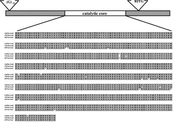
The predicted Fks1p protein is 1,902 amino acids and has a predicted molecular mass of 217 kDa and a pI of 8.72. The proteins that contain the highest degree of similarity to the predicted Fks1p protein of C. posadasii are Fks1p of P. brasiliensis (84% identity) and Fks1p of A. fumigatus (81% identity). Much higher degrees of identity were found in the large hydrophilic domain of approximately 577 amino acids (Fig. 1) that is hypothesized to contain the catalytic site: C. posadasii Fks1p and P. brasiliensis Fks1p (96% identity) and C. posadasii Fks1p and A. fumigatus Fks1p (93% identity) (26). Fks1p also has the putative UDP-glucose binding consensus sequence RXTG (Fks1p; RITG, amino acids 1569 to 1572). Curiously, there is a stretch of 12 glycine residues in the N-terminal portion of the polypeptide (amino acids 9 to 20) that does not appear in any other fungal glucan synthase proteins characterized to date. It is present in both the genomic and cDNA sequences of strain Silveira that were generated and analyzed for this project and in the C735 genomic sequence (TIGR).
FIG. 1.
The C. posadasii Fks1 protein sequence is similar to Fks protein sequences from related pathogenic fungi. A graphic representation of C. posadasii Fks1p is shown. Features indicated are the glycine repeat region, the catalytic core domain, and the UDP-glucose consensus binding site (RITG). A multiple sequence alignment of selected fungal FKS catalytic domains is shown in the bottom half of the figure (26). The alignment was generated using ClustalW (version 1.4) and the BLOSUM (blocks substitution matrix) similarity matrix with an open gap penalty of 10, an extended gap penalty of 0.05, and a delay divergent of 40%. Shaded residues indicate amino acid identities. The predicted protein sequences of P. brasiliensis Fks1p (amino acids 754 to 1331) (accession number AF148715) and A. fumigatus Fks1p (amino acids 755 to 1332) (accession number U79728) are aligned with the C. posadasii Fks1p predicted protein sequence (amino acids 760 to 1337).
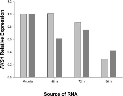
Real-time RT-PCR was used to examine the FKS1 message levels in both liquid-grown mycelial and spherulating cultures of wild-type C. posadasii strain Silveira (Fig. 2). FKS1 was found to be expressed at all time points tested. The levels of FKS1 expression in two separate experiments (biological replicates) relative to the levels in the mycelial RNA are shown in Fig. 2. The results of both experiments taken together indicate that FKS1 expression at 48 and 72 h of spherule formation is similar to or slightly less than its levels in mycelia and that its levels decrease as spherules mature at 96 h (Fig. 2).
FIG. 2.
FKS1 RNA levels decrease during spherule maturation relative to the level in mycelia. FKS1 RNA levels were determined using real-time RT-PCR, normalized to actin as an internal control, and then expressed as severalfold changes during spherule development relative to the levels found in mycelia. The two differently shaded columns for each time point represent biological replicates.
Transformation of C. posadasii with a gene replacement cassette.
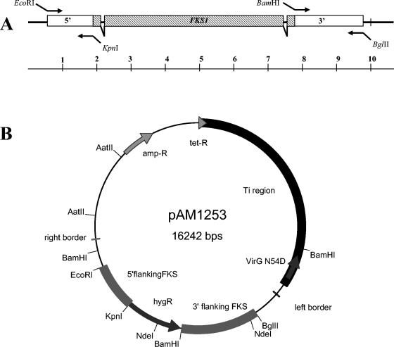
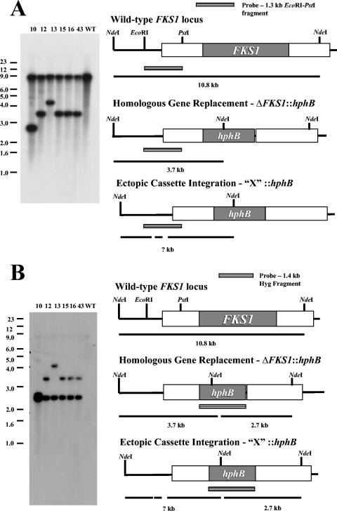
To determine whether 1,3-β-glucan synthase activity is essential for viability of C. posadasii, as it is for other fungal species, we sought to create a strain lacking FKS1. C. posadasii germlings were transformed by A. tumefaciens-mediated transformation by use of a strain containing a gene replacement construct composed of the Escherichia coli hphB gene flanked by 1 kb of FKS1 5′ sequence upstream and 2 kb of FKS1 3′ sequence downstream. This construct was cloned between the T-DNA border sequences of the broad-host-range plasmid pAM1145 (Fig. 3). A total of 24 hygromycin-resistant transformants were isolated. In an attempt to obtain homokaryons, arthroconidia were streaked on agar plates and individual colonies were subcultured three times. Multiple passages were performed due to the multinucleate nature of arthroconidia, which usually contain two nuclei and occasionally contain more (6). In our experience, three passages are sufficient to purify a homokaryotic strain. A combination of PCR and Southern blot analyses was used to examine the nature of the insertions present. Six of the 24 transformants had patterns consistent with the presence of a FKS1 replacement event, but they also contained a wild-type allele of FKS1. The remaining 18 transformants appeared to have resulted from a nonhomologous insertion event, and two of these strains, VFC10 and VFC13, were chosen as control strains for further analysis. Representative Southern analyses are shown in Fig. 4A for selected transformants. Southern blots containing NdeI-digested genomic DNA from the transformants VFC10, VFC12, VFC13, VFC15, VFC16, and VFC43 as well as the wild-type strain Silveira are shown in Fig. 4A. When hybridized with a fragment from the 5′ upstream sequence of FKS1, strains VFC12, VFC15, VFC16, and VFC43 each contained a 3.7-kb NdeI fragment indicative of a homologous gene replacement event and a 10.8-kb NdeI fragment from the wild-type FKS1 allele (Fig. 4A).
FIG. 3.
FKS1 gene structure and deletion plasmid. (A) The genomic structure of the FKS1 locus is depicted, with a scale bar below in 1-kb increments. Primers used to generate PCR fragments containing the upstream and downstream flanking sequences with added restriction sites for cloning are indicated. (B) pAM1253 is the broad-host-range plasmid used to transform C. posadasii and create the FKS1 deletion.
FIG. 4.
Southern analysis of C. posadasii pAM1253 transformants. (A) Genomic DNA (10 μg) from transformant strains VFC10, VFC12, VFC13, VFC15, VFC16, and VFC43 and wild-type strain Silveira was digested with NdeI and separated on a 0.8% agarose gel. The gel was transferred to nitrocellulose, and the blot was probed with a radiolabeled 1.3-kb EcoRI-PstI fragment from the upstream region of FKS1. An autoradiograph of the resulting blot is shown on the left, and the predicted fragment sizes and genomic structures of possible recombinants are shown on the right. (B) The blot depicted in panel A was stripped of the 1.3-kb EcoRI-PstI FKS1 probe and reprobed with a radiolabeled 1.4-kb fragment containing the hphB gene, as shown on the diagram at the right side of the figure.
These results were consistent with the idea that these transformants were heterokaryotic, containing both the ΔFKS1::hphB null allele and the FKS1WT allele in separate nuclei. Digests of VFC10 and VFC13 exhibited a hybridizing 10.8-kb NdeI fragment and additional hybridizing fragments of approximately 2.9 and 4.7 kb, respectively. These patterns were predicted to arise from a FKS1WT allele and an ectopic integration event of the gene replacement cassette at an unknown genomic locus. These data were insufficient to determine whether VFC10 and VFC13 were homokaryotic or heterokaryotic for the ectopic loci where the replacement cassette was inserted but do provide support for the idea that these strains arose from a single integration event, due to the presence of only one band in addition to that of the wild-type fragment. The digest of strain Silveira DNA showed only the single predicted 10.8-kb NdeI fragment of the wild-type FKS1 locus. The blots were stripped and reprobed with a radioactively labeled 1.4-kb hphB gene (Fig. 4B). The probe detected a 2.7-kb NdeI fragment internal to the gene replacement cassette in all six transformants but not in the wild-type Silveira strain. The 3.7-kb NdeI fragment predicted to arise from strains with a homologous gene replacement hybridized with the hphB probe only in strains VFC12, VFC15, VFC16, and VFC43. The hphB probe hybridized with the same 2.9- and 4.7- fragments as the FKS1-upstream probe in lanes containing digests from VFC10 and VFC13, respectively. No hphB-hybridizing bands were present in the lane containing the NdeI-digested genomic DNA from wild-type strain Silveira. These data support the conclusion that each transformant strain arose through a single integration event with the FKS1 deletion cassette, with VFC12, VFC15, VFC16, and VFC43 arising from a homologous integration event and VFC10 and VFC13 arising from an ectopic, nonhomologous integration, likely at the T-DNA border regions.
Growth rates, conidiation, and mitotic stability of nuclei in ΔFKS1::hphB transformants.
No defects in growth rates, with or without hygromycin selection, were observed during propagation of the four putative heterokaryotic strains VFC12, VFC15, VFC16, and VFC43 (ΔFKS1::hphB; FKS1WT) or the control strains VFC10 and VFC13 (FKS1WT; ectopic hphB) on GYE agar medium at 28°C (Table 1, column 3). Total arthroconidial yields were somewhat higher when VFC12, VFC15, VFC16, and VFC43 were grown without hygromycin selection for the ΔFKS1::hphB allele (Table 1, column 4). In contrast, the control strains, VFC10 and VFC13, showed more similar yields of arthroconidia, regardless of the presence or absence of hygromycin in the growth medium (Table 1, column 4). More striking was that among the arthroconidia produced by the putative heterokaryotic strains VFC12, VFC15, VFC16, and VFC43 (ΔFKS1::hphB; FKS1WT) grown in the absence of selection, only a small fraction were hygromycin resistant, i.e., contained the ΔFKS1::hphB allele. In contrast, the control strains VFC10 and VFC13, within the margin of error, produced only hygromycin-resistant arthroconidia. These findings supported the idea that the strains carrying the FKS1 null allele were heterokaryotic, containing wild-type nuclei in addition to nuclei with the gene replacement. The data suggest that when selection pressure was removed, those nuclei with the FKS1 null allele were readily lost or did not produce viable conidia. In contrast, since the control strains VFC10 and 13 did not lose the hygromycin marker even without selection they are likely to be mitotically stable homokaryons. In fact, in all cases conidia from VFC10 and VFC13 taken from nonselective plates and scored for hygromycin resistance retained resistance (data not shown). Arthroconidia from VFC12, a strain with a FKS1 deletion allele, and VFC13, the control strain, were capable of forming spherules in vitro, and a single round of spherule formation did not decrease the number of hygromycin-resistant CFU (data not shown).
TABLE 1.
Growth rates, conidiation, and mitotic stability of ΔFKS1::hphB transformantsa
| Strain | Hyg selection in growth medium | Growth rate (mm/day) | Total arthroconidium yield (CFU/mm2 × 103) | HygR arthrocondium yield (CFU/mm2 × 103) | HygR CFU/total CFU (102) |
|---|---|---|---|---|---|
| VFC12 | − | 2.8 ± 0.24 | 50 ± 11 | 1.3 ± 0.0 | 2.7 |
| VFC12 | + | 2.7 ± 0.24 | 7.8 ± 1.2 | 4.0 ± 0.9 | 52 |
| VFC15 | − | 2.8 ± 0.22 | 25 ± 1.4 | 0.6 ± 0.1 | 2.3 |
| VFC15 | + | 2.9 ± 0.13 | 12 ± 6.3 | 15 ± 2.1 | 140 |
| VFC16 | − | 2.7 ± 0.26 | 20 ± 5.6 | 0.6 ± 0.0 | 3.4 |
| VFC16 | + | 2.5 ± 0.10 | 6.6 ± 1.1 | 6 ± 2.0 | 90 |
| VFC43 | − | 2.6 ± 0.13 | 28 ± 7.8 | 0.6 ± 0.1 | 2.4 |
| VFC43 | + | 2.7 ± 0.13 | 9.4 ± 0.8 | 11 ± 0.0 | 120 |
| VFC10 | − | 2.8 ± 0.16 | 37 ± 8.5 | 32 ± 2.1 | 87 |
| VFC10 | + | 2.8 ± 0.13 | 32 ± 19 | 42 ± 4.9 | 160 |
| VFC13 | − | 2.7 ± 0.24 | 34 ± 13 | 28 ± 6.4 | 86 |
| VFC13 | + | 2.7 ± 0.19 | 54 ± 18 | 58 ± 12 | 110 |
The table shows the radial colony growth rates (in millimeters per day) on 2 × GYE agar both in the absence of hygromycin selection and under hygromycin selection (50 μg/ml) for transformant strains. Growth rates shown represent the average of three colonies for each strain with standard deviations. Arthroconidia were prepared from two of colonies for each strain (propagated both with and without hygromycin). For each preparation, the total number of viable arthroconidia and hygromycin-resistant arthroconidia, expressed as CFU per square millimeter with standard deviations, was determined and is shown in columns 4 and 5. The ratio of HygR CFU/total CFU is shown in column 6.
Heterokaryotic strains with FKS1 null alleles exhibit abnormal swelling of some hyphal elements.
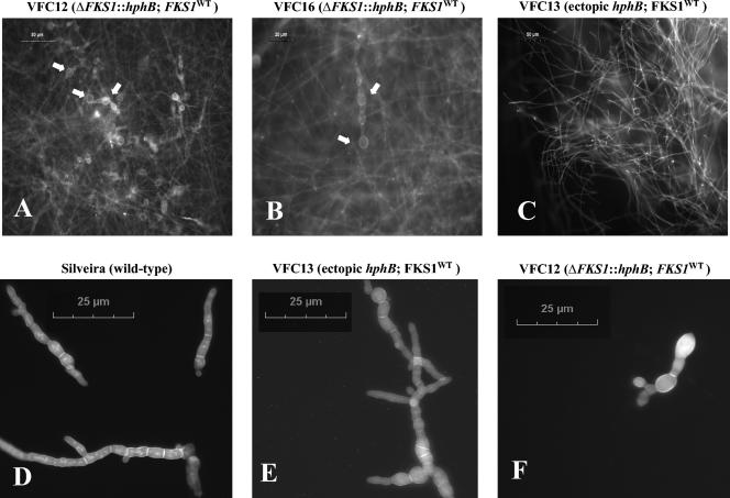
To determine whether the FKS1 gene deletion caused an observable phenotype when present in a heterokaryon, both the null allele-containing heterokaryon strains (VFC12, VFC15, VFC16, and VFC43) and the ectopic insertion control strains (VFC10 and VFC13) were examined microscopically. Arthroconidia were germinated on thin layers of 2× GYE agar medium and allowed to grow at 28°C for 72 h. Hyphae were stained with Calcofluor White and DAPI, mounted on slides, and examined using fluorescent microscopy. Most of the hyphal elements in strains VFC12, VFC15, VFC16, and VFC43 were indistinguishable from wild-type hyphae with respect to hyphal diameter, length, and septation. Upon closer inspection, however, certain regions of the mycelium displayed aberrant conformations, such as blowing out of cell wall elements at hyphal tips and internal compartments (Fig. 5A and B). These aberrant structures were completely absent from mycelia of VFC10 and VFC13, the control strains (Fig. 5C), and of wild-type strains (data not shown). The growth of FKS1 deletion strains and control strains in the presence of caspofungin, the 1,3-β-glucan synthase inhibitor, was also examined, and typical results are shown in Fig. 5D, E, and F. As has been observed previously with related cilofungin treatments in C. posadasii studies, caspofungin inhibited growth (compare Fig. 5D, E, and F with 5C) and produced a swelling of hyphae in wild-type strain Silveira as well as VFC12 (ΔFKS1, FKS1WT) and VFC13 (ectopic hphB, FKS1WT) (17).
FIG. 5.
Transformant strains with ΔFKS1::hphB alleles show hyphal defects. (A to C) Transformant strains were grown for 72 h in 2× GYE agar medium with hygromycin at 50 μg/ml, stained with DAPI and calcofluor, and visualized using a Leica compound microscope with fluorescent optics. The strains in the micrographs are as follows: VFC12 (ΔFKS1::hphB; FKS1WT) (A), VFC16 (ΔFKS1::hphB; FKS1WT) (B), and VFC13 (ectopic hphB; FKS1WT) (C). Scale bars are indicated in the upper left corners of the micrographs. Arrows indicate abnormally shaped hyphal compartments. (D to F) Transformant strains and the wild-type Silveira strain were grown as described above, with the inclusion of caspofungin (20 μg/ml) and the omission of hygromycin. The strains shown are as follows: Silveira (D), VFC13 (E), and VFC12 (F).
DISCUSSION
Genes encoding the 1,3-β-glucan synthase catalytic subunit have been isolated from several fungi and have been studied best in S. cerevisiae experiments (11, 23, 24, 32, 33, 37). β-glucan synthesis has been found to be essential to life in the human pathogenic fungi examined to date (3, 10, 41). Thus, this function is an attractive target for antifungal therapeutics. The 1,3-β-glucan synthase catalytic subunit is targeted by the echinocandin class of antifungal drugs which are effective against several pathogenic fungi in a clinical setting. We sought to determine whether there was a molecular basis for such therapy to be effective in treating patients with coccidioidomycosis. To begin to address this question, we looked for genes in C. posadasii that shared similarity to known fungal FKS genes.
In this study, we identified a single gene, FKS1, in C. posadasii that showed significant similarity to known fungal FKS genes. Analysis of the predicted protein product, Fks1p, revealed the highest degree of similarity to Fks1p of P. brasiliensis (84% identity). P. brasiliensis is a dimorphic human pathogenic fungus and, like C. posadasii, is a member of the order Onygenales. Fks1p of A. fumigatus and FksAp of A. nidulans also showed a high degree of similarity to Fks1p of C. posadasii (81 and 80%, respectively) and, among fungi whose genomes have been sequenced, are the most closely related to C. posadasii, all belonging to the class Eurotiomycetes. Curiously, the predicted Fksp protein homologs in C. posadasii strain Silveira (this work) and strain C735 (TIGR) contain a unique stretch of 12 glycine residues in their amino termini after amino acid residue 8. These glycine residues are encoded by six GGA codons followed by five GGC codons and a single GGT codon. It will be interesting to see whether this region is present in the sequence of C. immitis strain RS presently being sequenced by The Broad Institute.
Several different approaches have been used to demonstrate the essential nature of FKS genes in other human pathogenic fungi (10, 12, 41). In C. albicans, sequential targeted disruption experiments yielded heterozygotic diploid strains with one FKS1 allele disrupted, but no strains that contained disruptions of both FKS1 alleles were obtained (10). Thompson et al. approached the problem of potential FKS1 essentiality by employing a plasmid that could integrate by homologous recombination in two possible orientations, one that leaves an intact FKS1 gene and one that results in a loss of FKS1 function (41). They found that all transformants obtained had integration events in a single orientation that maintained a functional FKS1 gene; this finding thus supports the idea of its essential nature under the growth conditions examined. Parasexual genetics was applied to determine the essential nature of FKS1 of A. fumigatus (12). A diploid strain was constructed that contained a disrupted FKS1 allele and a wild-type FKS1 allele. Subsequent haploidization of this strain yielded no haploid segregants that contained the disrupted allele.
Initial transformants of C. posadasii obtained by A. tumefaciens-mediated transformation are often heterokaryotic, but homokaryons can be isolated through repeated conidial passage (K. Orsborn, M. J. Orbach, A. Das, M.L Lewis, and J. Galgiani, Abstr. 101st Gen. Meet. Am. Soc. Microbiol., abstr. 48526, 2001). The heterokaryotic nature of primary transformants was considered an advantage in our attempt to disrupt a potentially essential gene. Transformation of C. posadasii to hygromycin resistance using a knockout cassette designed to replace the FKS1 coding sequences with the bacterial hphB gene by a double-crossover event resulted in two classes of transformants. Southern analysis of the isolates from the first class of transformants, after multiple conidial passages, showed patterns of hybridization that indicated the presence of the replacement allele at FKS1 and a wild-type copy of FKS1 (ΔFKS1::hphB, FKS1WT). When grown in the absence of hygromycin selection, this class gave rise to predominantly HygS arthroconidia. Subsequent DNA hybridization analysis of these segregants showed a loss of the FKS1 replacement allele, leaving only the wild-type FKS1 (data not shown). Our interpretation of this result is that a defect in the nuclei containing the null FKS1 allele results in their rapid loss from a growing mycelial colony in the absence of selection. These results support the hypothesis that these transformants are heterokaryotic for alleles of FKS1 (ΔFKS1::hphB, FKS1WT) and that the inability to purify strains containing only the replacement allele is due to the requirement of Fks1p for viability. This work also demonstrates the utility of the use of Agrobacterium-mediated transformation to generate a gene replacement in C. posadasii.
A second class of transformants was obtained that appeared to result from an ectopic insertion of the FKS1::hphB knockout cassette at random locations in the C. posadasii genome. In contrast to the first class of transformants, after initial passaging for purification, these strains exhibited no loss of the HygR marker in the absence of hygromycin selection. This result supports the idea that these strains are homokaryons and that all nuclei present harbored a wild-type FKS1 allele and an ectopic insertion of the hygromycin resistance marker.
Determining whether FKS1 is essential during spherule development is more complicated due to the multinucleate character of the developing spherule and the finding that in our hands, strain Silveira only undergoes a single round of spherule development from arthroconidia, with the mature endospore-containing spherules remaining largely intact. In preliminary studies we found no obvious defects in spherules formed by the FKS1 null allele-containing strains (data not shown). We are thus unable to make a determination about the requirement for Fks1p during spherule development. Measurements of FKS1 expression using real-time RT-PCR showed that FKS1 is expressed during 48 and 72 h of spherule development at levels similar to or slightly less than its levels in mycelia but that its levels decrease at 96 h, when spherules are mature. The finding that FKS1 is expressed during spherule development, along with previous biochemical analyses of the 1,3-β-glucan content of spherules and developing endospores, supports the idea of a role for Fks1p in spherule morphogenesis (21, 42).
Heterokaryotic (ΔFKS1::hphB, FKS1WT) strains exhibited wild-type colonial growth rates on 2× GYE agar plates at 28°C, both with or without hygromycin selection, suggesting that the wild-type nuclei are able to provide enough 1,3-β-glucan synthase activity to complement the ΔFKS1::hphB-containing nuclei. Arthroconidiation appeared to be slightly less efficient in the heterokaryotic strains under hygromycin selection compared to the results seen with control strains. This may indicate a decrease in fitness due to the presence of the null FKS1 allele. Microscopic examination of mycelia of transformant strains demonstrated that although most hyphae appeared normal, some were swollen or exhibited blowing out of cell walls in VFC12 and VFC16 (ΔFKS1::hphB, FKS1WT) but not in VFC10 or VFC13, the ectopic insertion strains, or the wild-type strain Silveira. It is likely that those hyphae that showed phenotypic defects in the heterokaryotic strains were the ones that contained disproportionate numbers of FKS1 null allele-containing nuclei and thus lacked sufficient 1,3-β-glucan synthase activity to maintain cell wall integrity. These morphologies are similar to what we observed in germlings of the wild-type strain Silveira grown in the presence of 20 μg of caspofungin/ml. A similar response of Silveira to cilofungin, the related echinocandin drug, was observed by our group in a previous study (17). Related inhibitors of (1,3)-β-d-glucan synthase have been demonstrated to cause hyphal swelling to occur in A. fumigatus germlings (30). Further examination of the heterokaryons with respect to growth at high temperature (37°C), increased salt concentrations, and MICs for cell wall-perturbing drugs such as caspofungin, nikkomycin Z, Zeocin, and calcofluor showed little or no differences from the control strain or wild-type strain results (data not shown). These experiments were performed both with and without hygromycin selection (at 15 μg/ml), and the results were identical. These data indicate the FKS1 null allele is largely complemented by the wild-type allele in the heterokaryons and that no gross defects in growth or cell wall integrity are apparent.
The data presented in this report support the use of 1,3-β-glucan synthase inhibitors in the treatment of coccidioidomycosis. FKS1 appears to be essential for viability of Coccidioides spp., and although we were unable to create a strain homokaryotic for a null allele of FKS1, the heterokaryotic strain exhibited abnormalities in cell wall integrity and was hypersensitive to inhibitors of 1,3-β-glucan synthase activity. Combination therapy of echinocandins with other classes of antifungals such as polyenes, azoles, or nikkomycin may also be effective approaches to treatment and warrant investigation.
Acknowledgments
This work was supported in part by the U.S. Department of Veterans Affairs, by Merck Pharmaceuticals, and by NIH research grant 1PO1AIO-61310-01.
We thank Myra Kurtz and Cameron Douglas for their helpful advice during the course of this work.
REFERENCES
- 1.Abuodeh, R. O., M. J. Orbach, M. A. Mandel, A. Das, and J. N. Galgiani. 2000. Genetic transformation of Coccidioides immitis facilitated by Agrobacterium tumefaciens. J. Infect. Dis. 181:2106-2110. [DOI] [PubMed] [Google Scholar]
- 2.Bartizal, K., C. J. Gill, G. K. Abruzzo, A. M. Flattery, L. Kong, P. M. Scott, J. G. Smith, C. E. Leighton, A. Bouffard, J. F. Dropinski, and J. Balkovec. 1997. In vitro preclinical evaluation studies with the echinocandin antifungal MK-0991 (L-743,872). Antimicrob. Agents Chemother. 41:2326-2332. [DOI] [PMC free article] [PubMed] [Google Scholar]
- 3.Beauvais, A., J. M. Bruneau, P. C. Mol, M. J. Buitrago, R. Legrand, and J. P. Latge. 2001. Glucan synthase complex of Aspergillus fumigatus. J. Bacteriol. 183:2273-2279. [DOI] [PMC free article] [PubMed] [Google Scholar]
- 4.Cabib, E., B. Bowers, A. Sburlati, and S. J. Silverman. 1988. Fungal cell wall synthesis: the construction of a biological structure. Microbiol. Sci. 5:370-375. [PubMed] [Google Scholar]
- 5.Carroll, A., J. Sweigard, and B. Valent. 1994. Improved vectors for selecting resistance to hygromycin. Fungal Genet. Newsl. 41:22. [Google Scholar]
- 6.Cole, G. T., and S. H. Sun. 1985. Arthroconidium-spherule-endospore transformation in Coccidioides immitis, p. 281-333. In P. J. Szaniszlo (ed.), Fungal dimorphism. Plenum Publishing Corp., New York, N.Y.
- 7.Converse, J. L., and A. R. Besemer. 1959. Nutrition of the parasitic phase of Coccidioides immitis in a chemically defined liquid medium. J. Bacteriol. 78:231-239. [DOI] [PMC free article] [PubMed] [Google Scholar]
- 8.Das, A., L. Anderson, and Y.-H. Xie. 1997. Delineation of the interaction domains of Agrobacterium tumefaciens VirB7 and VirB9 by use of the yeast two-hybrid assay. J. Bacteriol. 179:3404-3409. [DOI] [PMC free article] [PubMed] [Google Scholar]
- 9.Debono, M., and R. S. Gordee. 1994. Antibiotics that inhibit fungal cell wall development. Annu. Rev. Microbiol. 48:471-497. [DOI] [PubMed] [Google Scholar]
- 10.Douglas, C. M., J. A. D'Ippolito, G. J. Shei, M. Meinz, J. Onishi, J. A. Marrinan, W. Li, G. K. Abruzzo, A. Flattery, K. Bartizal, A. Mitchell, and M. B. Kurtz. 1997. Identification of the FKS1 gene of Candida albicans as the essential target of 1,3-β-d-glucan synthase inhibitors. Antimicrob. Agents Chemother. 41:2471-2479. [DOI] [PMC free article] [PubMed] [Google Scholar]
- 11.Douglas, C. M., F. Foor, J. A. Marrinan, N. Morin, J. B. Nielsen, A. M. Dahl, P. Mazur, W. Baginsky, W. Li, M. El-Sherbeini, J. A. Clemas, S. M. Mandala, B. R. Frommer, and M. B. Kurtz. 1994. The Saccharomyces cerevisiae FKS1 (ETG1) gene encodes an integral membrane protein which is a subunit of 1,3-β-d-glucan synthase. Proc. Natl. Acad. Sci. USA 91:12907-12911. [DOI] [PMC free article] [PubMed] [Google Scholar]
- 12.Firon, A., A. Beauvais, J. P. Latge, E. Couve, M.-C. Grosjean-Cournoyer, and C. Enfert. 2002. Characterization of essential genes by parasexual genetics in the human fungal pathogen Aspergillus fumigatus: impact of genomic rearrangements associated with electroporation of DNA. Genetics 161:1077-1087. [DOI] [PMC free article] [PubMed] [Google Scholar]
- 13.Fisher, M. C., G. L. Koenig, T. J. White, and J. W. Taylor. 2002. Molecular and phenotypic description of Coccidioides posadasii sp nov., previously recognized as the non-California population of Coccidioides immitis. Mycologia 94:73-84. [PubMed] [Google Scholar]
- 14.Fontaine, T., I. Mouyna, R. P. Hartland, S. Paris, and J. P. Latge. 1996. From the surface to the inner layer of the fungal cell wall. Biochem. Soc. Trans. 25:194-199. [DOI] [PubMed] [Google Scholar]
- 15.Friedman, L., C. E. Smith, and L. E. Gordon. 1955. The assay of virulence of Coccidioides in white mice. J. Infect. Dis. 97:311-316. [DOI] [PubMed] [Google Scholar]
- 16.Galgiani, J. N., K. O. Dugger, N. M. Ampel, S. H. Sun, and J. H. Law. 1988. Extraction of serologic and delayed hypersensitivity antigens from spherules of Coccidioides immitis. Diagn. Microbiol. Infect. Dis. 11:65-80. [DOI] [PubMed] [Google Scholar]
- 17.Galgiani, J. N., S. H. Sun, K. V. Clemons, and D. A. Stevens. 1990. Activity of cilofungin against Coccidioides immitis: differential in vitro effects on mycelia and spherules correlated with in vivo studies. J. Infect. Dis. 162:944-948. [DOI] [PubMed] [Google Scholar]
- 18.Galgiani, J. N., S. H. Sun, K. O. Dugger, N. M. Ampel, G. G. Grace, J. Harrison, and M. A. Wieden. 1992. An arthroconidial-spherule antigen of Coccidioides immitis: differential expression during in vitro fungal development and evidence for humoral response in humans after infection or vaccination. Infect. Immun. 60:2627-2635. [DOI] [PMC free article] [PubMed] [Google Scholar]
- 19.Gonzalez, G. M., R. Tijerina, L. K. Najvar, R. Bocanegra, M. Luther, M. G. Rinaldi, and J. R. Graybill. 2001. Correlation between antifungal susceptibilities of Coccidioides immitis in vitro and antifungal treatment with caspofungin in a mouse model. Antimicrob. Agents Chemother. 45:1854-1859. [DOI] [PMC free article] [PubMed] [Google Scholar]
- 20.Gubba, S., Y.-H. Xie, and A. Das. 1995. Regulation of Agrobacterium tumefaciens virulence gene expression: isolation of a mutation that restores virGD52E function. Mol. Plant Microb. Interact. 8:788-791. [DOI] [PubMed] [Google Scholar]
- 21.Hector, R. F., and D. Pappagianis. 1982. Enzymatic degradation of the walls of spherules of Coccidioides immitis. Exp. Mycol. 6:136-152. [Google Scholar]
- 22.Inoue, S. B., H. Qadota, M. Arisawa, Y. Anraku, T. Watanabe, and Y. Ohya. 1996. Signaling toward yeast 1,3-β-glucan synthesis. Cell Struct. Funct. 21:395-402. [DOI] [PubMed] [Google Scholar]
- 23.Inoue, S. B., H. Qadota, M. Arisawa, T. Watanabe, and Y. Ohya. 1999. Prenylation of Rho1p is required for activation of yeast 1,3-β-glucan synthase. J. Biol. Chem. 274:38119-38124. [DOI] [PubMed] [Google Scholar]
- 24.Inoue, S. B., N. Takewaki, T. Takasuka, T. Mio, M. Adachi, Y. Fujii, C. Miyamoto, M. Arisawa, Y. Furuichi, and T. Watanabe. 1995. Characterization and gene cloning of 1,3-β-d-glucan synthase from Saccharomyces cerevisiae. Eur. J. Biochem. 231:845-854. [DOI] [PubMed] [Google Scholar]
- 25.Iwata, K., Y. Yamamoto, H. Yamaguchi, and T. Hiratani. 1982. In vitro studies of aculeacin A, a new antifungal antibiotic. J. Antibiot. 35:203-209. [DOI] [PubMed] [Google Scholar]
- 26.Kelly, R., E. Register, H. Ming-Jo, M. B. Kurtz, and J. Nielsen. 1996. Isolation of a gene involved in 1,3-β-glucan synthesis in Aspergillus nidulans and purification of the corresponding protein. J. Bacteriol. 178:4381-4391. [DOI] [PMC free article] [PubMed] [Google Scholar]
- 27.Klee, H., M. F. Yanofsky, and E. W. Nester. 1985. Vectors for transformation of higher plants. Bio/Technology 3:637-642. [Google Scholar]
- 28.Kottom, T. J., and A. H. Limper. 2000. Cell wall assembly by Pneumocystis carinii. J. Biol. Chem. 275:40628-40634. [DOI] [PubMed] [Google Scholar]
- 29.Krishnarao, T. V., and J. N. Galgiani. 1997. Comparison of the in vitro activities of the echinocandin LY303366, the pneumocandin MK-0991, and fluconazole against Candida species and Cryptococcus neoformans. Antimicrob. Agents Chemother. 41:1957-1960. [DOI] [PMC free article] [PubMed] [Google Scholar]
- 30.Kurtz, M. B., I. B. Heath, J. Marrinan, S. Dreikorn, J. Onishi, and C. Douglas. 1994. Morphological effects of lipopeptides against Aspergillus fumigatus correlate with activities against (1,3)-β-d-glucan synthase. Antimicrob. Agents Chemother. 38:1480-1489. [DOI] [PMC free article] [PubMed] [Google Scholar]
- 31.Livak, K., and T. Schmittgen. 2001. Analysis of relative gene expression data using real-time quantitative PCR and the 2−DDCT method. Methods 25:402-408. [DOI] [PubMed] [Google Scholar]
- 32.Mazur, P., and W. Baginsky. 1996. In vitro activity of 1,3-β-d-glucan synthase requires the GTP-binding protein Rho1. J. Biol. Chem. 271:14604-14609. [DOI] [PubMed] [Google Scholar]
- 33.Mazur, P., N. Morin, W. Baginsky, M. El-Sherbeini, J. A. Clemas, J. B. Nielsen, and F. Foor. 1995. Differential expression and function of two homologous subunits of yeast 1,3-β-d-glucan synthase. Mol. Cell. Biol. 15:5671-5681. [DOI] [PMC free article] [PubMed] [Google Scholar]
- 34.Peng, T., K. I. Orsborn, M. J. Orbach, and J. N. Galgiani. 1999. Proline-rich vaccine candidate antigen of Coccidioides immitis: conservation among isolates and differential expression with spherule maturation. J. Infect. Dis. 179:518-521. [DOI] [PubMed] [Google Scholar]
- 35.Pereira, M., M. S. S. Felipe, M. M. Brigido, C. M. A. Soares, and M. O. Azevedo. 2000. Molecular cloning and characterization of a glucan synthase gene from the human pathogenic fungus Paracoccidioides brasiliensis. Yeast 16:451-462. [DOI] [PubMed] [Google Scholar]
- 36.Pfaller, M. A., S. A. Messer, and S. Coffman. 1997. In vitro susceptibilities of 435 clinical yeast isolates to a new echinocandin derivative, LY303366, and other antifungal agents. Antimicrob. Agents Chemother. 41:763-766. [DOI] [PMC free article] [PubMed] [Google Scholar]
- 37.Qadota, H., C. P. Python, S. B. Inoue, M. Arisawa, Y. Anraku, Y. Zheng, T. Watanabe, D. E. Levin, and Y. Ohya. 1996. Identification of yeast Rho1p GTPase as regulatory subunit of 1,3-b-glucan synthase. Science 272:279-281. [DOI] [PubMed] [Google Scholar]
- 38.Ruiz-Herrera, J. 1992. Fungal cell wall: structure, synthesis and assembly. CRC Press, Boca Raton, Fla.
- 39.Smith, C. E., R. R. Beard, E. G. Whiting, and H. G. Rosenberger. 1946. Varieties of coccidioidal infection in relation to the epidemiology and control of the disease. Am. J. Public Health 36:1394-1402. [DOI] [PMC free article] [PubMed] [Google Scholar]
- 40.Stevens, D. A. 1995. Current concepts: Coccidioidomycosis. N. Engl. J. Med. 332:1077-1082. [DOI] [PubMed] [Google Scholar]
- 41.Thompson, J. R., C. M. Douglas, W. Li, C. K. Jue, B. Pramanik, X. Yuan, T. H. Rude, D. L. Toffaletti, J. R. Perfect, and M. B. Kurtz. 1999. A glucan synthase FKS1 homolog in Cryptococcus neoformans is single copy and encodes an essential function. J. Bacteriol. 181:444-453. [DOI] [PMC free article] [PubMed] [Google Scholar]
- 42.Wheat, R., T. Terai, A. Kiyomoto, N. F. Conant, E. P. Lowe, and J. Converse. 1967. Studies on the composition and structure of Coccidioides immitis cell walls, p. 237-242. In L. Ajello (ed.), Coccidioidomycosis. Papers from the Second Symposium on Coccidioidomycosis. University of Arizona Press, Tucson, Ariz.


