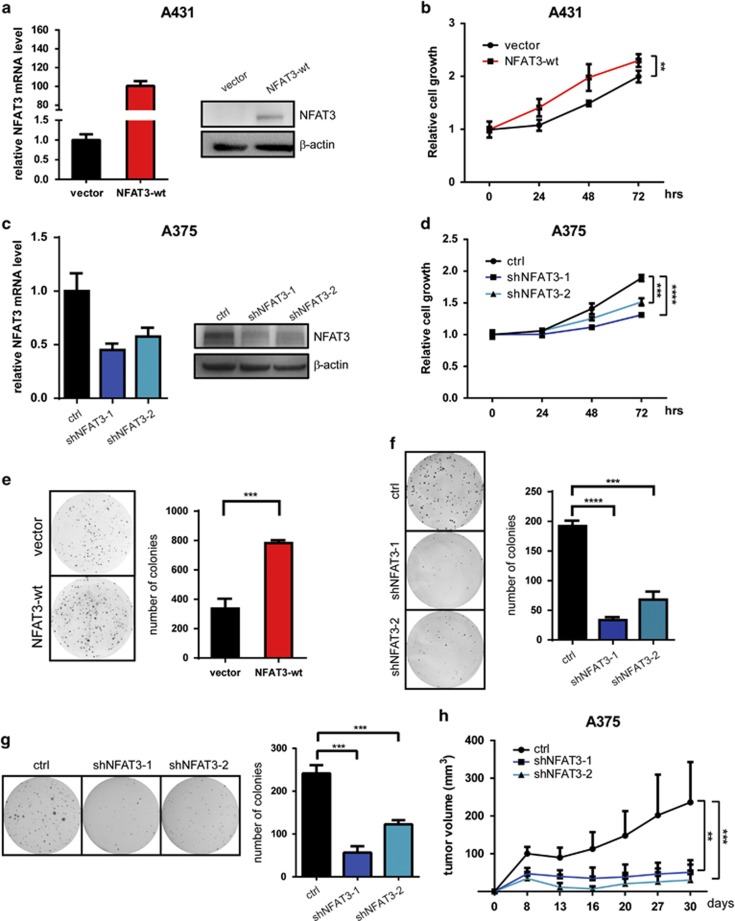
Figure 2.
NFAT3 has an oncogenic role in skin cancer cells. (a) OE of NFAT3 in A431 cells was detected by qPCR (left) or western blot (right). (b) Proliferation of stable A431 cells was measured by MTS assay. Results are expressed as mean values±s.d. from six replicate wells. (c) KD efficiency of NFAT3 in A375 cells was detected by qPCR (left) or western blot (right). (d) Proliferation of stable A375 cells was measured by MTS assay. (e and f) Representative photos and statistical analysis of 2-D colony formation in A431 stable cells with or without (e) NFAT3-OE and A375 stable cells with or without (f) NFAT3-KD. (g) Soft agar assay analysis of anchorage-independent growth of A375 stable cells with or without NFAT3-KD. Left panel shows representative photos. Right panel shows statistical analysis of colony number. (h) Xenograft tumor growth of A375 stable cells with or without NFAT3-KD in nude mice. Points indicate mean values (n=6); bars indicate s.d. *P<0.05, **P<0.01, ***P<0.001 and ****P<0.0001.

