

A 23-year-old man with known chronic pulmonary thromboembolic disease was admitted with large-volume hemoptysis. Computed tomography angiogram showed an aneurysm of the right middle lobe pulmonary artery and an enlarged right bronchial artery with extensive collateral vessels. The bronchial artery was successfully occluded (Figures 1A and 1B) with injection of 300- to 700-μm Embosphere Microspheres (Biosphere Medical, Rockland, MA). However, hemoptysis persisted and a pulmonary endarterectomy was performed to relieve pulmonary vascular obstruction and decrease additional collateral bronchial vascular flow. A significant amount of material, composed of organizing thrombus and nonpolarizable, brightly eosinophilic microspheres (Figure 2), was removed from the right pulmonary artery. Unfortunately, the aneurysmal right middle lobe pulmonary artery subsequently ruptured, necessitating surgical resection of the right middle lobe. Pathologic examination demonstrated eosinophilic-appearing microspheres in the vessel (Figure 3). Connections between the bronchial and pulmonary circulations exist and were reported 65 years ago in surgical lobectomy specimens from patients with bronchiectasis (1). This case confirms previous observations and demonstrates that material injected in vivo into the bronchial circulation can reach the pulmonary circulation.
Figure 1.

Enlarged right bronchial artery with two branches (arrow) before (A) and after (B) embolization, resulting in complete occlusion of both branches of the vessel.
Figure 2.

Microsphere fragments (arrows) embedded in organizing thrombus (asterisks) in a large pulmonary artery. L = lumen. Original magnification, ×42.5.
Figure 3.
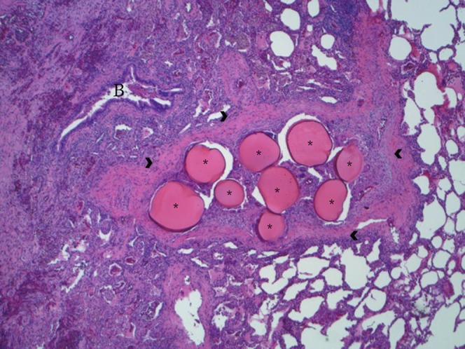
Microspheres (asterisks) embedded in intraparenchymal pulmonary artery (arrowheads). Note hemorrhagic atelectatic parenchyma (left) and normal alveolar tissue (right). B = companion bronchiole. Original magnification, ×42.5.
Footnotes
Author Contributions: Agreement to be accountable for all aspects of the work in ensuring that questions related to the accuracy or integrity of any part of the work are appropriately investigated and resolved: I.M.R. Substantial contributions to the conception or design of the work or the acquisition, analysis, or interpretation of data for the manuscript, and final approval of the version to be published: all authors. Drafting the work or revising it critically for important intellectual content: I.M.R., J.J., M.E.P., and A.R.H.
Author disclosures are available with the text of this article at www.atsjournals.org.
Reference
- 1.Liebow AA, Hales MR, Lindskog GE. Enlargement of the bronchial arteries, and their anastomoses with the pulmonary arteries in bronchiectasis. Am J Pathol. 1949;25:211–231. [PMC free article] [PubMed] [Google Scholar]