Leishmaniases are a spectrum of diseases caused by protozoans from the genus Leishmania (Kinetoplastida: Trypanosomatidae) and are divided into 2 main clinical forms: tegumentary leishmaniasis (TL) and visceral leishmaniasis (VL). Transmission occurs after the bite of sandfly vectors (Diptera: Phlebotominae) when females take a blood meal from the vertebrate host [1].
In the New World, several species of Leishmania (~20) cause disease to man, the symptoms and epidemiology of which vary depending on species. However, there are species that are nonpathogenic to humans, such as L. enriettii. In 1946, Medina observed ear lesions in 2 farm-reared guinea pigs (Cavia porcellus [Rodentia: Cavida]) from the neighboring state of São Paulo. After lesion analysis, Leishmania was confirmed as the pathogen. The complete L. enriettii description was published by Muniz and Medina in 1948 at the Federal University of Paraná, Brazil [2]. Although this species has been used as a model for cutaneous leishmaniasis (CL), many aspects of its biology remain unknown. In the past 6 years, an increased interest has emerged after the finding of a similar isolate in the red kangaroo (Macrofus rufus) in Australia [3]. This article aims to summarize some of the most important publications on this unique pathogen. It demonstrates a high phenotypic plasticity, being able to infect different vertebrate hosts and vectors. It also discusses recent human and veterinary infections due to other L. enriettii complex members.
L. enriettii: Vertebrate hosts
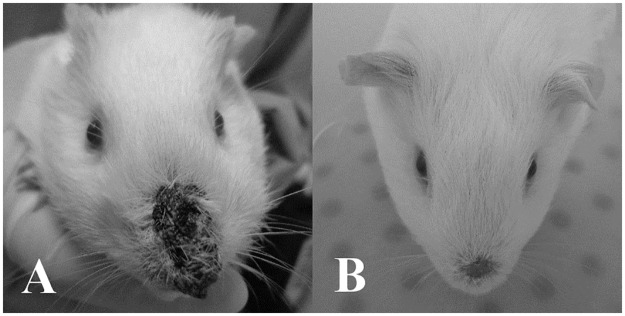
After L. enriettii discovery in C. porcellus in the 1940s [4], the authors failed to infect monkeys, dogs, and wild guinea pigs (C. aperea). They succeeded in infecting only 1 hamster out of 8 animals, and its lesion was poorly infected [2]. This is very likely to occur with wild reservoirs of Leishmania, such as opossums and armadillos, which, in nature, harbor low parasite densities without visible infection. At that time, no molecular approaches were available, opening the possibility of detecting L. enriettii in wild reservoirs other than C. aperea (Fig 1).
Fig 1. Cavia porcellus infected with Leishmania enriettii in the nose.
(A) Four to 7 weeks of infection and (B) healed lesion after 8–10 weeks of infection.
In spite of that, L. enriettii epidemiological studies involving hosts in Brazil are scarce, and new information on this parasite did not emerge until almost 50 years later. Two infected guinea pigs from the city of Capão Bonito (São Paulo state) and 3 from Campina Grande do Sul (Paraná state) were found. Species status was confirmed by isoenzyme analysis, with the description of a zymodeme polymorphism in the strains from Paraná [5]. Those data suggest that L. enriettii seems located in the Southern parts of Brazil, but its presence in other states should be prospected. A recent interesting study on Leishmania detection in road-killed wild animals in São Paulo found 1 C. aperea (1/4) infected with Leishmania. Although this study did not type the species, it provided some evidence that sylvatic C. aperea could harbor Leishmania [6]. Although the above-mentioned rodents have been considered the most common reservoirs for L. enriettii in Brazil, the finding of a putative similar isolate in captive red kangaroos (M. rufus), northern wallaroos (Macropus robustus woodwardii), black wallaroos (M. bernardus), and agile wallabies (M. agilis) was a landmark in the leishmaniasis epidemiology in Australia. The complete identification at species level only came in 2011 [3], classifying this isolate as a member of the L. enriettii complex [7].
L. enriettii: Possible invertebrate vectors
Only 20 years after the description of L. enriettii (1967), evidence appeared regarding its invertebrate host [8]. A survey of phlebotomine fauna in Paraná state identified Lutzomyia monticola and Lutzomyia correalimai in the vicinities where the guinea pigs were infected. Experimental sandfly infections on those guinea pigs were performed where 60% (6/10) of L. monticula exhibited a very high infection in their midguts. For this reason, L. enriettii was considered a suprapylarian species and included in the subgenus Leishmania. The authors recovered those parasites from sandflies and inoculated naive guinea pigs. However, no development of infection was observed, and transmission through L. monticola bite has yet to be determined.
Since no L. monticola laboratory colony is available, transmission experiments are difficult to perform. For this reason, it is a challenge to ascertain its current status as a L. enriettii vector. This vector is widely spread from southern to northern Brazilian states. Although L. monticola has no human medical importance, it is very anthropophilic and is often captured together with vectors of CL and VL. More importantly, its distribution overlaps with domestic C. porcellus and wild C. aperea, reinforcing further studies with this vector.
In spite of having many sandfly species in Australia, the first evidence that midges from the subgenus Forcipomyia (Lasiohelea) (Diptera: Ceratopogonidae) could be an alternative vector of Leishmania has emerged. Although this vector fell into most of the Killick-Kendrick criteria, transmission still needs to be demonstrated after its colonization [3]. It is important to mention that this work triggered a recent increase in studying L. enriettii by many groups. Recently, 2 other ceratopogonids (Culicoides nubeculosus and Culicoides sonorensis) and Lutzomyia longipalpis were tested for L. enriettii infection using 2 strains (Brazilian and Australian) [9]. Only C. sonorensis was able to sustain infection, whereas L. longipalpis (permissive vector) developed moderate infections. Those data remarkably demonstrated L. enriettii’s ability to sustain and develop infection in different invertebrate hosts. Also, those studies stimulate further epidemiological investigations to identify other potential/alternative non–sandfly vectors.
The L. enriettii complex members in human and veterinary infections
A detailed and historical review on Leishmania and sandflies was recently reported. However, a universal consensus regarding Leishmania classification is yet to be achieved [1], especially for the “L. enrietti complex.” In addition to L. enriettii, it may include Leishmania martiniquensis [10], “Leishmania siamensis” [11], and the Australian isolate [12]. In spite of the molecular techniques in establishing phylogenetic relationships among Leishmania species, a few studies have included L. enriettii complex members. The L. martiniquensis (strain MAR1) was isolated in Martinique Island, French Antilles, in 1995, causing diffuse CL in an HIV-infected patient [13]. A similar human isolate of L. martiniquensis causing VL was also reported from Thailand [14,15]. Finally, “L. siamensis” was first reported in Thailand as causing VL [16] and disseminated CL/VL in a patient with HIV. The isolate from this patient was named Trang strain and had 100% identity with L. enrietti after molecular analysis [11]. More recently, some isolates of L. enriettii complex members, probably “L. siamensis” and L. martiniquensis, were isolated from CL lesions in Ghana [15]. Detection of this species was also reported from a low number of cases in horses from Florida (2) [17] and Central Europe (6) [18] and in 1 cow from Switzerland [19]. However, as mentioned by Kwakye-Nuako et al. [15], although it appeared in the literature several times, the species “L. siamensis” was not formally described, and its name should be used in quotation marks.
The above-mentioned data suggest that members of L. enriettii complex are able to infect a wide range of different hosts, from rodents to humans, and to cause many clinical manifestations. In Table 1, we summarized some of the main isolates and reference strains of the L. enriettii complex members. Therefore, an international consortium for whole genome sequencing could be useful to ascertain the taxonomic status of this complex using those strains. This is of importance since the real status of “L. siamensis” and L. martiniquensis are still the subject of debate.
Table 1. Available isolates and strains of the Leishmania enriettii complex.
| Species | World Health Organization code | Reference |
|---|---|---|
| L. enriettii | MCAV/BR/1945/L88 MCAV/BR/95/CUR3 MCAV/BR/1985/COBAIA_SP MCAV/BR/45/LV90 AM-2004 |
[20–23] [22] [23] [9] [12] |
| “L. siamensis” | Trang MHOM/GH/2012/GH5; LV757 MHOM/GH/2012/GH10; LV758 MHOM/GH/2012/GH11; LV759 |
[11] [15] [15] [15] |
| L. martiniquensis | MHOM/MQ/92/MAR 1 MHOM/MQ/92/MAR 2 MHOM/TH/2012/LSCM1 |
[10, 13, 24] [10] [14] |
Concluding remarks
Since its discovery in the 1940s, L. enriettii studies have been occurring in pulses. In the 1950s and 1960s, most of the studies were focused on its biology, transmission, and epidemiology. Later on, in the 1970s, some immunopathological and immunological studies appeared. In the 1990s, its use as a model for molecular biology was successfully employed. More recently, the findings of other putative L. enriettii complex members such as L. martiniquensis and “L. siamensis” have generated great interest in understanding the taxonomical relationships among those different isolates. Whole genome sequencing would be a very important tool to investigate such relationships and help to establish their real species status. The species of this complex exhibit a high phenotypic plasticity in being able to infect a wide range of vertebrate hosts, including humans and other vectors. They may also cause different symptoms, ranging from CL to VL. However, many aspects of their epidemiology are still unknown in the geographic areas where those parasites were isolated. Although there is strong evidence of some suspected vectors, another important gap yet to be demonstrated is vectorial transmission by either ceratopogonids or L. monticula.
In this context, almost 70 years after L. enriettii discovery, many questions and uncertainties about its biology, epidemiology, classification, and immunology remain unanswered. The finding of members of this complex on different continents and its veterinary and human medical importance, especially in patients with HIV, reinforces the need for more studies. Several groups in the world are now investigating those parasites, and an increase in the published papers in the past years has demonstrated that this parasite is here to stay.
Funding Statement
This work was supported by Fundação de Amparo do Estado de Minas Gerais Programa Pesquisador Mineiro-X, PPM-00102-16 and Conselho Nacional de Pesquisa e Desenvolvimento CNPq. The funders had no role in study design, data collection and analysis, decision to publish, or preparation of the manuscript.
References
- 1.Akhoundi M, Kuhls K, Cannet A, Votýpka J, Marty P. A Historical Overview of the Classification, Evolution, and Dispersion of Leishmania Parasites and Sandflies. PLoS Negl Trop Dis. 2016;10: 1–40. [DOI] [PMC free article] [PubMed] [Google Scholar]
- 2.Muniz J, Medina H. [Cutaneous leishmaniasis of the guinea pig, Leishmania enriettii n. sp]. Hospital (Rio J). 1948;33: 7–25. Portuguese. [PubMed] [Google Scholar]
- 3.Dougall AM, Alexander B, Holt DC, Harris T, Sultan AH, Bates PA, et al. Evidence incriminating midges (Diptera: Ceratopogonidae) as potential vectors of Leishmania in Australia. Int J Parasitol. Australian Society for Parasitology Inc.; 2011;41: 571–579. [DOI] [PubMed] [Google Scholar]
- 4.Medina H. Estudos Sobre Leishmaniose: I. Primeiros Casos de Leishmaniose Espontânea Observados em Cobáios. Brazilian Arch Biol Technol. 2001;jubilee: 13–55. [Google Scholar]
- 5.Thomaz-Soccol V, Pratlong F, Langue R, Castro E, Luz E, Dedet JP. New isolation of Leishmania enriettii Muniz and Medina, 1948 in Paranástate, Brazil, 50 years after the first description, and isoenzymatic polymorphism of the L. enriettii taxon. Ann Trop Med Parasitol. 1996;90: 491–5. [DOI] [PubMed] [Google Scholar]
- 6.Richini-Pereira VB, Marson PM, Hayasaka EY, Victoria C, da Silva RC, Langoni H. Molecular detection of Leishmania spp. in road-killed wild mammals in the Central Western area of the State of São Paulo, Brazil. J Venom Anim Toxins Incl Trop Dis. 2014;20: 27 10.1186/1678-9199-20-27 [DOI] [PMC free article] [PubMed] [Google Scholar]
- 7.Dougall A, Shilton C, Low Choy J, Alexander B, Walton S. New reports of Australian cutaneous leishmaniasis in Northern Australian macropods. Epidemiol Infect. 2009;137: 1516–1520. 10.1017/S0950268809002313 [DOI] [PubMed] [Google Scholar]
- 8.Luz E, Giovannoni M, Borba A. Infecção de Lutzomyia monticola por Leishmania enriettii. An da Fac Med da Univ Fed do Paraná. 1967;9: 121–128. [Google Scholar]
- 9.Seblova V, Sadlova J, Vojtkova B, Votypka J, Carpenter S, Bates PA, et al. The Biting Midge Culicoides sonorensis (Diptera: Ceratopogonidae) is Capable of Developing Late Stage Infections of Leishmania enriettii. PLoS Negl Trop Dis. 2015;9. [DOI] [PMC free article] [PubMed] [Google Scholar]
- 10.Desbois N, Pratlong F, Quist D, Dedet J-P. Leishmania (Leishmania) martiniquensis n. sp. (Kinetoplastida: Trypanosomatidae), description of the parasite responsible for cutaneous leishmaniasis in Martinique Island (French West Indies). Parasite. 2014;21: 1–4. [DOI] [PMC free article] [PubMed] [Google Scholar]
- 11.Bualert L, Charungkiattikul W, Thongsuksai P, Mungthin M, Siripattanapipong S, Khositnithikul R, et al. Case report: Autochthonous disseminated dermal and visceral leishmaniasis in an AIDS patient, Southern Thailand, caused by Leishmania siamensis. Am J Trop Med Hyg. 2012;86: 821–824. [DOI] [PMC free article] [PubMed] [Google Scholar]
- 12.Rose K, Curtis J, Baldwin T, Mathis A, Kumar B, Sakthianandeswaren A, et al. Cutaneous leishmaniasis in red kangaroos: Isolation and characterisation of the causative organisms. Int J Parasitol. 2004;34: 655–664. 10.1016/j.ijpara.2004.03.001 [DOI] [PubMed] [Google Scholar]
- 13.Dedet JP, Roche B, Pratlong F, Caies-Quist D, Jouannelle J, Benichou JC, et al. Diffuse cutaneous infection caused by a presumed monoxenous trypanosomatid in a patient infected with HIV. Trans R Soc Trop Med Hyg. 1995;89: 644–646. [DOI] [PubMed] [Google Scholar]
- 14.Pothirat T, Tantiworawit A, Chaiwarith R, Jariyapan N, Wannasan A, Siriyasatien P, et al. First Isolation of Leishmania from Northern Thailand: Case Report, Identification as Leishmania martiniquensis and Phylogenetic Position within the Leishmania enriettii Complex. PLoS Negl Trop Dis. 2014;8: 1–8. [DOI] [PMC free article] [PubMed] [Google Scholar]
- 15.Kwakye-Nuako G, Mosore MT, Duplessis C, Bates MD, Puplampu N, Mensah-Attipoe I, et al. First isolation of a new species of Leishmania responsible for human cutaneous leishmaniasis in Ghana and classification in the Leishmania enriettii complex. Int J Parasitol. Australian Society for Parasitology Inc.; 2015;45: 679–684. [DOI] [PubMed] [Google Scholar]
- 16.Sukmee T, Siripattanapipong S, Mungthin M, Worapong J, Rangsin R, Samung Y, et al. A suspected new species of Leishmania, the causative agent of visceral leishmaniasis in a Thai patient. Int J Parasitol. 2008;38: 617–622. 10.1016/j.ijpara.2007.12.003 [DOI] [PubMed] [Google Scholar]
- 17.Reuss SM, Dunbar MD, Calderwood Mays MB, Owen JL, Mallicote MF, Archer LL, et al. Autochthonous Leishmania siamensis in horse, Florida, USA. Emerg Infect Dis. 2012;18: 1545–1547. 10.3201/eid1809.120184 [DOI] [PMC free article] [PubMed] [Google Scholar]
- 18.Muller N, Welle M, Lobsiger L, Stoffel MH, Boghenbor KK, Hilbe M, et al. Occurrence of Leishmania sp. in cutaneous lesions of horses in Central Europe. Vet Parasitol. 2009;166: 346–351. 10.1016/j.vetpar.2009.09.001 [DOI] [PubMed] [Google Scholar]
- 19.Lobsiger L, Muller N, Schweizer T, Frey CF, Wiederkehr D, Zumkehr B, et al. An autochthonous case of cutaneous bovine leishmaniasis in Switzerland. Vet Parasitol. 2010;169: 408–414. 10.1016/j.vetpar.2010.01.022 [DOI] [PubMed] [Google Scholar]
- 20.Thomaz-Soccol V, Lanotte G, Rioux JA, Pratlong F, Martini-Dumas A, Serres E. Phylogenetic taxonomy of New World Leishmania. Ann Parasitol Hum Comp. 1993;68: 104–106. [PubMed] [Google Scholar]
- 21.Asato Y, Oshiro M, Myint CK, Yamamoto Y, Kato H, Marco JD, et al. Phylogenic analysis of the genus Leishmania by cytochrome b gene sequencing. Exp Parasitol. Elsevier Inc.; 2009;121: 352–361. [DOI] [PubMed] [Google Scholar]
- 22.Noyes H, Pratlong F, Chance M, Ellis J, Lanotte G, Dedet JP. A previously unclassified trypanosomatid responsible for human cutaneous lesions in Martinique (French West Indies) is the most divergent member of the genus Leishmania. Parasitology. 2002;124: 17–24. [DOI] [PubMed] [Google Scholar]
- 23.Paranaíba LF, de Assis RR, Nogueira PM, Torrecilhas AC, Campos JH, de O Silveira AC, et al. Leishmania enriettii: biochemical characterisation of lipophosphoglycans (LPGs) and glycoinositolphospholipids (GIPLs) and infectivity to Cavia porcellus. Parasit Vectors. 2015;8: 31 10.1186/s13071-015-0633-8 [DOI] [PMC free article] [PubMed] [Google Scholar]
- 24.Liautaud B, Vignier N, Miossec C, Plumelle Y, Kone M, Delta D, et al. First case of visceral leishmaniasis caused by Leishmania martiniquensis. Am J Trop Med Hyg. 2015;92: 317–319. 10.4269/ajtmh.14-0205 [DOI] [PMC free article] [PubMed] [Google Scholar]


