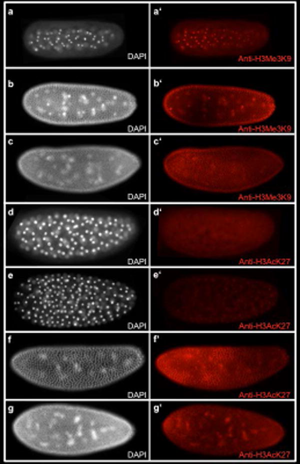
Fig. 2. Patterns of Histone modification in the Nasonia embryo.

a-g White, DAPI (DNA). Fig a′-c′ red, anti-H3Me3K9 marks repressive chromatin. (a-a′) Embryo in division cycle 5. Anti-H3Me3K9 staining is seen in all nuclei. (b-b′) Embryo in division cycle 9. (c-c′) Embryo in division cycle 12. Fig D-G White, DAPI (DNA) Fig d′-g′ red, anti-H3AcK27 marks active enhancers and promoters. (d-d′) Embryo in division cycle 6 does not show any Anti-H3AcK27 staining. (e-e′) Embryo in division cycle 7 shows nuclear staining of Anti-H3AcK27. (f-f′) Embryo in division cycle 11. (g-g′) Embryo in division cycle 12 shows increased levels of of Anti-H3AcK27 nuclear staining at the anterior and posterior poles of the embryo.
