

Abstract
Background:
Paracoccidioidomycosis is a systemic mycosis of significant importance in some Latin American countries. The widespread use of neuroimaging methods has shown that involvement of the central nervous system was more frequent than previously reported. The most common form of occurrence of neuroparacoccidioidomycosis is the pseudotumoral one. The authors report a case of pseudotumoral neuroparacoccidioidomycosis localized in the posterior fossa.
Case Description:
A 49-year-old single man, rural worker, born and raised in Laranjal Paulista-SP, was admitted to the hospital with 3 months history of bilateral occipital headache every day. Along with a history of active smoking and previous use of alcohol, the patient reported personal history of mild occipitotemporal injury 3 months ago. The patient was submitted to computed tomography in a 16-row multidetector scanner, which revealed a nodular hypodense lesion with a ring-enhancement and associated perilesional edema in the left cerebellar hemisphere. Radiological workup was initiated to investigate the eventual primary neoplastic site.
Conclusion:
The analysis of the lipid peak by spectroscopy of proton magnetic resonance may indicate the neurological involvement by paracoccidioidomycosis, notably in patients with concomitant risk and pulmonary involvement signals.
Keywords: Central nervous system, computed tomography, magnetic resonance spectroscopy, neuroparacoccidioidomycosis, paracoccidioidomycosis
INTRODUCTION
Paracoccidioidomycosis (PCM) is a systemic mycosis caused by a temperature-dependent dimorphic fungus, the Paracoccidioides brasiliensis. This fungus has significant importance in some countries of Latin America. Humans and some animals, such the armadillo, act as the host. In the natural habitat, the fungus present as the conidia infective form (infective propagules), and once inhaled they give rise to yeast from fungi in the host tissues.[12]
Males are the most affected, especially rural workers aged 30–50 years. In adults, the most common chronic form is the multifocal one, in which there is a pulmonary manifestation in approximately 90% of the cases. The lungs are considered the primary sites of infection. PCM relates to immunosuppressive conditions, and smoking and alcohol use are associated with this disease.[12] Neuroparacoccidioidomycosis (NPCM) can occur in parenchymal and/or meningeal forms, and parenchymal pseudotumoral is the most frequently observed.[3,6,10] Neurological involvement usually occurs in the context of chronic infection, however, it can also occur alone.[1,7] Although literature reports NPCM with variable incidence,[1,7] in recent years, with the widespread use of neuroimaging methods, it has become more common than earlier expected; in some samples, it occurs in up to 36% of the cases.[3,8,9] A systematic literature review of Pedroso et al. has shown that this form of infection is also prevalent in male individuals, especially farm laborers with a mean age of 43 years. The mean period of evolution is 4.9 months. The characteristic symptoms were motor deficits or intracranial hypertension. The chronic pseudotumoral form is predominately characterized by granulomas, abscesses, lumps, or intraparenchymal cysts. The lesions are mainly in the supratentorial compartment of the skull, especially in the frontal and parietal lobes. In the posterior fossa, the cerebellum is the most affected organ. In majority of NPCM cases, simultaneous lung impairment and common abnormalities are seen on chest radiographs.[1,8,9] Although NPCM lesions have nonspecific patterns on neuroimaging,[3] the literature reports the importance of considering this diagnosis in patients of the risk group, such as rural workers, especially when there are signs of infection outside the central nervous system.[7] P. Brasiliensis is sensitive to several antifungals. The treatment is based on their mild and moderate forms involving oral itraconazole or trimethoprim-sulfamethoxazole. Cases of severe forms, antifungal therapy is based on the use of amphotericin B or trimethoprim–sulfamethoxazole intravenously. In antifungal therapy, the treatment includes supportive care and management of eventual complications, such as stricture airways and pulmonary fibrosis.[12]
CASE REPORT
A 49-year-old single man (rural worker), born and raised in Laranjal Paulista-SP, was admitted to our Emergency Room at the Botucatu Medical School University hospital with 3 months history of bilateral occipital headache. Along with a history of active smoking and previous use of alcohol, the patient reported a personal history of mild occipitotemporal injury 3 months ago. On physical examination, left dysdiadochokinesia and three palpable cervical lymph nodes were noted on the left side; one of them with increased dimensions. The patient was submitted to computed tomography (CT) in a 16-row multidetector scanner, which revealed, after iodinated contrast infusion, a nodular hypodense lesion with ring enhancement and associated perilesional edema in the left cerebellar hemisphere [Figure 1]. In light of these findings, hypothetic diagnoses of cerebellar abscess and neoplasy (primary or metastatic) were made. Radiological workup was initiated to investigate the eventual primary neoplastic site. Chest radiography [Figure 2] showed reticular opacities, perihilar and poorly defined in the right hemithorax. The CT in a 16-row multidetector scanner [Figure 3] and high-resolution chest protocol were done, showing pulmonary nodules in the right, some of them with excavated wall, sometimes thin and smooth, sometimes asymmetrical, and occasionally associated with perilesional ground-glass opacities. Magnetic resonance imaging (MRI) scans of the brain revealed similar finding found on CT of the anterior skull, in T1-weighted sequences [Figure 4a], showed peripheral hyperintense rim surrounding a hypointense center and ring enhancement after intravenous infusion of paramagnetic contrast [Figure 4d]. The T2-weigthted sequences and fluid-attenuated inversion recovery (FLAIR) [Figure 4b and c] showed perilesional edema in the left cerebellar hemisphere and peripheral hypointense in the center of the respective lesion. The diffusion-weighted (DWI) sequences and apparent diffusion coefficient (ADC), respectively, showed hyperintense and hypointense lesions with restricted diffusion of water molecules [Figure 4e and f]. The proton spectroscopy curves for MR of cerebellar lesions showed an increase in lactate and lipids peaks and reduction of N-acetyl-aspartate (NAA) peak, without increase in choline peak [Figure 5]. Fine needle aspiration of the lymph node was performed, and cytological analysis revealed a nonspecific reactive lymphadenopathy. Microsurgery biopsy of cerebellar lesion was performed, and the histological analysis concluded that it was paracoccidioidomycosis without neoplasia signals [Figure 6].
Figure 1.

Chest X-ray from posteroanterior projections (a) and left lateral (b) showing discrete perihilar opacities poorly defined at right
Figure 2.

Protocol for high-resolution CT scanning of the chest, axial sections showing lesions with excavated walls, sometimes thin and smooth (a), sometimes thicker and asymmetric (b), and peripheral nodular solid lesions (c and d) in lung parenchyma, as well as area of lowest vascularity and not well defined in both upper and lower regions of the lungs
Figure 3.
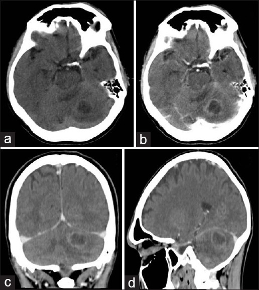
Axial sections of computed tomography before (a) and after (b) the infusion of iodinated contrast agent, revealing nodular hypodense lesion with ring- enhancement, in the left cerebellar hemisphere. Reformation in the coronal (c) and sagittal (d) confirmed the lesion was located in the posterior cranial fossa
Figure 4.

Brain MRI. (a) T1W nodular lesion with peripheral hyperintense rim surrounding a hypointense white matter in the left cerebellar hemisphere. Axial images in (b) T2W sequences and (c) FLAIR-perilesional edema, heterogeneity with peripheral hypointense white matter, center hyperintense. (d) T1W revealed ring-enhancement after intravenous infusion of paramagnetic contrast (gadolinium) in axial section. Axial image of diffusion- DWI sequences (e) lesion hyperintense center; and apparent diffusion coefficient (f), hypointense lesion with restricted diffusion of water molecules
Figure 5.

Proton spectroscopy curvs for magnetic resonance of cerebellar lesions showing increased peaks of lactate and lipids and reduction of N-acetyl-aspartate (NAA) peak, without increasing of choline peak
Figure 6.

(a) Cerebellar lesion fragment Grocott-Gomori staining revealing the typical histological findings of paracoccidioidomycosis forms: “ship's steering wheel” (arrow in a) and “Mickey Mouse” (arrow in b)
DISCUSSION
A review of national and international literature regarding evaluation of NPCM by neuroimaging methods shows the most common occurrence of NPCM, usually as with multiple granulomas, is the pseudotumoral form. They are commonly seen in CT as supra or infratentorial hypodense injuries, rounded, with variable perilesional edema, ring enhancement on iodinated contrast media. Therefore, CT differential diagnosis include primary or metastatic tumors and pyogenic abscesses.[1,3,8,9,11] The pulmonary form of this disease with NCPM is common, even with detectable changes on chest X-ray, such as hyperinflation and gross lesions of interstitial pattern.[8,9,11] MRI is the method of choice in the diagnosis of neurological involvement of paracoccidioidomycosis, in parenchymal or in meningeal lesions. Parenchymal lesions of the pseudotumoral form are usually characterized by peripheral hyperintense on T1-weighted sequence, and hypersignal on T2-weighted sequence with variable perilesional edema and peripheral enhancement after intravenous infusion of paramagnetic contrast.[8,10] In a recent retrospective study of NPCM cases by MRI, Reis et al. showed that the majority of parenchyma lesions had no restricted diffusion of water molecules, and the spectroscopy identified a common increased lipid peaks; in one case, there was an increased choline peak. Even as the meningea of NPCM lesion is considered exclusively rare, studies reveal meningea lesion extension at 42.9% when done by CT. In all cases, the study highlights the occurrence of lung lesions detectable on CT.[10] In literature, the majority of NPCM cases mention the cases of NPCM where the first diagnosis was cancer of the primary central nervous system[5] or metastatic,[2] similar to the case reported in this paper. In the other cases cited, there was a large proportion of the cases were associated with the pulmonary form of the disease.[2,5]
CONCLUSION
NPCM is more frequent than it was expected with the advent and the widespread of neuroimaging methods, especially MRI. The patterns for this entity by CT and MRI, in conventional sequences, are considered nonspecific and require differential diagnosis with other lesions such as primary or metastatic tumors and pyogenic abscesses. However, the presence of low signal intensity on the T2-weighted sequence suggests the presence of hemoglobin degradation products.[4,6] The analysis of the lipid peak by spectroscopy of proton MR may indicate the neurological involvement by paracoccidioidomycosis,[4] notably in patients with concomitant risk and pulmonary involvement signals.
Financial support and sponsorship
Nil.
Conflicts of interest
There are no conflicts of interest.
Footnotes
Contributor Information
Luiz Antonio Jorge, Jr, Email: lajjunior@gmail.com.
Seizo Yamashita, Email: seizo_eid@uol.com.br.
André Petean Trindade, Email: andreptrindade@hotmail.com.
Luiz Antonio Lima Resende, Email: ladlr@gmail.com.
Marco Antonio Zanini, Email: mzanini@fmb.unesp.br.
José Cândido Caldeira Xavier, Jr, Email: josecandidojr@yahoo.com.br.
Marcelo Padovani de Toledo Moraes, Email: mmoraes@fmb.unesp.br.
REFERENCES
- 1.Almeida SM, Queiroz-Telles F, Teive HAG, Ribeiro CEL, Werneck LC. Central nervous system paracoccidioidomycosis: Clinical features and laboratorial findings. J Infect. 2004;48:193–8. doi: 10.1016/j.jinf.2003.08.012. [DOI] [PubMed] [Google Scholar]
- 2.Cunha MLV, Castro CAO, Piekala C, Neto JFA, Pletz ALB. Neuroparacoccidioidomycosis simulating cerebral metastasis: Case report and literature review. J Bras Neurocirurg. 2012;23:226–33. [Google Scholar]
- 3.Elias J, Jr, dos Santos AC, Carlotti CG, Colli BO, Canheu A, Matias C, et al. Central nervous system paracoccidioidomycosis: Diagnosis and treatment. Surg Neurol. 2005;63:13–21. doi: 10.1016/j.surneu.2004.09.019. [DOI] [PubMed] [Google Scholar]
- 4.Faria AV, Dabus GC, Zanardi VA, Cendes F. Proton magnetic resonance spectroscopy and magnetic resonance imaging findings in a patient with central nervous system paracoccidioidomycosis. J Neuroimaging. 2004;14:377–9. doi: 10.1177/1051228404267053. [DOI] [PubMed] [Google Scholar]
- 5.Lambertucci JR, Lana-Peixoto MA, Pitella JEH. Paracoccidioidomycosis of the central nervous system. Rev Soc Bras Med Trop. 2001;34:395–6. doi: 10.1590/s0037-86822001000400016. [DOI] [PubMed] [Google Scholar]
- 6.Magalhaes AC, Caramelli P, Silva ED, Bacheschi LA, Lo LS, Menezes JR, et al. Magnetic resonance imaging in intracranial paracoccidioidomycosis. J Neuroimaging. 1993;3:216–9. doi: 10.1111/jon199334216. [DOI] [PubMed] [Google Scholar]
- 7.Paniago AM, de Oliveira PA, Aguiar ES, Aguiar JI, da Cunha RV, Leme LM, et al. Neuroparacoccidioidomycosis: Analysis of 13 cases observed in an endemic area in Brazil. Arq Neuropsiquiatr. 1989;47:224–9. doi: 10.1016/j.trstmh.2006.07.006. [DOI] [PubMed] [Google Scholar]
- 8.Pedroso VSP, Vilela MC, Pedroso ERP, Teixeira AL. Paracoccidioidomycosis compromising the central nervous system: a systematic review of the literature. Rev Soc Bras Med Trop. 2009;42:691–7. doi: 10.1590/s0037-86822009000600016. [DOI] [PubMed] [Google Scholar]
- 9.Pedroso VSP. Experimental studies and clinical prospects of paracoccidioidomycosis. [Dissertation] Belo Horizonte: Universidade Federal de. Minas Gerais; 2009. [Google Scholar]
- 10.Reis F, Collier PP, Souza TF, Lopes GP, Bronzatto E, Silva Junior NA, Pereira RM, Appenzeller S. Neuroparacoccidioidomycosis (NPCM): Magnetic Resonance Imaging (MRI) findings. Mycopathologia. 2013;175:181–6. doi: 10.1007/s11046-012-9607-y. [DOI] [PubMed] [Google Scholar]
- 11.Rodacki MA, De Toni G, Borba LA, Oliveira GG. Paracoccidioidomycosis of the central nervous system: CT findings. Neuroradiology. 1995;37:636–41. doi: 10.1007/BF00593377. [DOI] [PubMed] [Google Scholar]
- 12.Shikanai-Yasuda MA, Telles Filho FQ, Mendes RP, Colombo AL, Moretti ML. Consensus in paracoccidioidomycosis. Rev Soc Bras Med Trop. 2006;39:297–310. doi: 10.1590/s0037-86822006000300017. [DOI] [PubMed] [Google Scholar]