Abstract
Background:
Due to rapid advances in modern technologies such as telecommunication technology, the world has witnessed an exponential growth in the use of digital handheld devices (e.g. smartphones and tablets). This drastic growth has resulted in increased global concerns about the safety of these devices. Smartphones, tablets, laptops, and other digital screens emit high levels of short-wavelength visible light (i.e. blue color region in the visible light spectrum).
Material and Methods:
At a dark environment, Staphylococcus aureus bacteria were exposed to the light emitted from common tablets/smartphones. The control samples were exposed to the same intensity of light generated by a conventional incandescent light bulb. The growth rate of bacteria was examined by measuring the optical density (OD) at 625 nm by using a spectrophotometer before the light exposure and after 30 to 330 minutes of light exposure.
Results:
The growth rates of bacteria in both smartphone and tablet groups were higher than that of the control group and the maximum smartphone/control and tablet/control growth ratios were observed in samples exposed to digital screens’ light for 300 min (ratios of 3.71 and 3.95, respectively).
Conclusion:
To the best of our knowledge, this is the first study that investigates the effect of exposure to light emitted from digital screens on the proliferation of Staphylococcus aureus and its association with acne pathogenesis. Our findings show that exposure to short-wavelength visible light emitted from smartphones and tablets can increase the proliferation of Staphylococcus aureus.
Keywords: Smartphones , Tablets , Blue Light , Staphylococcus aureus , Acne
Introduction
The exponential rise in the use of handheld devices such as smartphones and tablets has raised global concerns about the safety of these devices [1-6]. Smartphones, tablets, laptops, and other LED screens can emit high levels of short-wavelength visible light (blue region in the light spectrum). Over the past several years, the biological effects of exposure to short-wavelength visible light emitted from smartphones and tablets on the eye and skin (disorders such as premature skin aging) have been widely studied. However, to the best of our knowledge, the effect of exposure to light emitted from these devices on the proliferation of Staphylococcus aureus and the possible association of these exposures with acne pathogenesis have not been investigated yet. Some studies show that S. aureus can intensify the symptoms in chronic inflammatory skin diseases. Acne vulgaris has been reported to be the most common human skin disorder. This skin disorder was reported to be persistent in 80% of the women (58% of these women had an ongoing need for treatment). Over the past several years, our laboratories at the non-ionizing department of the Ionizing and Non-ionizing Radiation Protection Research Center (INIRPRC) have conducted experiments on the health effects of exposure to different sources of electromagnetic fields such as cellular phones [7-14], mobile base stations [15,16], mobile phone jammers [17,18] and laptop computers [19]. We have also focused on the possible interactions between either ionizing or non-ionizing radiations as well as mechanical waves (e.g. ultrasound) and bacteria [20-23].
It has been revealed that exposure to bright light at night suppresses the secretion of melatonin and it has also been shown that human circadian system is susceptible to the biological effects of the short-wavelength part of the visible light spectrum [24,25]. The application of blue light in different medical fields such as phototherapy [26,27] or antibacterial treatment of plaque-induced periodontal pathologies [28] is well documented. It has recently been suggested to use screens with the emission peak of 470-480 nm instead of using screens with emission peaks below 450 nm. This suggestion is due to known risks associated with the exposure to blue light [29]. In this study, the effect of exposure to visible light generated by the screens of a commercial smartphone (Sony Xperia) and a commercial tablet (Samsung Galaxy Note 10.1) on the growth rate of S. aureus bacteria (ATCC No. 25923) is investigated.
Material and Methods
All experiments were performed at 37°C in a separate incubator and bacteria were grown in 20 ml Brain Heart Infusion Broth (BHI) 10 cm plates. Then, in a dark environment, bacteria were exposed to the light of the tablet and smartphone at a distance of 2-3 mm (common distance between the smartphones and facial skin). The brightness of the displays of these devices was set at 50%. The control samples were exposed to the same intensity of light generated by a conventional incandescent light bulb. The growth rate of bacteria was examined by measuring the optical density (OD) at 625 nm by using a spectrophotometer (UNICO UV-2100 spectrophotometer) before the light exposure and after 30, 60, 90, 120, 150, 180, 210, 240, 300, and 330 minutes of light exposure.
Results
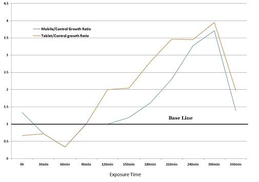
The growth rates of bacteria in both smartphone and tablet groups were higher than that of the control group. Optical density values in both smartphone and tablet groups before and after exposure to short wavelength visible light are summarized in Table 1. The maximum smartphone/control and tablet/control growth ratios were observed in samples exposed to digital screens’ light for 300 min (ratios of 3.71 and 3.95, respectively). These ratios declined in samples exposed to screens’ light for durations higher than 300 min. The growth rates of bacteria in both smartphone and tablet groups are shown in Figure 1.
Table 1.
OD values before and after exposure to short wavelength visible light
| Time | Optical Density (OD) | ||||
|---|---|---|---|---|---|
| Control | Smartphone | Smartphone/ Control ratio | Tablet | Tablet/ Control ratio | |
| 0 min | 0.003 | 0.004 | 1.33 | 0.002 | 0.66 |
| 30 min | 0.007 | 0.005 | 0.71 | 0.005 | 0.71 |
| 60 min | 0.003 | 0.001 | 0.33 | 0.001 | 0.33 |
| 90 min | 0.005 | 0.005 | 1 | 0.005 | 1 |
| 120 min | 0.008 | 0.008 | 1 | 0.016 | 2 |
| 150 min | 0.026 | 0.031 | 1.19 | 0.053 | 2.03 |
| 180 min | 0.021 | 0.034 | 1.61 | 0.059 | 2.80 |
| 210 min | 0.013 | 0.03 | 2.30 | 0.045 | 3.46 |
| 240 min | 0.029 | 0.095 | 3.27 | 0.1 | 3.44 |
| 300 min | 0.038 | 0.141 | 3.71 | 0.15 | 3.95 |
| 330 min | 0.137 | 0.19 | 1.38 | 0.27 | 1.97 |
Figure1.

The growth rates of bacteria in both smartphone and tablet groups were higher than that of the control group. This chart shows that the maximum smartphone/control and tablet/control growth ratios were observed in samples exposed to digital screens’ light for 300 min (3.71 and 3.95, respectively).
Discussion
Altogether, these findings show that exposure to short-wavelength visible light emitted from smartphones and tablets can increase the proliferation of Staphylococcus aureus. These findings are generally in line with the findings of our previous studies which showed the potential interactions between either mechanical waves or electromagnetic radiation and bacteria [21-23]. We have previously shown that exposure of Klebsiella pneumoniae (K. pneumonia) to electromagnetic radiation in the radiofrequency range can lead to a statistically significant rise in the susceptibility of this microorganism to different antibiotics. As in our study on K. pneumoniae, a minimum level of effect was needed for the induction of resistance after a pre-exposure, we postulated that the so-called “window theory” could be used for interpreting the findings [23]. This study was our first experiment on the effect of exposure to short wavelength visible light emitted from digital screens on the proliferation of S. aureus. Some studies have shown that treatment with visible light in the red region can be utilized as a therapeutic method to inactivate some pathogenic strains of porphyrin producing bacteria [30]. In this light, it seems that long wavelength red light and short wavelength blue light may have different effects on the growth rate of bacteria. This difference possibly reflects the role of visible photon energy on the proliferation of micro-organisms. This theory should be verified by further experiments.
It is worth noting that the results obtained in this study cannot be entirely extrapolated to daily applications of smartphones because in this condition there are intense light sources such as sunlight or high intensity artificial sources of light which are much stronger than the light emitted from digital screens. As it is discussed in the materials and methods section, this study was conducted in a dark environment. In such an environment, digital screen’s light can be the only source of light. As today there are many people who use their smartphones in bed in a relatively dark condition, our study helps scientists better evaluate the response of bacteria to screens’ light in such a specific environment. However, due to limitations of this study, further studies are needed to shed more light on the dark corners of the effect of digital screens’ light on the prolifration of different microorganisms and also to verify if these exposures can be linked to acne pathogenesis.
Acknowledgement
This study was supported by the Ionizing and Non-ionizing Radiation Protection Research Center (INIRPRC), Shiraz University of Medical Sciences (SUMS), Shiraz, Iran.
Conflict of Interest:None Declared.
References
- 1.Buckus R, Strukcinskiene B, Raistenskis J. The assessment of electromagnetic field radiation exposure for mobile phone users. Vojnosanit Pregl. 2014;71(12):1138–43. doi: 10.2298/vsp140119013b. [DOI] [PubMed] [Google Scholar]
- 2.Deshmukh PS, Nasare N, Megha K, Banerjee BD, Ahmed RS, Singh D, et al. Cognitive impairment and neurogenotoxic effects in rats exposed to low-intensity microwave radiation. International journal of toxicology. 2015;34(3):284–90. doi: 10.1177/1091581815574348. [DOI] [PubMed] [Google Scholar]
- 3.Medeiros LN, Sanchez TG. Tinnitus and cell phones: the role of electromagnetic radiofrequency radiation. Braz J Otorhinolaryngol. 2016;82(1):97–104. doi: 10.1016/j.bjorl.2015.04.013. [DOI] [PMC free article] [PubMed] [Google Scholar]
- 4.Paul B, Saha I, Kumar S, Samim Ferdows SK, Ghose G. Mobile phones: time to rethink and limit usage. Indian J Public Health. 2015;59(1):37–41. doi: 10.4103/0019-557X.152856. [DOI] [PubMed] [Google Scholar]
- 5.Yang L, Chen Q, Lv B, Wu T. Long-Term Evolution Electromagnetic Fields Exposure Modulates the Resting State EEG on Alpha and Beta Bands. Clin EEG Neurosci. 2016;25:1550059416644887. doi: 10.1177/1550059416644887. [DOI] [PubMed] [Google Scholar]
- 6.Zhou Y, Zhang H, Niu Z. [Analysis of Electric Stress in Human Head in High-frequency Low-power Electromagnetic Environment] Sheng Wu Yi Xue Gong Cheng Xue Za Zhi. 2015;32(2):295–9. [PubMed] [Google Scholar]
- 7.Mortazavi SMJ, Motamedifar M, Namdari G, Taheri M, Mortazavi AR, Shokrpour N. Non-Linear Adaptive Phenomena which Decrease the Risk of infection after Pre-Exposure to Radiofrequency Radiation. Dose-Response. 2014;12(2) doi: 10.2203/dose-response.12-055.Mortazavi. [DOI] [PMC free article] [PubMed] [Google Scholar]
- 8.Mortazavi SMJ, Taeb S, Dehghan N. Alterations of Visual Reaction Time and Short Term Memory in Military Radar Personnel. Iranian J Publ Health. 2013;42(4):428–35. [PMC free article] [PubMed] [Google Scholar]
- 9.Mortazavi SMJ, Rouintan MS, Taeb S, Dehghan N, Ghaffarpanah AA, Sadeghi Z, et al. Human short-term exposure to electromagnetic fields emitted by mobile phones decreases computer-assisted visual reaction time. Acta Neurologica Belgica. 2012;112(2):171–5. doi: 10.1007/s13760-012-0044-y. [DOI] [PubMed] [Google Scholar]
- 10.Mortazavi SMJ, Mosleh-Shirazi MA, Tavassoli AR, Taheri M, Mehdizadeh AR, Namazi SAS, et al. Increased Radioresistance to Lethal Doses of Gamma Rays in Mice and Rats after Exposure to Microwave Radiation Emitted by a GSM Mobile Phone Simulator. Dose-response : a publication of International Hormesis Society. 2013;11 (2):281–92. doi: 10.2203/dose-response.12-010.Mortazavi. [DOI] [PMC free article] [PubMed] [Google Scholar]
- 11.Mortazavi S, Mosleh-Shirazi M, Tavassoli A, Taheri M, Bagheri Z, Ghalandari R, et al. A comparative study on the increased radioresistance to lethal doses of gamma rays after exposure to microwave radiation and oral intake of flaxseed oil. Iranian Journal of Radiation Research. 2011;9(1):9–14. [Google Scholar]
- 12.Mortazavi SMJ, Habib A, Ganj-Karimi AH, Samimi-Doost R, Pour-Abedi A, Babaie A. Alterations in TSH and Thyroid Hormones Following Mobile Phone Use. OMJ. 2009;24:274–8. doi: 10.5001/omj.2009.56. [DOI] [PMC free article] [PubMed] [Google Scholar]
- 13.Mortazavi SMJ, Daiee E, Yazdi A, Khiabani K, Kavousi A, Vazirinejad R, et al. Mercury release from dental amalgam restorations after magnetic resonance imaging and following mobile phone use. Pakistan Journal of Biological Sciences. 2008;11(8):1142–6. doi: 10.3923/pjbs.2008.1142.1146. [DOI] [PubMed] [Google Scholar]
- 14.Mortazavi SMJ, Ahmadi J, Shariati Prevalence of subjective poor health symptoms associated with exposure to electromagnetic fields among University students. Bioelectromagnetics. 2007;28(4):326–30. doi: 10.1002/bem.20305. [DOI] [PubMed] [Google Scholar]
- 15.Mortazavi SMJ. Safety Issue of Mobile Phone Base Stations. Journal of biomedical physics & engineering. 2013;3(1):1–2. [PMC free article] [PubMed] [Google Scholar]
- 16.Mortazavi SA, Mortazavi G, Mortazavi SM. Comments on Meo et al. Association of Exposure to Radio-Frequency Electromagnetic Field Radiation (RF-EMFR) Generated by Mobile Phone Base Stations with Glycated Hemoglobin (HbA1c) and Risk of Type 2 Diabetes Mellitus. Int J Environ Res Public Health, 2015, 12, 14519-14528 Int J Environ Res Public Health 2016;13(3) doi: 10.3390/ijerph13030261. [DOI] [PMC free article] [PubMed] [Google Scholar]
- 17.Parsanezhad ME, Mortazavi SMJ, T D. Exposure to Radiofrequency Radiation Emitted from Mobile Phone Jammers Adversely Affects the Quality of Human Sperm. International Journal of Radiation Research (IJRR) in press. [Google Scholar]
- 18.Mortazavi SMJ, Parsanezhad ME, Kazempour M, Ghahramani P, Mortazavi AR, Davari M. Male reproductive health under threat: Short term exposure to radiofrequency radiations emitted by common mobile jammers. Journal of Human Reproductive Sciences. 2013;6(2):124–8. doi: 10.4103/0974-1208.117178. [DOI] [PMC free article] [PubMed] [Google Scholar]
- 19.Mortazavi SMJ, Tavasoli AR, Ranjbari F, Moamaei P. Effects of Laptop Computers’ Electromagnetic Field on Sperm Quality. Journal of Reproduction and Infertility. 2011;11(4):251–8. [Google Scholar]
- 20.Mortazavi SM. Isolation a new strain of Kocuria rosea capable of tolerating extreme conditions. J Environ Radioact. 2015 Sep;147:153–4. doi: 10.1016/j.jenvrad.2015.05.010. [DOI] [PubMed] [Google Scholar]
- 21.Mortazavi SM, Darvish L, Abounajmi M, Zarei S, Zare T, Taheri M, et al. Alteration of Bacterial Antibiotic Sensitivity After Short-Term Exposure to Diagnostic Ultrasound. Iranian Red Crescent medical journal. 2015;17(11) doi: 10.5812/ircmj.26622. [DOI] [PMC free article] [PubMed] [Google Scholar]
- 22.Mortazavi SM, Motamedifar M, Namdari G, Taheri M, Mortazavi AR, Shokrpour N. Non-linear adaptive phenomena which decrease the risk of infection after pre-exposure to radiofrequency radiation. Dose-response : a publication of International Hormesis Society. 2013;12(2):233–45. doi: 10.2203/dose-response.12-055.Mortazavi. [DOI] [PMC free article] [PubMed] [Google Scholar]
- 23.Taheri M, Mortazavi SM, Moradi M, Mansouri S, Nouri F, Mortazavi SA, et al. Klebsiella pneumonia, a Microorganism that Approves the Non-linear Responses to Antibiotics and Window Theory after Exposure to Wi-Fi 2. 4 GHz Electromagnetic Radiofrequency Radiation. Journal of biomedical physics & engineering 2015;5(3):115–20. [PMC free article] [PubMed] [Google Scholar]
- 24.Kozaki T, Kubokawa A, Taketomi R, Hatae K. Light-induced melatonin suppression at night after exposure to different wavelength composition of morning light. Neurosci Lett. 2016 Mar 11;616:1–4. doi: 10.1016/j.neulet.2015.12.063. [DOI] [PubMed] [Google Scholar]
- 25.Figueiro MG, Plitnick BA, Lok A, Jones GE, Higgins P, Hornick TR, et al. Tailored lighting intervention improves measures of sleep, depression, and agitation in persons with Alzheimer’s disease and related dementia living in long-term care facilities. Clinical interventions in aging. 2014;9:1527–37. doi: 10.2147/CIA.S68557. [DOI] [PMC free article] [PubMed] [Google Scholar]
- 26.Ebbesen F, Vandborg PK, Madsen PH, Trydal T. Effect of phototherapy with turquoise vs. blue LED light of equal irradiance in jaundiced neonates. 2016 Mar;79(2):308–12. doi: 10.1038/pr.2015.209. [DOI] [PubMed] [Google Scholar]
- 27.Uchida Y, Morimoto Y, Uchiike T, Kamamoto T, Hayashi T, Arai I, et al. Phototherapy with blue and green mixed-light is as effective against unconjugated jaundice as blue light and reduces oxidative stress in the Gunn rat model. Early Hum Dev. 2015 Jul;91(7):381–5. doi: 10.1016/j.earlhumdev.2015.04.010. [DOI] [PubMed] [Google Scholar]
- 28.Mahdi Z, Habiboallh G, Mahbobeh NN, Mina ZJ, Majid Z, Nooshin A. Lethal effect of blue light-activated hydrogen peroxide, curcumin and erythrosine as potential oral photosensitizers on the viability of Porphyromonas gingivalis and Fusobacterium nucleatum. Laser therapy. 2015 Mar 31;24(2):103–11. doi: 10.5978/islsm.15-OR-09. [DOI] [PMC free article] [PubMed] [Google Scholar]
- 29.Tosini G, Ferguson I, Tsubota K. Effects of blue light on the circadian system and eye physiology. Molecular vision. 2016;22:61–72. [PMC free article] [PubMed] [Google Scholar]
- 30.Konig K, Teschke M, Sigusch B, Glockmann E, Eick S, Pfister W. Red light kills bacteria via photodynamic action. Cell Mol Biol. 2000;46(7):1297–303. [PubMed] [Google Scholar]


