“Dermatoscopy” is considered to be the stethoscope or the third eye to a dermatologist to detect sub-surface features not visible to the unaided eye. The device was evolved primarily to detect melanomas and to get biopsy of suspicious lesions. Recently it has found application in diagnosis of papulosquamous disorders, pigmentary disorders other than melanoma and other benign conditions.[1] However, there is paucity of data in literature regarding image differences between various dermatoscopes.
This short communication aims to compare images between four commonly used standard dermatoscopes, viz. Heine Delta 20 (Heine optotechnik, Herrsching, Germany), Heine Delta 20 plus (Heine optotechnik, Herrsching, Germany), Dermlite DL II multispectral (3Gen Inc, USA), and Dermlite DL3 (3Gen Inc, USA) in three common skin conditions, namely, acanthosis nigricans (AN), psoriasis and melasma. Images were captured using the same setting and zoom using a Nikon AW1 14.1 MP mirrorless camera (Nikon Corp., Tokyo, Japan). Heine Delta 20 plus and Dermlite DL3 have both polarized and non-polarized modes. Heine Delta 20 has only non-polarized mode, which requires a contact fluid, whereas Dermlite DL II has only cross-polarized mode. Non-polarized dermatoscope helps in the detection of superficial structures in the skin, whereas a polarized dermatoscope allows deeper visualization. Though polarized dermatoscopes do not require contact fluid, images captured by them are sharper in the presence of interface fluid.[2] Hence, all images were taken with liquid paraffin as contact fluid with all 4 dermatoscopes.
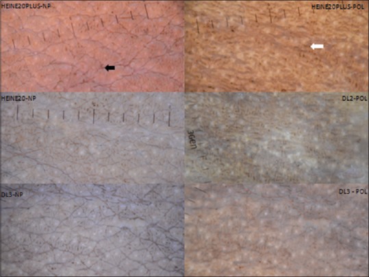
Images taken with non-polarized mode offer better visualization of dermatoscopic findings in AN as the pathology is in the superficial layers of skin. Images taken in the non-polarized and polarized modes with all 4 devices were comparable. Dermatoscopy images taken with Heine Delta 20 plus were appreciably warmer compared to the other 3 dermatoscopes. Dermlite DL3 polarized images were the brightest because of high illumination with 21 LEDs for the polarized mode [Figure 1].
Figure 1.

Dermatoscopy of Acanthosis nigricans over the neck (10×) – Non-polarized images show multiple cristae and sulci clearly (black arrow) whereas polarized imaging allows better visualization of hyperpigmented dots and streaks (white arrow). Polarized images taken with Heine Delta 20 plus have a warmer tone whereas DL3 polarized images are the brightest
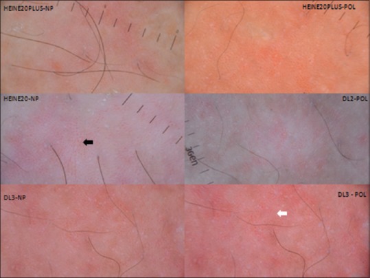
Vascular lesions are better characterized under the polarized mode as the light in this mode penetrates deeper. Regular distribution of red-dotted vessels on a light red background with diffuse white scales are suggestive of psoriasis under dermatoscopy.[3] Visualization of regular red dots in psoriasis is superior with earlier devices, viz. Heine Delta 20 and Dermlite II, where images are less bright and relatively have a white tone [Figure 2]. However, these devices are less efficient in picking up the background erythema as compared to Heine Delta 20 plus and Dermlite DL3.
Figure 2.

Dermatoscopy of Psoriasis over the trunk (10×) – Heine Delta 20 and Dermlite DLII offer maximum contrast for the regular red dots (black arrow), whereas Heine delta 20 plus and Dermlite DL3 help in the better visualization of background erythema (white arrow)
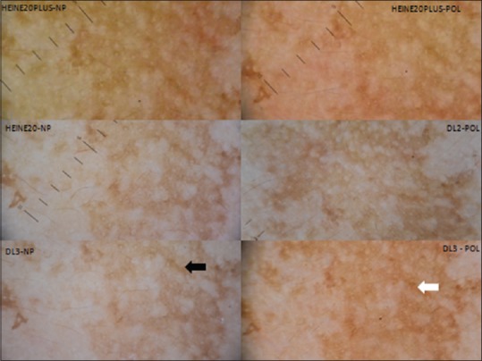
Dermatoscopy of melasma shows accentuation of the normal pseudoreticular pattern of face, sparing the follicular and sweat gland openings with dark brown globules and granules depending on the depth of melasma.[4] Pigmentation in melasma is more dark and brown in the polarized mode as compared to non-polarized images. The warmer tone in images of Heine Delta 20 plus dermatoscope is attributed to higher color rendering index values of the device [Figure 3].
Figure 3.

Dermatoscopy of Melasma over the face (10×) – Polarized images show the accentuation of pseudoreticular pattern, which is more brownish (white arrow) than nonpolarized images (black arrow). Heine delta 20 plus images are warmer
As outlined above, a particular mode in a dermatoscope can better highlight the dermatoscopic findings compared to other modes. Future studies can be undertaken to analyze the features of other dermatological diseases with different dermatoscopes.
Financial support and sponsorship
Nil.
Conflicts of interest
There are no conflicts of interest.
References
- 1.Nischal KC, Khopkar U. Dermoscope. Indian J Dermatol Venereol Leprol. 2005;71:300–3. doi: 10.4103/0378-6323.16633. [DOI] [PubMed] [Google Scholar]
- 2.Pan Y, Gareau DS, Scope A, Rajadhyaksha M, Mullani NA, Marghoob AA. Polarized and nonpolarized dermoscopy: The explanation for the observed differences. Arch Dermatol. 2008;144:828–9. doi: 10.1001/archderm.144.6.828. [DOI] [PubMed] [Google Scholar]
- 3.Lallas A, Kyrgidis A, Tzellos TG, Apalla Z, Karakyriou E, Karatolias A, et al. Accuracy of dermoscopic criteria for the diagnosis of psoriasis, dermatitis, lichen planus and pityriasis rosea. Br J Dermatol. 2012;166:1198–205. doi: 10.1111/j.1365-2133.2012.10868.x. [DOI] [PubMed] [Google Scholar]
- 4.Mahajan SA. Melasma. In: Khopkar US, editor. Dermoscopy and trichoscopy in diseases of the brown skin-atlas and short text. 1st ed. New Delhi: Jaypee Brothers Med Publishers; 2012. pp. 50–62. [Google Scholar]


