Abstract
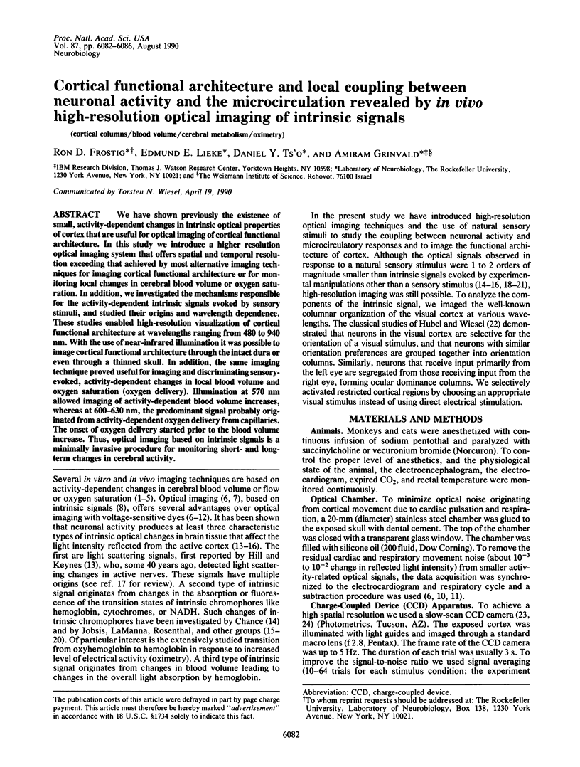
We have shown previously the existence of small, activity-dependent changes in intrinsic optical properties of cortex that are useful for optical imaging of cortical functional architecture. In this study we introduce a higher resolution optical imaging system that offers spatial and temporal resolution exceeding that achieved by most alternative imaging techniques for imaging cortical functional architecture or for monitoring local changes in cerebral blood volume or oxygen saturation. In addition, we investigated the mechanisms responsible for the activity-dependent intrinsic signals evoked by sensory stimuli, and studied their origins and wavelength dependence. These studies enabled high-resolution visualization of cortical functional architecture at wavelengths ranging from 480 to 940 nm. With the use of near-infrared illumination it was possible to image cortical functional architecture through the intact dura or even through a thinned skull. In addition, the same imaging technique proved useful for imaging and discriminating sensory-evoked, activity-dependent changes in local blood volume and oxygen saturation (oxygen delivery). Illumination at 570 nm allowed imaging of activity-dependent blood volume increases, whereas at 600-630 nm, the predominant signal probably originated from activity-dependent oxygen delivery from capillaries. The onset of oxygen delivery started prior to the blood volume increase. Thus, optical imaging based on intrinsic signals is a minimally invasive procedure for monitoring short- and long-term changes in cerebral activity.
Full text
PDF


Images in this article
Selected References
These references are in PubMed. This may not be the complete list of references from this article.
- Blasdel G. G., Salama G. Voltage-sensitive dyes reveal a modular organization in monkey striate cortex. Nature. 1986 Jun 5;321(6070):579–585. doi: 10.1038/321579a0. [DOI] [PubMed] [Google Scholar]
- Buchweitz E., Weiss H. R. Alterations in perfused capillary morphometry in awake vs anesthetized brain. Brain Res. 1986 Jul 2;377(1):105–111. doi: 10.1016/0006-8993(86)91195-9. [DOI] [PubMed] [Google Scholar]
- CHANCE B., COHEN P., JOBSIS F., SCHOENER B. Intracellular oxidation-reduction states in vivo. Science. 1962 Aug 17;137(3529):499–508. doi: 10.1126/science.137.3529.499. [DOI] [PubMed] [Google Scholar]
- Chance B., Leigh J. S., Miyake H., Smith D. S., Nioka S., Greenfeld R., Finander M., Kaufmann K., Levy W., Young M. Comparison of time-resolved and -unresolved measurements of deoxyhemoglobin in brain. Proc Natl Acad Sci U S A. 1988 Jul;85(14):4971–4975. doi: 10.1073/pnas.85.14.4971. [DOI] [PMC free article] [PubMed] [Google Scholar]
- Cohen L. B. Changes in neuron structure during action potential propagation and synaptic transmission. Physiol Rev. 1973 Apr;53(2):373–418. doi: 10.1152/physrev.1973.53.2.373. [DOI] [PubMed] [Google Scholar]
- Connor J. A. Digital imaging of free calcium changes and of spatial gradients in growing processes in single, mammalian central nervous system cells. Proc Natl Acad Sci U S A. 1986 Aug;83(16):6179–6183. doi: 10.1073/pnas.83.16.6179. [DOI] [PMC free article] [PubMed] [Google Scholar]
- Egger M. D., Petrăn M. New reflected-light microscope for viewing unstained brain and ganglion cells. Science. 1967 Jul 21;157(3786):305–307. doi: 10.1126/science.157.3786.305. [DOI] [PubMed] [Google Scholar]
- Eggert H. R., Blazek V. Optical properties of human brain tissue, meninges, and brain tumors in the spectral range of 200 to 900 nm. Neurosurgery. 1987 Oct;21(4):459–464. doi: 10.1227/00006123-198710000-00003. [DOI] [PubMed] [Google Scholar]
- Fox P. T., Mintun M. A., Raichle M. E., Miezin F. M., Allman J. M., Van Essen D. C. Mapping human visual cortex with positron emission tomography. 1986 Oct 30-Nov 5Nature. 323(6091):806–809. doi: 10.1038/323806a0. [DOI] [PubMed] [Google Scholar]
- Fox P. T., Raichle M. E., Mintun M. A., Dence C. Nonoxidative glucose consumption during focal physiologic neural activity. Science. 1988 Jul 22;241(4864):462–464. doi: 10.1126/science.3260686. [DOI] [PubMed] [Google Scholar]
- Grinvald A., Anglister L., Freeman J. A., Hildesheim R., Manker A. Real-time optical imaging of naturally evoked electrical activity in intact frog brain. 1984 Apr 26-May 2Nature. 308(5962):848–850. doi: 10.1038/308848a0. [DOI] [PubMed] [Google Scholar]
- Grinvald A., Frostig R. D., Lieke E., Hildesheim R. Optical imaging of neuronal activity. Physiol Rev. 1988 Oct;68(4):1285–1366. doi: 10.1152/physrev.1988.68.4.1285. [DOI] [PubMed] [Google Scholar]
- Grinvald A., Lieke E., Frostig R. D., Gilbert C. D., Wiesel T. N. Functional architecture of cortex revealed by optical imaging of intrinsic signals. 1986 Nov 27-Dec 3Nature. 324(6095):361–364. doi: 10.1038/324361a0. [DOI] [PubMed] [Google Scholar]
- Grinvald A., Manker A., Segal M. Visualization of the spread of electrical activity in rat hippocampal slices by voltage-sensitive optical probes. J Physiol. 1982 Dec;333:269–291. doi: 10.1113/jphysiol.1982.sp014453. [DOI] [PMC free article] [PubMed] [Google Scholar]
- HUBEL D. H., WIESEL T. N. Receptive fields, binocular interaction and functional architecture in the cat's visual cortex. J Physiol. 1962 Jan;160:106–154. doi: 10.1113/jphysiol.1962.sp006837. [DOI] [PMC free article] [PubMed] [Google Scholar]
- Hertz M. M., Paulson O. B. Transfer across the human blood-brain barrier: evidence for capillary recruitment and for a paradox glucose permeability increase in hypocapnia. Microvasc Res. 1982 Nov;24(3):364–376. doi: 10.1016/0026-2862(82)90023-1. [DOI] [PubMed] [Google Scholar]
- Hill D. K., Keynes R. D. Opacity changes in stimulated nerve. J Physiol. 1949 May 15;108(3):278–281. [PMC free article] [PubMed] [Google Scholar]
- Jöbsis F. F., Keizer J. H., LaManna J. C., Rosenthal M. Reflectance spectrophotometry of cytochrome aa3 in vivo. J Appl Physiol Respir Environ Exerc Physiol. 1977 Nov;43(5):858–872. doi: 10.1152/jappl.1977.43.5.858. [DOI] [PubMed] [Google Scholar]
- Jöbsis F. F. Noninvasive, infrared monitoring of cerebral and myocardial oxygen sufficiency and circulatory parameters. Science. 1977 Dec 23;198(4323):1264–1267. doi: 10.1126/science.929199. [DOI] [PubMed] [Google Scholar]
- Kreisman N. R., Sick T. J., Rosenthal M. Importance of vascular responses in determining cortical oxygenation during recurrent paroxysmal events of varying duration and frequency of repetition. J Cereb Blood Flow Metab. 1983 Sep;3(3):330–338. doi: 10.1038/jcbfm.1983.48. [DOI] [PubMed] [Google Scholar]
- LASSEN N. A., INGVAR D. H. The blood flow of the cerebral cortex determined by radioactive krypton. Experientia. 1961 Jan 15;17:42–43. doi: 10.1007/BF02157946. [DOI] [PubMed] [Google Scholar]
- LaManna J. C., Pikarsky S. M., Sick T. J., Rosenthal M. A rapid-scanning spectrophotometer designed for biological tissues in vitro or in vivo. Anal Biochem. 1985 Feb 1;144(2):483–493. doi: 10.1016/0003-2697(85)90145-9. [DOI] [PubMed] [Google Scholar]
- LaManna J. C., Sick T. J., Pikarsky S. M., Rosenthal M. Detection of an oxidizable fraction of cytochrome oxidase in intact rat brain. Am J Physiol. 1987 Sep;253(3 Pt 1):C477–C483. doi: 10.1152/ajpcell.1987.253.3.C477. [DOI] [PubMed] [Google Scholar]
- Lou H. C., Edvinsson L., MacKenzie E. T. The concept of coupling blood flow to brain function: revision required? Ann Neurol. 1987 Sep;22(3):289–297. doi: 10.1002/ana.410220302. [DOI] [PubMed] [Google Scholar]
- Orbach H. S., Cohen L. B. Optical monitoring of activity from many areas of the in vitro and in vivo salamander olfactory bulb: a new method for studying functional organization in the vertebrate central nervous system. J Neurosci. 1983 Nov;3(11):2251–2262. doi: 10.1523/JNEUROSCI.03-11-02251.1983. [DOI] [PMC free article] [PubMed] [Google Scholar]
- Raichle M. E., Martin W. R., Herscovitch P., Mintun M. A., Markham J. Brain blood flow measured with intravenous H2(15)O. II. Implementation and validation. J Nucl Med. 1983 Sep;24(9):790–798. [PubMed] [Google Scholar]
- Roy C. S., Sherrington C. S. On the Regulation of the Blood-supply of the Brain. J Physiol. 1890 Jan;11(1-2):85–158.17. doi: 10.1113/jphysiol.1890.sp000321. [DOI] [PMC free article] [PubMed] [Google Scholar]
- Schuette W. H., Whitehouse W. C., Lewis D. V., O'Connor M., Van Buren J. M. A television fluorometer for monitoring oxidative metabolism in intact tissue. Med Instrum. 1974 Nov-Dec;8(6):331–333. [PubMed] [Google Scholar]
- Sokoloff L. Local cerebral energy metabolism: its relationships to local functional activity and blood flow. Ciba Found Symp. 1978 Mar;(56):171–197. doi: 10.1002/9780470720370.ch10. [DOI] [PubMed] [Google Scholar]
- Svaasand L. O., Ellingsen R. Optical properties of human brain. Photochem Photobiol. 1983 Sep;38(3):293–299. doi: 10.1111/j.1751-1097.1983.tb02674.x. [DOI] [PubMed] [Google Scholar]


