Abstract
Onchocerca gibsoni subcutaneous nodules in four cross bred Jersey cows aged 5–6 years brought for post mortem with calcified and caseated skin nodules in the lateral flank region is reported. Examination and dissection of these nodules revealed that they were “worm nests” of Onchocerca sp. of filarid nematodes. The worm nests were carefully dissected and filarid worms were extracted out. Multiple numbers of worms were recovered from each nodule ranging from 15 to 20. Female worms were found inside the worm nests and were found to be filled with microfilariae. The nodules weighed 2–3 g. Based on the morphological features the worms were identified as O. gibsoni.
Keywords: Onchocerca gibsoni, Skin nodules, Worm nest, Cross bred cows
Introduction
Onchocerca sp. is one among the important filarid nematodes affecting cattle. Among the various species affecting cattle, Onchocerca gibsoni is found in subcutaneous tissue and causing nodules in the chest, abdomen, brisket and hind legs, These worms have an indirect life cycle with blood sucking insects acting as vectors. Midges of the genus Culicoides are intermediate hosts of O. gibsoni. Adult female worms in final hosts release microfilaria that reach the blood stream. The bloodsucking insects get the microfilariae with its blood meal when feeding on infected cattle, which transmits them further to another final host. Affected animals usually do not elicit any clinical signs. Sometimes subcutaneous nodules may be palpable in affected areas. Mostly infections are observed only during post mortem examination or slaughter. Economic loss occurs mainly due to the trimming of parts of carcass and carcass rejection at slaughter due to the repugnant nodules when the infection is severe. The incidence of infection can be very high in endemic areas. This paper deals with the incidence of O. gibsoni subcutaneous nodules in cross bred Jersey cows observed during post mortem examination.
Case report
In the present study, four cross bred Jersey cows were examined during post mortem examination at the Department of Veterinary Pathology, Madras Veterinary College which had subcutaneous swellings in the flank and lower abdomen regions. On removal of skin these swellings were found to be subcutaneous nodules attached to the fatty tissue. Inflammatory reactions in the areas of nodules were also observed. The nodules carefully removed along with the subcutaneous fat in normal saline were gently cut with a surgical scalpel to find worm nests inside. Female round worms were found inside the worm nests and they were carefully removed for morphological identification. The nodules were also weighed and counted.
Results
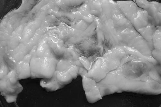
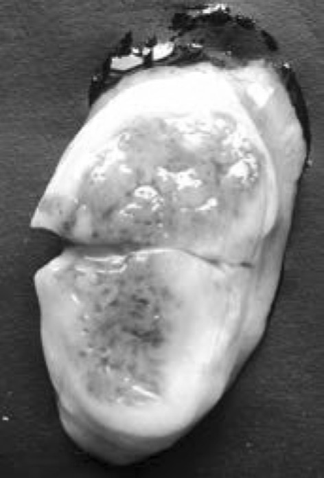
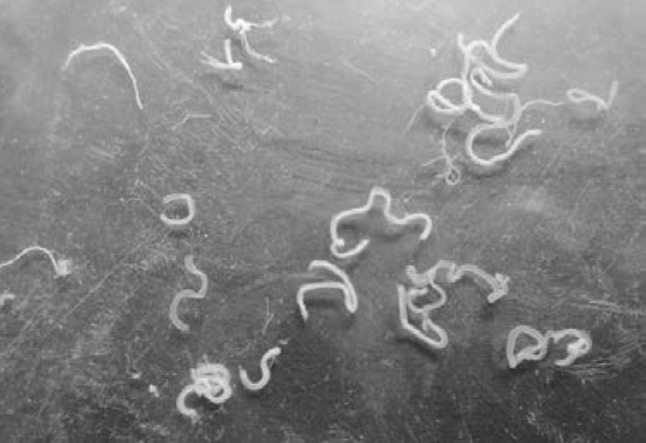
The subcutaneous nodules were identified as worm nests belonging to Onchocerca sp. The number of nodules ranged from 6 to 8 with a diameter of 1–2 cm. The nodules were ovoid in structure with an irregular outline, weighed 2–3 g and were pinkish red in colour embedded to the subcutaneous fascia and connective tissue (Fig. 1). Some of the nodules were found to have undergone calcification and caseation. Dissection of the nodules revealed worm nests with numerous slender white coloured thread like worms inside (Fig. 2) showing pinpoint tunnels in different directions (Fig. 3). The worms were slowly extracted out from the nodules. The worms were found to be female O. gibsoni (Fig. 4) according to the description given by Soulsby 1982. The cuticle was striated transversely forming ring like structures. The uterus was fully filled with microfilariae. The size of the nodules was found to be directly proportional to the number of female worms. Larger nodules contained more number of worms compared to smaller ones. The number of worms recovered from the nodules ranged from 15 to 20. All the worms recovered from the worm nests were found to be females. Microfilaria from the uterus of the worms were dissected out and measured. The length of the microfilaria ranged from 250 to 270 µm and was found to be unsheathed.
Fig. 1.

Onchocerca gibsoni worm nests embedded in fatty tissue
Fig. 2.

Onchocerca gibsoni worm nests showing worms embedded inside
Fig. 3.

Cut section of O. gibsoni worm nests showing grayish white matrix and pin-point size tunnels
Fig. 4.

Onchocerca gibsoni female worms recovered from worm nests
Discussion
Onchocerca sp. is one among the important filarid nematodes affecting cattle. These worms live in the connective tissue often giving rise to firm nodules in which the worms lie coiled up. The cuticle is transversely striated and in addition bears characteristic spiral thickenings, which are usually interrupted in the lateral fields. The worms are viviparous and microfilaria are found in the skin in lymph spaces and connective tissue spaces. Inside worm nests the worms occur in groups and provoke a fibrous reaction around the coiled worms and these nodules need to be trimmed and is one of the major economic losses due to carcass trimming. Insects of family Ceratopogonidae act as intermediate hosts (Soulsby 1982). O. gibsoni occurs in cattle and the worms are usually found in nodules which may occur especially on the brisket and the external surfaces of the hind limbs.
Ladds et al. 1979 conducted epidemiological and gross pathological studies on O. gibsoni infection in cattle and found that 86 % of cattle were infected with more worm nests found in the brisket region. Most nodules (>70 %) were 1–2 cm in diameter and weighed less than 2 g and 20–30 % of nodules were found to be calcified and caseated showing advanced degenerative changes. In the present observation most of the nodules were found in the abdomen and flank regions with 1–2 cm diameter weighing 2–3 g. Pathological changes in concurrent O. gibsoni and Onchocerca armillata infection in a cow was reported by Balachandran et al. 2005. They observed hard movable scattered marble sized nodules in the subcutaneous tissue measuring about 3 cm in diameter on both the sides of lower rib cages and at the brisket region in a 7-year-old crossbred cow carcass. On dissection, O. gibsoni worms were recovered from the worm nests. The worms also had a transversely striated cuticle reinforced externally by spiral thickenings. Similar observations were also made in the present study.
Beveridge et al. 1980 made observations on O. gibsoni and nodule development in cattle at Townsville, Australia. They extracted worms from 185 nodules of a total of 370 worm nodules collected from 94 naturally infected cattle. 183 of the 185 nodules contained a single female worm, 60 % of them contained a single male worm and 7 % contained two male worms. Small nodules (<0.2 g) contained immature female worms without microfilariae and no males. As nodule size and female worm size increased, the number of female worms with microfilariae and the number of nodules with males increased reaching almost 100 % in nodules weighing greater than 3.0 g.
Female worms normally become encapsulated when young, grow within the nodule and the male enters the nodule later, fertilizes the female and remains in the nodule (Soulsby 1982). In the present study also only female worms were recovered from the worm nests and the size of the worm nests was found to be directly proportional to the number of female worms with larger worm nests harbouring more worms compared to smaller ones.
Affected animals are not clinically ill and show no presenting signs other than the subcutaneous nodules at the predilection sites. Treatment is highly effective with a single dose of ivermectin. With the ubiquity of the insect vectors, there is little possibility of effective control, though insect repellents will help to reduce attack by midges. Due to the innocuous nature of the infection, there is unlikely to be any demand for control (Taylor et al. 2015; Levine 1980).
References
- Balachandran C, Pazhanivel N, Vairamuthu S, Raman M, Sreekumar C, Manohar BM. Pathological changes in concurrent Onchocerca gibsoni and Onchocerca armillata infestation in a Cow. Indian J Vet Pathol. 2005;29(2):131–132. [Google Scholar]
- Beveridge I, Kummerow EL, Wilkinson P. Observations on Onchocerca gibsoni and nodule development in naturally-infected cattle in Australia. Tropenmed Parasitol. 1980;31(1):75–81. [PubMed] [Google Scholar]
- Ladds PW, Nitisuwirjo S, Goddard ME. Epidemiological and gross pathological studies of Onchocerca gibsoni infection in cattle. Aust Vet J. 1979;55(10):455–462. doi: 10.1111/j.1751-0813.1979.tb00367.x. [DOI] [PubMed] [Google Scholar]
- Levine ND. Nematode parasites of domestic animals and man. 2. New York: Minnesota Burgess Publishing Company; 1980. pp. 360–365. [Google Scholar]
- Soulsby EJL. Helminths, arthropods and protozoa of domesticated animals. 7. London: Balliere Tindall; 1982. pp. 323–324. [Google Scholar]
- Taylor MA, Coop RL, Wall RL. Veterinary parasitology. 4. London: Wiley; 2015. pp. 127–128. [Google Scholar]


