Abstract
The fact that rabbit hemorrhagic disease virus (RHDV), an important member of the Caliciviridae family, cannot be propagated in vitro has greatly impeded the progress of investigations into the mechanisms of pathogenesis, translation, and replication of this and related viruses. In this study, we have successfully bypassed this obstacle by constructing a mutant RHDV (mRHDV) by using a reverse genetics technique. By changing two amino acids (S305R,N307D), we produced a specific receptor-recognition motif (Arg-Gly-Asp; called RGD) on the surface of the RHDV capsid protein. mRHDV was recognized by the intrinsic membrane receptor (integrin) of the RK-13 cells, which then gained entry and proliferated as well as imparted apparent cytopathic effects. After 20 passages, the titers of RHDV reached 1 × 104.3 50% tissue culture infectious dose (TCID50)/ml at 72 h. Furthermore, mRHDV-infected rabbits showed typical rabbit plague symptoms and died within 48–72 h. After immunization with inactivated mRHDV, the rabbits survived wild-type RHDV infection, indicating that mRHDV could be a candidate virus strain for producing a vaccine against RHDV infection. In summary, this study offers a novel strategy for overcoming the challenges of proliferating RHDV in vitro. Because virus uptake via specific membrane receptors, several of which specifically bind to the RGD peptide motif, is a common feature of host cells, we believe that this the strategy could also be applied to other RNA viruses that currently lack suitable cell lines for propagation such as hepatitis E virus and norovirus.
Keywords: integrin, viral DNA, viral protein, viral replication, virus entry, RK-13 cells, rabbit hemorrhagic disease virus, reverse genetics technique
Introduction
Viruses are the major pathogens threatening human health. To investigate viral pathogenesis and develop schemes for the prevention and treatment of viral diseases, studies on the life cycles of specific viruses are essential. Furthermore, the possibility of culturing virus in vitro may greatly facilitate viral research studies. Extensive research studies have determined that various viruses can be stably propagated using specific host cell lines in vitro, but there are still many important viruses for which this is not possible such as hepatitis E virus (1) and norovirus (2). Therefore, a solution to this problem would be of great significance to perform further studies on these viruses.
Rabbit hemorrhagic disease virus (RHDV)2 belongs to the family Caliciviridae (3, 4), a group of non-enveloped animal viruses that cause a highly contagious and lethal disease in rabbits that is strongly associated with liver degeneration and diffuse hemorrhages and has a high mortality rate of up to 90%. The disease was first described in 1984 in China (5). To date, RHDV has rapidly spread across rabbit populations throughout Asia (6), Europe (7), Australia (8), and the Americas (9), causing the deaths of millions of wild and domestic adult rabbits worldwide. The RHDV genome consists of a positive-sense single-stranded RNA molecule of 7,437 nucleotides with a virus-encoded protein (VPg) covalently attached to its 5′-end (3, 10) that acts as a novel cap substitute during the initiation of RHDV translation (11). Its genomic RNA contains two slightly overlapping open reading frames (ORFs): ORF1 encodes a hypothetical primary translation product that gives rise to a major structural protein (VP60), the capsid protein, and several nonstructural proteins by proteolytic processes, and ORF2 encodes a minor structural protein (VP10) (3, 12). Flanking the coding regions of RHDV is a 5′-terminal non-coding region of 9 nucleotides and a 3′-terminal non-coding region of 59 nucleotides, respectively. In the past 30 years, a suitable cell culture capable of supporting authentic RHDV has not been established, which in turn has greatly impeded the progress of investigations into the mechanisms of pathogenesis, translation, and replication of RHDV.
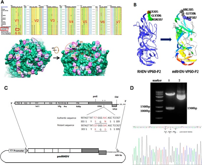
Integrins are a group of transmembrane heterodimeric proteins that are expressed on the surface of a variety of cells and mediate cell-cell and cell-extracellular matrix interactions by binding to corresponding ligands (13–15). Proteins containing the Arg-Gly-Asp (RGD) motif can be recognized and bound by nearly half of the over 20 known integrins; therefore, the RGD site of various proteins plays a central role in cell adhesion biology (16, 17). Numerous pathogenic microorganisms can also utilize the RGD-integrin recognition system to invade host cells (18), including human papillomavirus-16 (19), Kaposi's sarcoma-associated herpesvirus (20), reovirus (21), herpes simplex virus-2 (22), and adenovirus (18). The VP1 protein of the foot-and-mouth disease virus (FMDV) is a protein containing an RGD site with cell attachment activity. The interaction of VP1 with cells is important because VP1 is the capsid protein of FMDV that mediates the virus internalization into cells, thereby initiating the infection process (23). Based on these established facts, we hypothesize that the insertion of an RGD motif at the appropriate site of a mutated RHDV capsid protein for recognition by the natural receptors (integrins) expressed on the surface of host cells will allow its internalization and replication in host cells. Recently, the crystal structures of the S (shell) and P (protrusion) domains of RHDV VP60 have been revealed, and seven regions of high variation were also mapped onto the surface of the P2 subdomain (24) (Fig. 1A), which then served as the theoretical basis of our hypothesis.
Figure 1.
Location of variation regions on the capsomer surface and schematic diagrams of the organization of pmRHDV. A, the seven variation regions (V1–V7) are highlighted in yellow (24). JX/CHA/97 was used in this study, and the target amino acids are enclosed in a red box. Also shown are the locations of variation regions on the RHDV capsomer surface (24). B, protein structure of the RGD mutation region as predicted by SWISS-MODEL online tools (44). C, schematic diagrams of the organization of pmRHDV. The transcription of the mRHDV genome is driven by a T7 promoter, and the core sequence of the hepatitis delta virus ribozyme (HDV Rz) is placed immediately downstream of the viral poly(A) tract to ensure that the final transcripts have the correct 3′-end. The mutant nucleotide/amino acids are highlighted in red, and positions are also indicated (asterisks). D, the pmRHDV recombinant plasmid is identified by double enzyme digestion by PmlI and ClaI, and to ensure the presence of the RGD motif within the VP60 gene, sequence analysis was performed. Lane 1, pmRHDV recombinant plasmid digestion by pmlI and ClaI; lane 2, pmRHDV recombinant plasmid.
Here, we report a novel strategy for constructing an RHDV mutant that could be recognized by host cells and grow well in RK-13 cells. The rescued RHDV mutant not only showed similar biological characteristics to natural RHDV but also exhibited similar immunogenicity as the parent virus.
Results
Construction and characterization of pmRHDV
The overall strategy for the construction of the RHDV replicon is outlined in Fig. 1. In our previous study, a full-length cDNA clone of RHDV was constructed (25); the replicon was driven by the T7 promoter, and the core sequence of hepatitis delta virus ribozyme was placed immediately downstream of the viral poly(A) tract to ensure that the final transcripts had the correct 3′-end. To introduce a recognition site for integrins within the capsid protein of RHDV, two amino acids were mutated (S305R,N307D) through site-directed mutagenesis (Fig. 1B). As shown in Fig. 1C, the cDNA fragment containing the RGD sequence was inserted to displace the corresponding region (PmlI-to-ClaI) of pRHDV, and the final plasmid was named pmRHDV. Subsequently, the accuracy of the resulting pmRHDV plasmid was confirmed by sequencing (Fig. 1D).
Recovery of the RHDV mutant from RK-13 cells
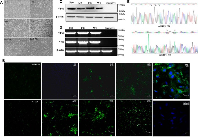
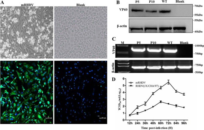
Transfected RK-13 cells that harbored the mutant RHDV exhibited cytopathic effects (CPEs) at 24 h post-transfection, which became more prominent at 72 h post-transfection (Fig. 2A). Antigen generation was subsequently confirmed by indirect immunofluorescence assay (IFA) as well as by Western blot assay using a VP60-specific antibody (Fig. 2, B and C). At 24 h post-transfection, the major capsid protein VP60 was clearly detected in the cytoplasm of cells transfected with pmRHDV, and the expression quantity reached its peak at 72 h post-transfection (Fig. 2B). To confirm that the rescued virus originated from the pmRHDV plasmid, after 20 passages in RK-13 cells, the viral RNA was detected by RT-PCR following by sequencing. The results showed that the target gene was not only present (Fig. 2D) but also contained the mutated RGD site (Fig. 2E).
Figure 2.
Construction and recovery of the mRHDV mutant. A, at 24 h post-transfection, the RK-13 cells showed cytopathic effects, which peaked at 72 h post-transfection. Blank, uninfected cells; WT, transfection of JX/CHA/97 RHDV plasmid. B, indirect immunofluorescence assay of RHDV VP60 expression in RK-13 cells transfected with pmRHDV. RHDV VP60 expression was detected at 24 h post-transfection and peaked at 72 h post-transfection. Blank, RK-13 cells not transfected with pmRHDV; WT; transfection of JX/CHA/97 RHDV plasmid. The monoclonal antibody specific to VP60 was utilized as the primary antibody. Cell nuclei were stained with DAPI (blue); scale bars, 26 μm. C, mRHDV capsid protein (VP60) was detected using a VP60-specific antibody. WT is the positive control, which was derived from a rabbit infected with the JX/CHA/97 RHDV strain. P10, P20, and P40 represent the 10th, 20th, and 40th generation of mRHDV that proliferated in the RK-13 cells, respectively. Blank RK-13 cells were used as the negative control. In addition, β-actin was used as an internal control. D, VP60 and VPg genes of mRHDV were amplified from mRHDV-infected RK-13 cells. β-Actin was also used as an internal control in this experiment. E, sequencing of PCR products showed that the RGD site was stably integrated into the genome of mRHDV after >40 passages of the RK-13 cells.
Growth kinetics
After 20 passages of the RK-13 cells, mRHDV titers were determined by using the 50% tissue culture infectious dose (TCID50), which was 1 × 10² TCID50/ml at 24 h.p.i.; after 48 h, the proliferation speed of mRHDV increased with a titer of 1 × 10³ TCID50/ml. Replication peaked at about 72 h (1 × 104.3 TCID50/ml), which was followed by a gradual decrease in mRHDV titers (Fig. 3A).
Figure 3.
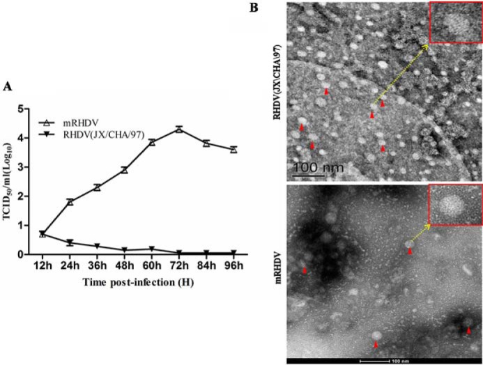
Growth characteristics and electron micrographs of mRHDV. A, the one-step growth curve of mRHDV replication in RK-13 cells. The RHDV JX/CHA/97 strain was used as the wild-type strain control. As the wild-type RHDV cannot proliferate in RK-13 cells, the level of TCID50 is very low. Error bars represent S.D. B, electron micrographs of negatively stained RHDV particles. RHDV JX/CHA/97 strain was used as a positive control. The mRHDV particles marked with red arrows have a diameter of about 35 nm, which is similar to that of the wild-type strain. Scale bars, 100 nm.
Electron microscopy
Electron microscopic observation of mRHDV particles negatively stained with phosphotungstate indicated that the mRHDV viral particles had a diameter of about 35 nm, which is similar to the wild-type strain (Fig. 3B). These results also demonstrate that we had successfully rescued the mRHDV mutant strain, and it was stably proliferating in RK-13 cells. The mRHDV mutant strain has been submitted to the China Center for Type Culture Collection (CCTCC) for preservation (Preservation Number V201501).
The mRHDV strains could infect rabbits and induce typical rabbit plague symptoms
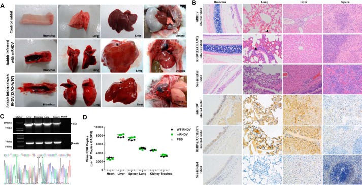
After infecting with the mRHDV strain, 8-week-old rabbits exhibited classic symptoms of RHDV (JX/CHA/97) strain infection; the first deaths were observed at 36h h.p.i., and by 72 h, all rabbit had expired. Fig. 4A shows the autopsy results in which typical pathological changes similar to those caused by wild-type RHDV such as diffuse bleeding of tissues and organs, ring hemorrhage of the trachea, and hepatomegaly were observed. Moreover, immunohistochemical analysis indicated large amounts of RHDV virions in the tissues (Fig. 4B). In addition, RT-PCR and sequencing of PCR products indicated the mRHDV RNA in post-mortem rabbit tissues (Fig. 4C). These findings show that the target gene was not only present but also contained the mutated RGD site. Lastly, the tissue distribution of the mRHDV was analyzed by quantitative PCR, which showed that it was similar to that of the wild-type strains (Fig. 4D).
Figure 4.
The pathogenicity of mRHDV mutant. A, post-mortem autopsy results. In the experiment, New Zealand White rabbits were used, and the RHDV JX/CHA/97 strain was used as the wild-type strain positive control. The autopsy results of the bronchus, lung, liver, and viscera are also shown. B, the results of post-mortem histopathology and immunohistochemical analysis. Large amounts of virus antigen are distributed throughout all tissues infected with mRHDV as well as rabbit tissues infected with wild-type RHDV (JX/CHA/97 strain). C, the VP60 gene of mRHDV was amplified from each rabbit post-mortem tissue that was infected with mRHDV. The β-actin gene was used as an internal control. Sequencing of PCR products indicated that the RGD site was stably integrated into the mRHDV genome. D, RHDV RNA copies in various tissues. Ten-milligram tissues were used in each assay, and the assay was performed in triplicate.
mRHDV strain is safe for other animals
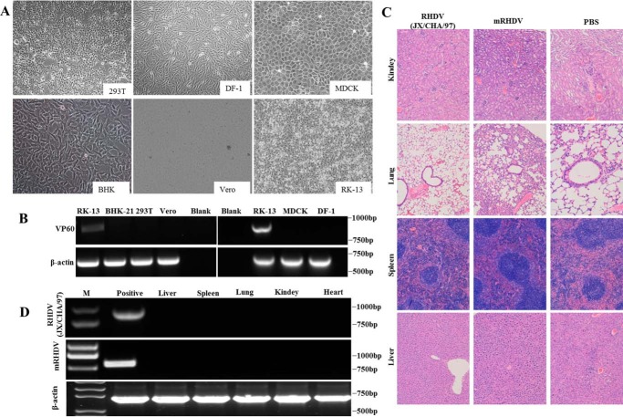
We have utilized mRHDV strains to infect 293T cells, Vero cells, BHK-21 cells, MDCK cells, and CRFK cells, and RT-PCR analysis indicated that the mRHDV strains do not infect other species of cells (Fig. 5, A and B). Moreover, mice infected with mRHDV or the wild-type RHDV strain did not develop the classic symptoms of RHD and instead remained healthy throughout the course of the study (Table 1). Furthermore, no histopathological changes were observed in the mRHDV-infected mice (Fig. 5C), and RT-PCR analysis indicated the absence of the RHDV genome in the mouse tissues (Fig. 5D), thereby demonstrating that the mRHDV strain does not proliferate in mice.
Figure 5.
The mRHDV mutant strain is safe to other animals. A, mRHDV has no ability to infect cells such as BHK-21 cells, 293T cells, Vero cells, MDCK cells, or DF-1 cells. The RK-13 cells were used as a positive control. B, mRHDV genome does not replicate in other species of cells such as BHK-21 cells, 293T cells, Vero cells, MDCK cells, or DF-1 cells. The β-actin gene was used as an internal control. C, mRHDV mutant strains have no ability to infect mouse. Histopathological examination of mouse tissues infected with mRHDV and wild-type RHDV (JX/CHA/97 strain) did not indicate symptoms of virus infection. Mouse tissues infected with PBS were used as a negative control. D, the RHDV and mRHDV genomes were not replicated in any mouse tissues. No VP60 gene was detected in mouse tissues using RT-PCR. Liver tissue of rabbits infected with RHDV JX/CHA/97 strain was used as a positive control, and the β-actin gene was used as an internal control; M, DNA ladder.
Table 1.
The details of mRHDV infection assay
| Groups | Animal | Number | Pathogen | Dosagea | Method | Clinical signs | Dead |
|---|---|---|---|---|---|---|---|
| ml | |||||||
| 1 | Rabbit | 3 | mRHDV | 1 | Intramuscular injection | + + + | 3/3 (100%) |
| 2 | Rabbit | 3 | RHDV (JX/CHA/97) | 1 | Intramuscular injection | + + + | 3/3 (100%) |
| 3 | Rabbit | 3 | PBS | 1 | Intramuscular injection | − − − | 0/3 (0) |
| 4 | Mouse | 5 | mRHDV | 0.1 | Intramuscular injection | − − − − − | 0/5 (0) |
| 5 | Mouse | 5 | RHDV (JX/CHA/97) | 0.1 | Intramuscular injection | − − − − − | 0/5 (0) |
| 6 | Mouse | 5 | PBS | 0.1 | Intramuscular injection | − − − − − | 0/5 (0) |
a mRHDV, 1 × 104.3 TCID50/ml; RHDV (JX/CHA/97), 1 × 10³ LD50/ml. +, positive; −, negative.
mRHDV strains proliferate in rabbit primary hepatocytes
Studies have shown that RHDV can infect rabbit primary hepatocytes, but only about 50% of these cells are infected (26). Therefore, we analyzed the proliferation of mRHDV in hepatocytes. Rabbit primary hepatocytes were first infected with mRHDV, which then resulted in the appearance of CPEs at 48 h postinfection (Fig. 6A). The generation of antigens was subsequently confirmed by IFA using a VP60-specific antibody. Fig. 6A shows that, at 48 h postinfection with mRHDV, VP60 was clearly detected in the cytoplasm of cells. After the mRHDV strains proliferated for 10 passages of the hepatocytes, VP60 was also detected by Western blot assay using a VP60-specific antibody (Fig. 6B). Moreover, the viral RNA of mRHDV was detected by RT-PCR in mRHDV-infected hepatocytes (Fig. 6C). After 10 passages of the hepatocytes, the virus titer of mRHDV, which was 1 × 105.8 TCID50/ml at 72 h.p.i., was higher than the virus titer of RK-13 cells (Fig. 6D). These findings indicate that mRHDV strains also have the ability to proliferate in primary hepatocytes. However, as the primary hepatocytes were not passaged in vitro, we selected the RK-13 cell line as the host cells for mRHDV proliferation.
Figure 6.
mRHDV has an ability to infect rabbit primary hepatocytes. A, at 48 h post-infection, cytopathic effects were observed in the rabbit primary hepatocytes infected with mRHDV. VP60 expression was also detected by IFA in those cells. The monoclonal antibody specific to VP60 was used as the primary antibody. Cell nuclei were stained with DAPI (blue). Rabbit hepatocytes not infected with mRHDV were used as blank. Scale bars, 26 μm. B, the mRHDV capsid protein (VP60) was detected and incubated with the specific antibody against VP60. WT, which was used as a positive control, was derived from a rabbit infected with the JX/CHA/97 RHDV strain. P5 and P10 represent for the 5th and 10th generation of mRHDV that proliferated in the rabbit hepatocytes, respectively. The blank hepatocytes were used as a negative control. β-Actin was used as an internal control. C, the VP60 gene of mRHDV was detected in mRHDV-infected hepatocyte cells by PCR. WT, which was used as a positive control, was derived from liver tissues of a rabbit infected with the JX/CHA/97 RHDV strain. M, DNA ladder. β-Actin was used as an internal control. D, the one-step growth curve of mRHDV in hepatocytes. The RHDV JX/CHA/97 strain was used as the wild-type strain control. Error bars represent S.D.
Mechanism of mRHDV interaction with integrin
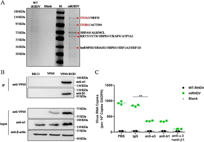
To analyze the mechanism underlying the entry of mRHDV into the host cell, we performed immunoprecipitation experiments with a VP60 antibody in RK-13 cells, and the resulting immunoprecipitated proteins were analyzed by mass spectrometry. The results indicated that the mRHDV capsid protein (VP60RGD) interacted with the α3β1 integrin (Fig. 7A). Subsequently, the interaction between VP60RGD and α3β1 integrin was verified by co-immunoprecipitation experiments (Fig. 7B). In addition, blocking α3β1 integrin resulted in a significant inhibition of viral replication in RK-13 cells (Fig. 7C). Therefore, we believe that mRHDV makes use of its interaction with α3β1 integrin to mediate entry into the RK-13 cells, which in turn subsequently allows viral replication and protein translation to proceed.
Figure 7.
Identification of integrins that interact with the mRHDV capsid protein. A, SDS-PAGE analysis of immunoprecipitation-purified host factors of RK-13 cells that interact with mRHDV capsid protein. The red arrows indicate the bands identified by mass spectrometry. In addition, M is protein marker, and NC is the negative control, RK-13 cells not infected with mRHDV. B, the interaction between mRHDV capsid protein and α3β1 integrin was confirmed by immunoprecipitation (IP). C, the effect of interrupting the interaction between VP60 and α3β1 integrin on the infectivity of mRHDV in RK-13 cells. Student's t test and analysis of variance were used in the statistical analyses. p < 0.01 indicates that the difference is highly statistically significant (**).The experiments were conducted in triplicate, and similar results were obtained from three independent experiments.
The inactivated mRHDV strain could induce high level antibody responses to RHDV
To investigate the immunogenicity of mRHDV, rabbits were immunized with inactivated mRHDV, which had undergone 20 passages in RK-13 cells, and antibody responses to RHDV were determined by indirect ELISA. Fig. 8 shows that all of the rabbits vaccinated with the inactivated mRHDV from groups I, II, and III produced antibodies against RHDV at 7 days after immunization, and the antibody levels were similar to those of the positive control animals, which were immunized with the commercial inactivated RHDV vaccine. Twenty-one days later, all of the rabbits were challenged intramuscularly with wild-type RHDV, which indicated that the protection rate of rabbit against wild-type RHDV of groups I, II, and IV was 40, 80, and 80%, respectively (Table 2). Most interesting is that all of the rabbits from group III were fully protected, whereas the rabbits from group V (negative control) died 48–72 h after being challenged with virulent RHDV. These negative control animals exhibited the clinical symptoms of RHD.
Figure 8.
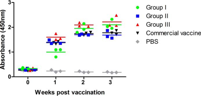
RHDV-specific antibody responses to mRHDV in rabbits. Rabbit sera from different groups were collected at 0, 7, 14, and 21 days postinfection, and absorbance values were measured in quadruplicate by indirect ELISA and recorded at a wavelength of 450 nm.
Table 2.
The details of protection assay after challenging with virulent RHDV
| Group | Number | Antigen | Dosage | Method of immunization | Challenge experiment |
|||
|---|---|---|---|---|---|---|---|---|
| Pathogen | Method of infection | Clinical signs | Protection level | |||||
| I | 5 | mRHDV | 1 mla | Subcutaneous | RHDV (JX/CHA/97) | Intramuscular injection | − − + + + | 2/5 (40%) |
| II | 5 | mRHDV | 2 mla | Subcutaneous | RHDV (JX/CHA/97) | Intramuscular injection | − − − − + | 4/5 (80%) |
| III | 5 | mRHDV | 3 mla | Subcutaneous | RHDV (JX/CHA/97) | Intramuscular injection | − − − − − | 5/5 (100%) |
| IV | 5 | Commercial vaccine | 1 dose | Subcutaneous | RHDV (JX/CHA/97) | Intramuscular injection | − − − − + | 4/5 (80%) |
| V | 5 | PBS | 2 ml | Subcutaneous | RHDV (JX/CHA/97) | Intramuscular injection | + + + + + | 0/5 (0%) |
a 1 × 104.3 TCID50/ml. +, positive; −, negative.
Discussion
Viruses are small in size; however, they are capable of inflicting some of the deadliest diseases around the world. An efficient pathogen is one that has evolved successful ways to colonize host cells using complicated but well orchestrated mechanisms to enter cells, which is critical for replication and sustenance. Due to the complex mechanism of virus entry into host cells, there are still several important pathogens that have not been fully investigated because no suitable in vitro culture system has been developed for research studies on their life cycle, pathogenicity, and control. It is well known that RHDV can infect almost all kinds of rabbits and is associated with high mortality (27). Thus, the rabbit and hare industries are seriously threatened by RHDV. Methods to elucidate the pathogenesis of RHDV as soon as possible and develop safe and effective vaccines against RHD are vital in the control and elimination of infectious diseases. However, no suitable cell line for proliferating RHDV in vitro has been identified, thereby impeding investigations on the pathogenesis of RHDV and the development of effective cell culture-derived vaccines. Indirect strategies that have been used in studying RHDV, including the identification of the histo-blood group antigen H type 2, A type 2, and B type 2 oligosaccharides as attachment factors (28, 29). Moreover, other attachment factors or receptors may play roles at the epithelial level because a low expression of the carbohydrate receptor at the site of viral entry confers only partial protection against infection (28). In addition, hepatocytes, the main cellular targets for viral replication, have been shown not to express histo-blood group antigen in young rabbits. However, the infection in young rabbits is accompanied by hepatic lesions due to viral replication, similar to that observed in adults (30–32). These findings indicate the existence of at least one additional hepatic cellular receptor(s) for RHDV. To date, no new receptors or factors for RHDV have been reported.
In the present study, we constructed a mutant RHDV containing two amino acid substitutions and allowed these to enter and grow in rabbit kidney epithelial cells (RK-13). The basis of our work is that the integrins of host cells can recognize the RGD peptide that is located at the surface of some microbial pathogens and thereby mediate entry of the pathogens into host cells. The integrin-RGD recognition system plays an important role in various cellular activities, including cell survival, proliferation, determination of morphology, attachment, migration, and angiogenesis (33). In addition, integrins are essential for numerous other important biological events such as embryonic development (17), tumor metastasis (34), wound healing (35), and inflammatory responses (36). Researchers have found that the integrin-RGD recognition system is utilized by various pathogenic microorganisms such as FMDV and adenoviruses to infect host cells (21, 23). In the present study, we demonstrated that mRHDV makes use of its interaction with α3β1 integrin to mediate entry of the mutant strain into RK-13 cells, thereby allowing viral replication and translation to begin. The α3β1 integrin also plays a crucial role in viral infection by Kaposi's sarcoma-associated herpesvirus (37). The successful determination of the atomic model of RHDV is the other important theoretical basis of our research; we found that the V1 region (amino acids 301–311) is situated on the surface of the capsid protein, and an RGD site could be produced in this region by mutating two amino acids (Fig. 1) without changing the biological characteristics of RHDV.
We successfully rescued the mRHDV, which not only was sensitive to RK-13 cells but could also be continuously passaged in the cell line. Our results showed that mRHDV grew well in RK-13 cells after at least 40 passages. We also assessed the pathogenicity and immunogenicity of mRHDV, which showed that mRHDV could infect adult rabbits and produce clinical symptoms similar to those of wild-type RHDV. More importantly, the inactivated mRHDV could induce a high level of humoral immune response in rabbits as well as protect the immunized animals from infection by the wild-type virus, thereby indicating that the mutant virus may be potentially utilized in the development of an RHDV vaccine. Currently, commercially available vaccines are produced by the chemical inactivation of crude virus preparations obtained from the organs of infected rabbits (38). Although highly effective for controlling RHD, the type of vaccine often raises serious concerns about biological safety, contaminant residues, and animal welfare (39). Over the past decade, several RHDV genetically engineered vaccines have been developed (40–43). However, these systems have problems such as a relatively low level of expression, high cost, or limitations related to the environmental release of genetically modified organisms. If mRHDV is eventually utilized as a vaccine strain, then this will significantly reduce the cost of vaccines as well as avoid animal welfare problems.
In conclusion, on the basis of the integrin-RGD recognition system, we have successfully obtained mRHDV strains that inherit the biological characteristics of the parent virus and have the ability to proliferate in vitro. The mutant strains will greatly facilitate molecular biological research of RHDV as well as the development of related vaccines. In addition, this novel strategy provides a new way of resolving the in vitro culture challenges associated with the proliferation of norovirus, hepatitis E virus, and other important RNA viruses.
Experimental procedures
Ethics statement
All experiments were performed in a biosafety level 2 laboratory. All experiments involving rabbits and mice were conducted in strict accordance with the recommendations in the Guide for the Care and Use of Laboratory Animals of the Ministry of Science and Technology of the People's Republic of China, and all efforts were made to minimize suffering. All animal procedures were approved by the Institutional Animal Care and Use Committee of the Shanghai Veterinary Research Institute, Chinese Academy of Agricultural Sciences, Shanghai, China (Permit Number SHVRIAU-16-0106).
Cells, virus, antibody, and plasmids
Rabbit kidney cells (RK-13, ATCC CCL37), primary hepatocytes of rabbit (PriCells, Wuhan, China), 293T cells (ATCC, CRL-3216), Vero cells (ATCC, CCL-81), BHK-21 cells (ATCC, CCL-10), MDCK cells (ATCC, CCL-34), and CRFK cells (ATCC, CCL-94) were grown in minimal essential medium (Life Technologies) or Dulbecco's modified Eagle's medium (DMEM) (Life Technologies) supplemented with 10% fetal bovine serum (FBS) in 5% CO2 at 37 °C, respectively. The parental RHDV JX/CHA/97 strain (GenBankTM accession number DQ205345) was isolated in 1997 from an RHDV outbreak in China and stored in our laboratory. The titer of the wild-type virus was 1 × 10³ LD50/ml. The RHDV JX/CHA/97 strain was used as the wild-type virus control in this study. Viral cDNA was synthesized as described elsewhere (25). The recombinant plasmid pRHDV that contained the full-length genome of RHDV was prepared in our previous study (25). mAb specific to VP60 was also prepared in our laboratory. mAbs to integrin α3 (ab140919) and integrin β1 (ab115146) were purchased from Abcam, UK. In addition, mAb to β-actin (MA5-15739) were purchased from Invitrogen.
Construction of the recombinant virus
The construction of RHDV was as described previously (25), and the construction of mRHDV mutant viruses is illustrated in Fig. 1C. The full-length cDNA clone of RHDV was assembled into a pBluescript II SK+ vector with unique restriction sites naturally found within the viral sequence, and a T7 RNA promoter was fused to the genome (25). To produce a specific RGD site in the capsid protein of RHDV, two amino acids within the first variation region (V1; amino acids 301–310) were mutated (S305R,N307D) using the following site-directed mutagenesis strategy. As shown in Fig. 1C, two fragments, covering nucleotides 6122–6226 and 6211–7378, respectively, were amplified from pRHDV using two pairs of specific primers: Pml-F (5′-TTCCACGTGCAACAGGCATTG-3′) and RGD-R (5′-GGTGGAGCTGTCCCCACGATA-3′) and RGD-F (5′-GCAAGTTATCGTGGGGACAGC-3′) and ClaI-R (5′-CCCATCGATTCAAACACTGGA-3′). Then the above two PCR products were fused together through overlap PCR using the primer set Pml-F and ClaI-R. Subsequently, the fused fragment containing the RGD site was substituted for the PmlI-to-ClaI region in pRHDV. The recombinant plasmid was named pmRHDV. Lastly, pmRHDV was transfected into RK-13 cells, which were infected with poxviruses expressing T7 RNA polymerase at an m.o.i. (based on the virus titer in chick embryo fibroblasts) of 0.5–1.0 pfu/cell using Lipofectamine 2000 according to the manufacturer's instructions (Invitrogen). To analyze protein expression, cells were harvested 24 h post-transfection for Western blot analysis and RT-PCR.
RT-PCR for mRHDV genome and sequencing
RNA from the recombinant and parental viruses was purified using an RNeasy extraction kit (Qiagen). Then a 900-bp fragment, which included the RGD nucleotide sequence, was amplified by RT-PCR using the primers RHDV-F (5′-ACTGCTACCACAGCATCAGTT-3′) and RHDV-R (5′-AAACTGGAGCACGTTGGT-3′). The RT-PCR products were sequenced by Jie-Li Biology, Shanghai, China.
RT-PCR quantification
The VP60 RNA copy number of RHDV in tissues or cells was estimated by quantitative real-time reverse transcription-polymerase chain reaction (qRT-PCR). For normalization of mRNA abundance, GAPDH in each sample was quantified and compared. Total RNA was isolated from the samples using TRIzol® reagent (Invitrogen) according to the manufacturer's instructions. The DNA was removed from the isolated RNA using DNase I (Takara, Japan), and then cDNA was produced using Moloney murine leukemia virus reverse transcriptase (Promega) and random hexamers. qRT-PCR was conducted in a 20-μl reaction system that consisted of 10 μl of SYBR Green PCR mixture (Takara), 1 μl of cDNA (100 ng), 0.4 μm qRHDV-F primer (5′-TTGTTTGTGATGGCMTCGGGT-3′), and 0.4 μm qRHDV-R primer (5′-GCGAACATGATGGGTGTGTTCTT-3′). The GAPDH gene was detected by using primers qGAPDH-F (5′-AGGGCTGCTTTTAACTCTGGTAAA-3′) and qGAPDH-R (5′-CATATTGGAACATGTAAACCATGTAGTTG-3′). Each qRT-PCR was conducted with three biological replicates. Amplification was conducted using a Mastercycler ep realplex real-time PCR system (Eppendorf, Germany) with the following conditions: 95 °C for 10 min followed by 40 cycles of 95 °C for 15 s, 58 °C for 45 s, and 72 °C for 45 s and a melting curve analysis of 95 °C for 15 s, 60 °C for 30 s, and 95 °C for 15 s.
IFA
IFAs were used to detect viral protein expression in RK-13 cells and rabbit hepatocytes, which were infected with mRHDV strain at an m.o.i. of 1. The cells were fixed in 3.7% paraformaldehyde in PBS, pH 7.5, at room temperature for 30 min and subsequently permeabilized by incubating in −20 °C methanol for 30 min. The fixed cells were blocked with 5% (w/v) nonfat milk in PBST buffer (137 mm NaCl, 2.7 mm KCl, 10 mm Na2HPO4, 2 mm KH2PO4, and 0.1% Tween 20, pH 7.4) for 3 h at 4 °C and then stained at 37 °C for 2 h with a specific mAb of VP60 (1:1,000 dilution). After washing three times for 10 min each, the cells were incubated with rabbit anti-mouse immunoglobulin G conjugated to fluorescein isothiocyanate (Sigma-Aldrich) (1:2,000 dilution) in PBST for 1 h at room temperature. Finally, after washing three times for 10 min each, the samples were observed under a fluorescence microscope equipped with a video documentation system (Nikon).
Detection of VP60 expression
The RK-13 cells and the rabbit primary hepatocytes were infected with the mRHDV mutant strain or mock-infected (PBS) at an m.o.i. of 1. Proteins were isolated from the cells using radioimmune precipitation assay lysis buffer (50 mm Tris, 150 mm NaCl, 1% Nonidet P-40, and protease inhibitor mixture; Beyotime, P0013F). Protein samples were separated by 12% sodium dodecyl sulfate-polyacrylamide gel electrophoresis (SDS-PAGE) and transferred to nitrocellulose membranes (Hybond-C, Amersham Biosciences) using a semidry transfer apparatus (Bio-Rad). Membranes were blocked with 5% (w/v) nonfat milk in TBST buffer (150 mm NaCl, 20 mm Tris, and 0.1% Tween 20, pH 7.6) for 3 h at 4 °C and then stained at 4 °C overnight with a VP60 mAb (1:2,000 dilution). After washing three times for 10 min each, the membranes were incubated with rabbit anti-mouse immunoglobulin G conjugated to horseradish peroxidase (HRP) (Sigma-Aldrich) (1:5,000 dilution) in PBST for 1 h at room temperature. Finally, after washing three times for 10 min each, the samples were detected by using an automatic chemiluminescence imaging analysis system (Tanon). In addition, the β-actin protein was used as an internal control.
Virus titration
To detect the growth kinetics of mRHDV, the RK-13 cells or primary hepatocyte cells were cultured in 96-well plates and then infected at an m.o.i. of 0.003 TCID50/cell with stocks of the viruses generated after 20 passages of RK-13 cells. After 2 h of incubation, the cells were washed twice, and fresh growth medium was added. The cells were incubated at 37 °C in a humidified 5% CO2 atmosphere and observed daily for the appearance of CPEs. From the onset of CPEs, the titers of the rescued viruses were determined by TCID50 at 12, 24, 36, 48, 60, 72, 84, and 96 h postinfection. In addition, the RHDV JX/CHA/97 strain was used as the wild-type virus control.
Electron microscopy
The mRHDV particles purified from the supernatant of infected RK-13 cells via sucrose gradient centrifugation were diluted in Tris and MgCl2 buffer. The samples were applied to glow-discharged carbon-coated grids for 2 min and negatively stained with 2% (w/v) aqueous uranyl acetate. Micrographs were recorded with a JEM-1230 electron microscope (Philips, Netherlands). The RHDV JX/CHA/97 strain was used as a positive control.
Animal infection assay
Nine eight-week-old New Zealand White rabbits that lacked anti-RHDV antibodies were purchased from the Qingdao Kangda Biological Technology, China and housed under specific pathogen-free conditions. Fifteen adult BALB/c mice between 6 and 8 weeks of age that also lacked anti-RHDV antibodies were purchased from the Laboratory Animal Centre of Shanghai Veterinary Research Institute, Chinese Academy of Agricultural Sciences, China and raised in specific pathogen-free isolation cages. The rabbits and mice received humane care in compliance with the guidelines of the Animal Research Ethics Board of Shanghai Veterinary Research Institute, Chinese Academy of Agricultural Sciences. The details of the animal experiment are presented in Table 1. All animals were continuously observed for clinical symptoms and maintained in appropriate care facilities for 2 weeks. Dead animals were immediately dissected, and each organ was stored at −30 °C. The surviving animals were euthanized via intravenous injection of sodium pentobarbital. All of the animals were necropsied, and the serum and organs were collected and stored at −30 °C. For histopathological examination, the tissues were fixed in 10% neutral buffered formalin, sectioned, and stained with hematoxylin and eosin. Alternatively, immunohistochemical analysis was performed using a VP60 mAb. In addition, the RHDV JX/CHA/97 strain was used as a positive control.
Biological safety assay
RK-13 cells, 293T cells, Vero cells, MDCK cells, BHK-21 cells, and CRFK cells were seeded into 6-well plates, and confluent monolayers (1 × 10⁶ cells) were mock-infected with PBS or infected with mRHDV at an m.o.i. of 1, respectively. All of the cells were continuously observed for CPEs and maintained in appropriate culture conditions for 96 h. RT-PCR was performed for the detection of RHDV using the method described earlier.
Immunization experiments and viral challenge
Twenty-five 8-week-old New Zealand male rabbits that were seronegative for RHDV were randomly distributed into five groups (n = 5 in each group) and raised in individual ventilated cages. All experimental protocols were reviewed and approved by a state ethics commission. Animals from groups I–III were subcutaneously vaccinated with 1, 2, and 3 ml of the inactivated mRHDV (1 × 104.3 TCID50), respectively, in Freund's complete adjuvant. Meanwhile, animals of groups IV and V were subcutaneously vaccinated with a dose of a commercial killed-RHDV vaccine (Nanjing Tianbang Bio-industry Co., Ltd., China) and PBS, respectively. All of the groups were immunized at day 0. Blood samples were collected from the marginal ear vein before each immunization and 3 weeks after the last injection to analyze the level and specificity of the antibody response against RHDV. Antibody titers were assessed using ELISA. Briefly, RHDV virus (JX/CHA/97 strain) (100 μl; 1 μg/ml; incubated overnight at 4 °C) were used to capture antibodies in the sera (incubated for 1 h at 37 °C), which then were detected with 100 μl of HRP-conjugated mouse anti-rabbit IgG (Sigma)/well (diluted 1:5,000 in PBS containing 0.5% Tween 20 and 10% FBS) followed by 100 μl of tetramethylbenzidine liquid substrate (Sigma)/well for 30 min at room temperature in the dark. End-point titers were defined as the highest plasma dilution that resulted in an absorbance value (at A450). Data were presented as log10 values. In addition, all of the rabbits then were challenged intramuscularly with 1,000 LD50 of RHDV 21 days after immunization. The rabbits were clinically examined daily for 7 days after the challenge.
Immunoprecipitation assay
Protein lysates from the RK-13 cells were infected with mRHDV at an m.o.i. of 1 or with RHDV at 1 LD50. The immunoprecipitation assay was performed with a co-immunoprecipitation kit (Pierce) according to the manufacturer's instructions.
Mass spectrometry
The Proteome Analysis Center of Shanghai Life Sciences Research Institute, Chinese Academy of Sciences, performed all mass spectrometry (MS) analysis.
Infection of mRHDV in the presence of blocking antibodies
RK-13 cells were incubated with α3 integrin antibodies (5 μg/ml), β3 integrin antibodies (5 μg/ml), α3 + β3 integrin antibodies (5 μg/ml), control serum (mouse IgG; 5 μg/ml), or cell culture medium (minimal essential medium), respectively, for 1 h at 37 °C, and mRHDV (an m.o.i. of 1) or RHDV (JX/CHA/97) (1 LD50) was added to these cells for attachment at 37 °C. After 2 h of incubation, the cells were washed extensively with PBS, fresh culture medium was added, and the cells were kept for another 24 h at 37 °C. Then qRT-PCR was performed to detect RNA copies of RHDV as described earlier.
Author contributions
J. Z. and G. L. conceived and designed the experiments. J. Z., Q. M., Y. T., and H. G. performed the experiments. J. Z. and G. L. analyzed the data. T. L., C. L., Z. C., and B. W. contributed reagents, materials, and analysis tools. J. Z., G. L., and Q. M. wrote the paper.
Acknowledgment
We thank LetPub for linguistic assistance during the preparation of this manuscript.
This work was supported by the key research project of National Science and Technology (Grant 2016YFD0500108) (to G. L.), the National Natural Science Foundation of China (Grant 31672572) (to G. L.), the key project of Agricultural Science and Technology of Shanghai (Grant 2016043) (to G. L.), the Fundamental Research Funds for the Central Institutes program (Grant 2016JB01) (to G. L.), and the Special Fund for Agro-scientific Research in the Public Interest (Grant 201303046) (to G. L.). The authors declare that they have no conflicts of interest with the contents of this article.
- RHDV
- rabbit hemorrhagic disease virus
- mRHDV
- mutant rabbit hemorrhagic disease virus
- FMDV
- foot-and-mouth disease virus
- CPE
- cytopathic effect
- IFA
- indirect immunofluorescence assay
- TCID50
- 50% tissue culture infectious dose
- qRT-PCR
- quantitative real-time reverse transcription-polymerase chain reaction
- h.p.i.
- hours postinfection
- MDCK
- Madin-Darby canine kidney
- CRFK
- Crandell-Rees feline kidney
- RHD
- rabbit hemorrhagic disease
- m.o.i.
- multiplicity of infection.
References
- 1. Córdoba L., Feagins A. R., Opriessnig T., Cossaboom C. M., Dryman B. A., Huang Y. W., and Meng X. J. (2012) Rescue of a genotype 4 human hepatitis E virus from cloned cDNA and characterization of intergenotypic chimeric viruses in cultured human liver cells and in pigs. J. Gen. Virol. 93, 2183–2194 [DOI] [PMC free article] [PubMed] [Google Scholar]
- 2. Thorne L. G., and Goodfellow I. G. (2014) Norovirus gene expression and replication. J. Gen. Virol. 95, 278–291 [DOI] [PubMed] [Google Scholar]
- 3. Meyers G., Wirblich C., and Thiel H. J. (1991) Rabbit hemorrhagic disease virus—molecular cloning and nucleotide sequencing of a calicivirus genome. Virology 184, 664–676 [DOI] [PubMed] [Google Scholar]
- 4. Parra F., and Prieto M. (1990) Purification and characterization of a calicivirus as the causative agent of a lethal hemorrhagic disease in rabbits. J. Virol. 64, 4013–4015 [DOI] [PMC free article] [PubMed] [Google Scholar]
- 5. Liu S. J., Xue H. P., Pu B. Q., and Qian S. H. (1984) A new viral disease in rabbits. Anim. Husb. Vet. Med. 16, 253–255 [Google Scholar]
- 6. CS L, and CK P. (1987) Aetiological studies on an acute fatal disease of Angora rabbits: co-called rabbit viral sudden death. Korean J. Vet. Res. 27, 277–290 [Google Scholar]
- 7. Cancellotti F. M., and Renzi M. (1991) Epidemiology and current situation of viral haemorrhagic disease of rabbits and the European brown hare syndrome in Italy. Rev. Sci. Tech. 10, 409–422 [DOI] [PubMed] [Google Scholar]
- 8. Morisse J. P., Le Gall G., and Boilletot E. (1991) Hepatitis of viral origin in Leporidae: introduction and aetiological hypotheses. Rev. Sci. Tech. 10, 269–310 [PubMed] [Google Scholar]
- 9. Gregg D. A., House C., Meyer R., and Berninger M. (1991) Viral haemorrhagic disease of rabbits in Mexico: epidemiology and viral characterization. Rev. Sci. Tech. 10, 435–451 [DOI] [PubMed] [Google Scholar]
- 10. Meyers G., Wirblich C., and Thiel H. J. (1991) Genomic and subgenomic RNAs of rabbit hemorrhagic disease virus are both protein-linked and packaged into particles. Virology 184, 677–686 [DOI] [PMC free article] [PubMed] [Google Scholar]
- 11. Zhu J., Wang B., Miao Q., Tan Y., Li C., Chen Z., Guo H., and Liu G. (2015) Viral genome-linked protein (VPg) is essential for translation initiation of rabbit hemorrhagic disease virus (RHDV). PLoS One 10, e0143467. [DOI] [PMC free article] [PubMed] [Google Scholar]
- 12. Nowotny N., Bascuñana C. R., Ballagi-Pordány A., Gavier-Widén D., Uhlén M., and Belák S. (1997) Phylogenetic analysis of rabbit haemorrhagic disease and European brown hare syndrome viruses by comparison of sequences from the capsid protein gene. Arch. Virol. 142, 657–673 [DOI] [PubMed] [Google Scholar]
- 13. Hynes R. O. (2002) Integrins: bidirectional, allosteric signaling machines. Cell 110, 673–687 [DOI] [PubMed] [Google Scholar]
- 14. Plow E. F., Haas T. A., Zhang L., Loftus J., and Smith J. W. (2000) Ligand binding to integrins. J. Biol. Chem. 275, 21785–21788 [DOI] [PubMed] [Google Scholar]
- 15. Hynes R. O. (1987) Integrins: a family of cell surface receptors. Cell 48, 549–554 [DOI] [PubMed] [Google Scholar]
- 16. Zevian S., Winterwood N. E., and Stipp C. S. (2011) Structure-function analysis of tetraspanin CD151 reveals distinct requirements for tumor cell behaviors mediated by α3β1 versus α6β4 integrin. J. Biol. Chem. 286, 7496–7506 [DOI] [PMC free article] [PubMed] [Google Scholar]
- 17. Francis S. E., Goh K. L., Hodivala-Dilke K., Bader B. L., Stark M., Davidson D., and Hynes R. O. (2002) Central roles of α5β1 integrin and fibronectin in vascular development in mouse embryos and embryoid bodies. Arterioscler. Thromb. Vasc. Biol. 22, 927–933 [DOI] [PubMed] [Google Scholar]
- 18. Wickham T. J., Filardo E. J., Cheresh D. A., and Nemerow G. R. (1994) Integrin αvβ5 selectively promotes adenovirus mediated cell membrane permeabilization. J. Cell Biol. 127, 257–264 [DOI] [PMC free article] [PubMed] [Google Scholar]
- 19. Yoon C. S., Kim K. D., Park S. N., and Cheong S. W. (2001) α6 integrin is the main receptor of human papillomavirus type 16 VLP. Biochem. Biophys. Res. Commun. 283, 668–673 [DOI] [PubMed] [Google Scholar]
- 20. Akula S. M., Pramod N. P., Wang F. Z., and Chandran B. (2002) Integrin α3β1 (CD 49c/29) is a cellular receptor for Kaposi's sarcoma-associated herpesvirus (KSHV/HHV-8) entry into the target cells. Cell 108, 407–419 [DOI] [PubMed] [Google Scholar]
- 21. Maginnis M. S., Forrest J. C., Kopecky-Bromberg S. A., Dickeson S. K., Santoro S. A., Zutter M. M., Nemerow G. R., Bergelson J. M., and Dermody T. S. (2006) β1 integrin mediates internalization of mammalian reovirus. J. Virol. 80, 2760–2770 [DOI] [PMC free article] [PubMed] [Google Scholar]
- 22. Cheshenko N., Trepanier J. B., González P. A., Eugenin E. A., Jacobs W. R. Jr, and Herold B. C. (2014) Herpes simplex virus type 2 glycoprotein H interacts with integrin αvβ3 to facilitate viral entry and calcium signaling in human genital tract epithelial cells. J. Virol. 88, 10026–10038 [DOI] [PMC free article] [PubMed] [Google Scholar]
- 23. Neff S., Sá-Carvalho D., Rieder E., Mason P. W., Blystone S. D., Brown E. J., and Baxt B. (1998) Foot-and-mouth disease virus virulent for cattle utilizes the integrin αvβ3 as its receptor. J. Virol. 72, 3587–3594 [DOI] [PMC free article] [PubMed] [Google Scholar]
- 24. Wang X., Xu F., Liu J., Gao B., Liu Y., Zhai Y., Ma J., Zhang K., Baker T. S., Schulten K., Zheng D., Pang H., and Sun F. (2013) Atomic model of rabbit hemorrhagic disease virus by cryo-electron microscopy and crystallography. PLoS Pathog. 9, e1003132. [DOI] [PMC free article] [PubMed] [Google Scholar]
- 25. Liu G., Zhang Y., Ni Z., Yun T., Sheng Z., Liang H., Hua J., Li S., Du Q., and Chen J. (2006) Recovery of infectious rabbit hemorrhagic disease virus from rabbits after direct inoculation with in vitro-transcribed RNA. J. Virol. 80, 6597–6602 [DOI] [PMC free article] [PubMed] [Google Scholar]
- 26. König M., Thiel H. J., and Meyers G. (1998) Detection of viral proteins after infection of cultured hepatocytes with rabbit hemorrhagic disease virus. J. Virol. 72, 4492–4497 [DOI] [PMC free article] [PubMed] [Google Scholar]
- 27. Abrantes J., van der Loo W., Le Pendu J., and Esteves P. J. (2012) Rabbit haemorrhagic disease (RHD) and rabbit haemorrhagic disease virus (RHDV): a review. Vet. Res. 43, 12. [DOI] [PMC free article] [PubMed] [Google Scholar]
- 28. Nyström K., Le Gall-Reculé G., Grassi P., Abrantes J., Ruvoën-Clouet N., Le Moullac-Vaidye B., Lopes A. M., Esteves P. J., Strive T., Marchandeau S., Dell A., Haslam S. M., and Le Pendu J. (2011) Histo-blood group antigens act as attachment factors of rabbit hemorrhagic disease virus infection in a virus strain-dependent manner. PLoS Pathog. 7, e1002188. [DOI] [PMC free article] [PubMed] [Google Scholar]
- 29. Ruvoën-Clouet N., Ganière J. P., André-Fontaine G., Blanchard D., and Le Pendu J. (2000) Binding of rabbit hemorrhagic disease virus to antigens of the ABH histo-blood group family. J. Virol. 74, 11950–11954 [DOI] [PMC free article] [PubMed] [Google Scholar]
- 30. Ferreira P. G., Costa-e Silva A., and Aguas A. P. (2006) Liver disease in young rabbits infected by calicivirus through nasal and oral routes. Res. Vet. Sci. 81, 362–365 [DOI] [PubMed] [Google Scholar]
- 31. Ferreira P. G., Costa-e-Silva A., Monteiro E., Oliveira M. J., and Aguas A. P. (2004) Transient decrease in blood heterophils and sustained liver damage caused by calicivirus infection of young rabbits that are naturally resistant to rabbit haemorrhagic disease. Res. Vet. Sci. 76, 83–94 [DOI] [PubMed] [Google Scholar]
- 32. Mikami O., Park J. H., Kimura T., Ochiai K., and Itakura C. (1999) Hepatic lesions in young rabbits experimentally infected with rabbit haemorrhagic disease virus. Res. Vet. Sci. 66, 237–242 [DOI] [PubMed] [Google Scholar]
- 33. Hussein H. A., Walker L. R., Abdel-Raouf U. M., Desouky S. A., Montasser A. K., and Akula S. M. (2015) Beyond RGD: virus interactions with integrins. Arch. Virol. 160, 2669–2681 [DOI] [PMC free article] [PubMed] [Google Scholar]
- 34. Holly S. P., Larson M. K., and Parise L. V. (2000) Multiple roles of integrins in cell motility. Exp. Cell Res. 261, 69–74 [DOI] [PubMed] [Google Scholar]
- 35. Carter R. T. (2009) The role of integrins in corneal wound healing. Vet. Ophthalmol. 12, Suppl. 1, 2–9 [DOI] [PubMed] [Google Scholar]
- 36. Ley K., Laudanna C., Cybulsky M. I., and Nourshargh S. (2007) Getting to the site of inflammation: the leukocyte adhesion cascade updated. Nat. Rev. Immunol. 7, 678–689 [DOI] [PubMed] [Google Scholar]
- 37. Feire A. L., Koss H., and Compton T. (2004) Cellular integrins function as entry receptors for human cytomegalovirus via a highly conserved disintegrin-like domain. Proc. Natl. Acad. Sci. U.S.A. 101, 15470–15475 [DOI] [PMC free article] [PubMed] [Google Scholar]
- 38. Argüello Villares J. L. (1991) Viral haemorrhagic disease of rabbits: vaccination and immune response. Rev. Sci. Tech. 10, 459–480 [PubMed] [Google Scholar]
- 39. Farnós O., Boué O., Parra F., Martín-Alonso J. M., Valdés O., Joglar M., Navea L., Naranjo P., and Lleonart R. (2005) High-level expression and immunogenic properties of the recombinant rabbit hemorrhagic disease virus VP60 capsid protein obtained in Pichia pastoris. J. Biotechnol. 117, 215–224 [DOI] [PubMed] [Google Scholar]
- 40. Cheng Y., Chen Z., Li C., Meng C., Wu R., and Liu G. (2013) Protective immune responses in rabbits induced by a suicidal DNA vaccine of the VP60 gene of rabbit hemorrhagic disease virus. Antiviral Res. 97, 227–231 [DOI] [PubMed] [Google Scholar]
- 41. Gil F., Titarenko E., Terrada E., Arcalís E., and Escribano J. M. (2006) Successful oral prime-immunization with VP60 from rabbit haemorrhagic disease virus produced in transgenic plants using different fusion strategies. Plant Biotechnol. J. 4, 135–143 [DOI] [PubMed] [Google Scholar]
- 42. Guo H., Zhu J., Tan Y., Li C., Chen Z., Sun S., and Liu G. (2016) Self-assembly of virus-like particles of rabbit hemorrhagic disease virus capsid protein expressed in Escherichia coli and their immunogenicity in rabbits. Antiviral Res. 131, 85–91 [DOI] [PubMed] [Google Scholar]
- 43. Pérez-Filgueira D. M., Resino-Talaván P., Cubillos C., Angulo I., Barderas M. G., Barcena J., and Escribano J. M. (2007) Development of a low-cost, insect larvae-derived recombinant subunit vaccine against RHDV. Virology 364, 422–430 [DOI] [PubMed] [Google Scholar]
- 44. Biasini M., Bienert S., Waterhouse A., Arnold K., Studer G., Schmidt T., Kiefer F., Gallo Cassarino T., Bertoni M., Bordoli L., and Schwede T. (2014) SWISS-MODEL: modelling protein tertiary and quaternary structure using evolutionary information. Nucleic Acids Res. 42, W252–W258 [DOI] [PMC free article] [PubMed] [Google Scholar]


