From the Column Editors,
Dr. Berardo Di Matteo and his colleagues at the Rizzoli Orthopaedic Institute in Bologna, Italy have again used the treasures of one the oldest and most-comprehensive medical libraries in the world to share with us a fascinating and beautifully documented time in the history of medical education. The clerical ban on the use of human dissection, lasting nearly a millennium, diminished physicians’ opportunities to learn about human health and sickness, and plunged medical knowledge into darkness. The story of how Mondino de’ Liuzzi boldly reintroduced the study of human anatomy is an important contribution to our Art in Science column.
Gary E. Friedlaender MD, Linda K. Friedlaender BA, MS
Introduction
The early Middle Ages (6th century to the 10th century) yielded few medical advances, primarily because of the church’s domineering influence on science in Europe during that time. This period of darkness was followed by the true revolution in art, philosophy, and science: The Renaissance.
However, the notion that the entire period we call the Middle Ages was scientifically barren has been revised by researchers and historians who have confirmed that the middle-to-late-Middle Ages indeed was marked by many scientific accomplishments [9]. The late 11th century and 12th century saw the establishment of a number of universities throughout Europe, including the University of Bologna [14]. Urban shelters affiliated with monasteries or churches housed local physicians who cared for the sick and injured. These shelters became popular and eventually evolved into the first established hospitals [7]. Perhaps most importantly, an imperial decree in 1231 by Frederick II, Emperor of the Holy Roman Empire, allowed those who studied surgery and anatomy to once again to dissect human bodies inside approved medical schools once every 5 years [13]. This decree established dissection as a scientific necessity, and—after centuries of interdiction—revolutionized the way scientists studied human anatomy.
Medieval anatomist Mondino de’ Liuzzi (1270–1326) lived and thrived when the “freeze” on medical and scientific advancement began to thaw. Known as the “Restorer of Anatomy”, de’ Liuzzi is considered the first to perform a dissection, document it, and publish his findings. His fundamental text, Anathomia (Anatomy) [2, 6], contained the collected experiences from the ancient anatomists, as well as de’ Liuzzi’s first-hand account of his own experiences with dissection.
Merely a Witness to History?
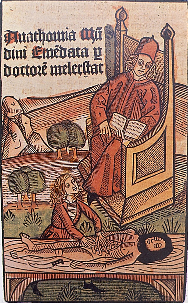
de’ Liuzzi was born into a wealthy medical family in Bologna, Italy, in approximately 1270. His father owned a pharmacy and his uncle was a medical professor at the University of Bologna. Pushed by his family legacy, de’ Liuzzi studied anatomy and practiced dissection as allowed under the new law [10]. In 1315, already a distinguished scientist and university professor, de’ Liuzzi performed the first documented public dissection in more than 1700 years [13]. de’ Liuzzi had already performed various “private” human dissections in his studies, and it is likely that he wanted to share his findings and experience with the public to generate a broader focus on the subject. In order to make these anatomic dissections more agreeable, a public dissection would be scheduled around festivities like a carnival. Food and wine would keep spectators warm and distracted from the rancid aroma of a decomposing body [9]. de’ Liuzzi’s first public dissection was performed in Bologna, in January, on the body of a female criminal, in the presence of medical students and other spectators, and with the full authorization by the church. As was customary at the time, de’ Liuzzi did not perform the dissection himself. Because of his distinguished status, the professor sat on a large, elevated chair above the dissection table, reading aloud from Galen’s book of anatomy and commenting on it to the audience. As he was reading, a demonstrator, often a barber-surgeon, performed the “dirty work” of the dissection itself following the instructions of the professor, while an ostensor (an exhibiter) pointed out for the attendees the specific parts of the body that were being examined [4].
Though he did not technically perform the dissection himself, the public dissection was a celebrated event that instantly resounded across the academic communities throughout Europe, eliciting many figurative representations of this symbol of scientific rejuvenation. An illustration of the event can be found inside the famous 1491 first edition [8] of Johannes de Ketham’s Fasciculus (Fig. 1).
Fig. 1.

Frontispiece of Johannes de Ketham’s book, Fasciculus is shown.
Anathomia
de’ Liuzzi’s approach to anatomy, as articulated in his book, Anathomia [12] requires videre ad sensum (“understanding through practice”). This novel critical approach to learning at the time defied previous generations of scientists who merely repeated the knowledge from the past [1], and was therefore essential reading for the up-to-date physician of the day.
Considered one of the most popular books in medical literature of the time (with 40 different editions, printings, and translations [12] in several languages), Anathomia described the segmental dissection of the abdomen, the thorax, the cranium, and the extremities—just as he described during his examination of human cadavers. The treatise consisted of six parts: (1) An introduction to the whole human body with comments to the authorities of the past; (2) the organs of the abdominal cavity; (3) the reproductive organs; (4) the organs from the thoracic cavity up to the mouth; (5) the skull, brain, eyes, ears; and (6) the spine and the upper and lower limbs. Leonardo da Vinci (1452–1518) used the Anathomia to learn about the human body for his work both as artist and scientist [1].
The original edition of his book carried no illustrations, but was later visually updated by followers of de’ Liuzzi, including the anatomist Berengario da Carpi (1466–1530) (Fig. 2) and the German anatomist Johannes Dryander in 1541. Dryander’s illustrations showed human bodies opening up like books to reveal their inner secrets (Fig. 3A–D). de’ Liuzzi was truly the first modern guide to a new generation of anatomists.
Fig. 2.

Frontispiece of Mondino de’ Liuzzi’s Anathomia is shown.
Fig. 3.
German anatomist Johannes Dryander’s illustrations show (A) a human body opening up like a book (B), a depiction of a bowel from the Anathomia (C), the female reproductive system (D), and Inevitable Fatum, (“Inevitable Fate”) showing the dissection of the torso.
The contributions provided by de’ Liuzzi go well beyond this one famous achievement. His work resulted in a long-lasting legacy, which shaped for centuries to come, the approach to anatomical dissection [3, 5, 11]—understanding through practice.
Footnotes
A note from the Editor-in-Chief:
I am pleased to present the next installment of our “Art in Science.” In this month’s column, Berardo Di Matteo MD and his colleagues from the Rizzoli Orthopaedic Institute in Bologna, Italy, profile Mondino de’ Liuzzi, the medieval anatomist widely credited with reestablishing the practice of dissection of human cadavers during the Middle Ages.
The authors certify that neither they, nor any members of their immediate families, have any commercial associations (such as consultancies, stock ownership, equity interest, patent/licensing arrangements, etc) that might pose a conflict of interest in connection with the submitted article.
All ICMJE Conflict of Interest Forms for authors and Clinical Orthopaedics and Related Research ® editors and board members are on file with the publication and can be viewed on request.
The opinions expressed are those of the writers, and do not reflect the opinion or policy of CORR ® or The Association of Bone and Joint Surgeons®.
The institution of one or more of the authors (BDM, VT, GF, AV, PT, MM) has received, during the study period, funding from Italian State through “5 per mille (year 2011)” project.
References
- 1.Crivellato E, Ribatti D. Mondino de’ Liuzzi and His Anathomia. Clinical Anatomy. 2006;19:581–587. doi: 10.1002/ca.20308. [DOI] [PubMed] [Google Scholar]
- 2.de’ Liuzzi M. Anathomia. Frankfurt, Germany: Marpurgi in officina Christian Egenolff; 1541.
- 3.Di Matteo B, Tarabella V, Filardo G, Tomba P, Viganò A, Marcacci M. Nicolaes Tulp: The overshadowed subject in The Anatomy Lesson of Dr. Nicolaes Tulp. Clin Orthop Relat Res. 2016;474:625–629. doi: 10.1007/s11999-015-4686-y. [DOI] [PMC free article] [PubMed] [Google Scholar]
- 4.Di Matteo B, Tarabella V, Filardo G, Viganò A, Tomba P, Bragonzoni L, Marcacci M. Art in science: The stage of the human body–The anatomical theatre of Bologna. Clin Orthop Relat Res. 2015;473:1873–1878. doi: 10.1007/s11999-015-4288-8. [DOI] [PMC free article] [PubMed] [Google Scholar]
- 5.Di Matteo B, Tarabella V, Filardo G, Viganò A, Tomba P, Kon E, Marcacci M. Art in science: Giovanni Paolo Mascagni and the art of anatomy. Clin Orthop Relat Res. 2015;473:783–788. doi: 10.1007/s11999-014-3909-y. [DOI] [PMC free article] [PubMed] [Google Scholar]
- 6.Infusino MH, Win D, O’Neill YV. Mondino’s book and the human body. Vesalius. 1995;1:71–76. [PubMed] [Google Scholar]
- 7.Kealy Edward J. Medieval Medicus - A Social History of Anglo-Norman Medicine. The Baltimore, MD: Johns Hopkins University Press; 1981. [Google Scholar]
- 8.Ketham J. Fasciculus Medicinae. Venice, Italy: Zuane & Gregorio di Gregorii; 1493.
- 9.Mavrodi A, Paraskevas G. Mondino de Luzzi: A luminous figure in the darkness of the Middle Ages. Croat Med J. 2014;55:50–53. doi: 10.3325/cmj.2014.55.50. [DOI] [PMC free article] [PubMed] [Google Scholar]
- 10.Mazzola RF, Mazzola IC. Treatise on skull fractures by Berengario da Carpi (1460-1530) J Craniofac Surg. 2009;20:1981–1984. doi: 10.1097/SCS.0b013e3181bd2ddc. [DOI] [PubMed] [Google Scholar]
- 11.Netter FM, Friedlaender GE. Frank H. Netter MD and a brief history of medical illustration. Clin Orthop Relat Res. 2014;472:812–819. doi: 10.1007/s11999-013-3459-8. [DOI] [PMC free article] [PubMed] [Google Scholar]
- 12.Olry R. Medieval neuroanatomy: The text of Mondino de’ Liuzzi and the plates of Guido da Vigevano. J Hist Neurosci. 1997;6:113–123. doi: 10.1080/09647049709525696. [DOI] [PubMed] [Google Scholar]
- 13.Rengachary SS, Colen C, Dass K, Guthikonda M. Development of anatomic science in the late middle ages: The roles played by Mondino de Liuzzi and Guido da Vigevano. Neurosurgery. 2009;65:787–793. doi: 10.1227/01.NEU.0000324991.45949.E4. [DOI] [PubMed] [Google Scholar]
- 14.University of Bologna. Our history. Available at: http://www.unibo.it/en/university/who-we-are/our-history/our-history. Accessed December 2, 2016.


