Abstract
Mercury (Hg) is a heavy metal pollutant, there is an urgent need to develop simple and sensitive methods for Hg(II) in water. In this article, a simple and sensitive resonance Rayleigh scattering (RRS) method was developed for determination of 0.008–1.33 µmol/L Hg, with a detection limit of 0.003 μmol/L, based on the Hg(II) regulation of gold nanoenzyme catalysis on the HAuCl4-H2O2 to form gold nanoparticles (AuNPs) with an RRS peak at 370 nm. Upon addition of molecular probes of Victoria blue B (VBB), the surface-enhanced Raman scattering (SERS) peak linearly decreased at 1612 cm−1 with the Hg(II) concentration increasing in the range of 0.013–0.5 μmol/L. With its good selectivity and good accuracy, the RRS method is expected to be a promising candidate for determining mercury ions in water samples.
Keywords: mercury ion, gold nanoparticle, nanocatalysis, resonance Rayleigh scattering, SERS
1. Introduction
Nanomaterials not only have unique optical, electrical, and magnetic properties, but also have bio-enzyme activity [1,2]. They are known as mimic nanoenzymes, and also called di-functional molecules or multifunctional molecules. Since Fe3O4 nanoparticles have been found to have intrinsic peroxidase-like activity [3], nanoenzyme have become interesting to people. They have vast application prospects with advantages of easy production process, good stability and recycling, low-cost of storage and transport, and high adaptability of heat, acid, and alkali [1,2,3,4,5,6,7,8,9,10,11]. It is a new study filed, that is, how to organically combine nanoenzyme catalysis with its physical and chemical properties to create more novel functions. In analytical chemistry, nanoenzymes have been used in absorption, fluorescence, and chemiluminescence analysis [3,4,5,6,7,8,9,10,11,12,13]. Gold nanoparticles have peroxidase activity that catalyze the colored redox reaction of 3,3′,5,5′-tetramethylbenzidine (TMB)-H2O2 [12]. Combining glucose peroxidase and gold nanoenzyme, a 18–1100 μmol/L glucose can be detected spectrophotometrically, with a detection limit of 4 μmol/L. Li et al. [13] constructed CdTe dots and gold nanoparticles into the SiO2 microball surface respectively to obtain a nanoenzyme that catalytically oxidized to form oxidant, and a 1.32 μmol/L glucose can be determined by fluorescence method.
Mercury is a highly toxic heavy metal element which is very widely distributed in nature. Therefore, the monitoring of Hg2+ becomes very important. At present, several methods have been reported to determine Hg2+, such as atomic fluorescence spectroscopy (AFS), atomic absorption spectrometry (AAS), and inductively coupled plasma mass spectrometry (ICP-MS) [14,15,16]. Although these methods have high selectivity and sensitivity for Hg2+ detection, however, these methods have a number of weaknesses such as expensive instruments, long analysis period, need for professional operators, and so on. It is of great significance to develop a simple, rapid, and low-cost method for mercury detection. In recent years, some new methods for the detection of Hg2+ have been reported such as aptamer and functionalized gold nanoparticle biosensors [17,18,19]. Resonance Rayleigh scattering (RRS) method is a simple and sensitive spectral analysis technique. It has been used for the analysis of proteins, nucleic acids, metal ions, and so on [20,21,22,23,24]. Early studies of our group have developed sensitive and selective nanogold catalysis, immunonanogold catalysis, aptamer-nanogold catalysis, and peptide modified-nanogold catalysis methods [25,26,27,28,29,30,31], including the silver nitrate-hydroquinone [28] and HAuCl4-ascorbic acid reactions [29]. The above nanoenzyme catalytic RRS methods were all based on the aggregations of nanoparticles that presented unstable analytical systems and complicated operations. In this study, we have observed that AuNPs have strong catalysis on the redox reaction of HAuCl4-H2O2, while Hg2+ exhibits strong inhibition on this nanocatalytic reaction, and the nanoreaction product of formed AuNPs demonstrated RRS effect and SERS activity in the presence of Victoria blue B (VBB) probes. Thus, two simple, rapid, low cost, high sensitivity, and good selectivity RRS and SERS methods were established for detection of Hg2+.
2. Results and Discussion
2.1. Principle
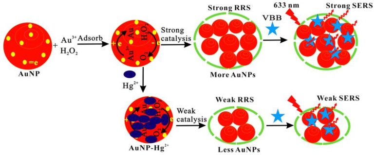
Nano-catalysis is an important way to amplify analysis signal. It has significance in developing a new nano-catalysis reaction. From the experiment, we found that, in hydrochloric acid medium, the reaction of H2O2-HAuCl4 was relatively slow. While nanocatalyst of AuNP was added, a large number of HAuCl4 adsorbed on the AuNP surface, and H2O2 was also adsorbed on the surface that there had many free electrons, which can enhance the redox electron-transfer between H2O2-HAuCl4. Thus, it enhanced the reduction of Au3+ to form elemental gold, and led to the gold nanoparticles forming greatly. The formed gold nanoparticles have strong SERS and RRS signal. Therefore, the nanogold catalytic reaction can be used to establish SERS and RRS detection methods for AuNPs (Figure 1).
Au3+ + AuNP = AuNP-Au3+ (adsorb)
H2O2 + AuNP-Au3+ = Au3+-AuNP-H2O2 (adsorb)
Au3+-AuNP-H2O2 = Au+-AuNP-H2O2 + O2
Au+-AuNP-H2O2 = AuNP + Au + O2
nAu = (Au)n = AuNPs
Figure 1.
Resonance Rayleigh scattering (RRS)/surface-enhanced Raman scattering (SERS) detection of trace Hg2+ based on the inhibition of gold nanoparticles (AuNP) catalysis.
It is known that Au and Hg have stronger affinities to form Au-amalgam than other metals such as Ag, Pb, and Cu. Thus, the stable AuNP-Hg2+ would form in the presence of Hg2+. Similarly, in the analytical system, the formed AuNP-HgCl42− can adsorb on the surface of AuNP, which greatly inhibits the red electron-transfer, and decreases the AuNP catalytic reaction. With the Hg2+ connection increased, the catalytic reaction rate slowed, and the RRS and SERS intensity decreased (Figure 1). Hereby, a RRS and SERS method was established for selective determination of Hg2+.
2.2. RRS Spectra
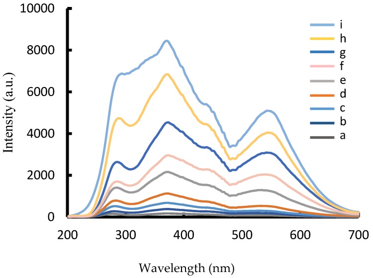
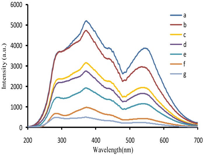
In HCl medium, nanocatalyst of AuNPb, AuNPc, and AgNP could catalyze H2O2 to reduced HAuCl4 to form AuNPs with three RRS peaks at 280, 370, and 550 nm. When nanocatalyst AuNP increased, the RRS intensity at 370 nm enhanced linearly (Figure 2 and Figure S1). The catalytic activity of AuNPb was higher than AuNPc, due to the smaller particle size, and the larger specific surface area. When Hg2+ was adsorbed on the surface of AuNPb to form AuNPb-HgCl42−, the catalytic activity of AuNPb was weakened. The RRS intensity at 370 nm decreased linearly with the Hg2+ concentration increased (Figure 3), and was selected for the determination of Hg(II). In addition, the RRS spectra of the Hg2+-AuNPc-HAuCl4-H2O2 system were recorded at room temperature. Results (Figure S2) show that the RRS signals were very weak and constant when Hg2+ concentration increased up to 12 µmol/L. This indicated that Hg2+ ions could strongly adsorb on the AuNPb surfaces and there are no aggregations in the system.
Figure 2.
RRS spectra of the AuNPb-HAuCl4-H2O2 nanocatalytic system. (a) 4.48 µmol/L HAuCl4 + 0.67 mmol/L HCl + 3.33 mmol/L H2O2; (b) a + 0.018 ng/mL AuNPb; (c) a + 0.095 ng/mL AuNPb; (d) a + 1.9 ng/mL AuNPb; (e) a + 5.7 ng/mL AuNPb; (f) a + 7.6 ng/mL AuNPb; (g) a + 1.9 ng/mL AuNPb; (h) a + 5.7 ng/mL AuNPb; (i) a + 7.6 ng/mL AuNPb.
Figure 3.
RRS spectra of the Hg2+-AuNPc-HAuCl4-H2O2 inhabited system. (a) 38 ng/mL AuNPb + 4.48 µmol/L HAuCl4 + 0.67 mmol/L HCl + 3.33 mmol/L H2O2; (b) a + 0.08 µmol/L Hg2+; (c) a + 0.5 µmol/L Hg2+; (d) a + 0.67 µmol/L Hg2+; (e) a + 0.83 µmol/L Hg2+; (f) a + 1.17 µmol/L Hg2+; (g) a + 1.33 µmol/L Hg2+.
2.3. SERS Spectra
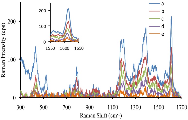
SERS is a sensitive molecular spectral technique [31,32,33], and has been used in trace analysis. Recently, it was selected to study some nanocatalysis reactions [32,33], with good results. In this article, it was used to examine the Hg(II)-AuNPb-H2O2-HAuCl4-molecular probe system. When RhS was used as SERS probe, it could adsorb on the generated gold nanoparticle surfaces that exhibited strong SERS peaks at the Raman shifts of 618, 732, 1199, 1277, 1356, 1507, 1527, and 1645 cm−1. Among them, the SERS peak at 1645 cm−1 was most sensitive and was selected for use. With the AuNPb increase, the intensity of SERS at 1645 cm−1 increased linearly (Figure S3). Using VBB as a SERS probe, it displayed SERS peaks at the Raman shifts of 795, 1167, 1200, 1364, 1394, and 1612 cm−1. Among them, the SERS peak at 1612 cm−1 was the most sensitive. As the AuNPb increased, the intensity of SERS at 1612 cm−1 linearly increased (Figure S4). Using safranin T as SERS probe, it displayed SERS peaks at the Raman shifts of 349, 612, 1240, 1372, 1551, and 1639 cm−1. Among them, the SERS peak at 1372 cm−1 was the most sensitive. With the AuNPb increased, the intensity of SERS at 1372 cm−1 linearly increased (Figure S5). Rhodamine 6G was tested as a SERS probe, but its Raman intensity was very weak. When Hg2+ adsorbed on the surface of AuNPb, the nanocatalytic activity weakened, the peak at 1612 cm−1 decreased linearly, and the AuNPb catalytic system was chosen for quantitative analysis of Hg(II) (Figure 4).
Figure 4.
SERS spectra of the Hg2+-AuNPb-HAuCl4-H2O2-Victoria blue B (VBB) system. (a) 38 ng/mL AuNPb + 4.48 µmol/L HAuCl4 + 0.67 mmol/L HCl + 3.33 mmol/L H2O2−1.3 µmol/L VBB; (b) a + 0.013 µmol/L Hg2+; (c) a + 0.17 µmol/L Hg2+; (d) a + 0.33 µmol/L Hg2+; (e) a + 0.5 µmol/L Hg2+.
2.4. Absorption Spectra
AuNPb exhibited a surface plasmon resonance (SPR) peak at 510 nm, it catalyzed H2O2 reduced HAuCl4 to form gold nanoparticles with a SPR peak at 570 nm. With AuNPb increased, the color changed from colorless to red (Figure S6) and the SPR peak value A570 nm increased (Figure S7). When AuNPc was used as nanocatalyst, it had a SPR peak at 590 nm. As AuNPc increased, the SPR peak increased (Figure S8). AgNP exhibited the nanocatalysis on the reaction, the SPR peak increased with the nanocatalyst increase (Figure S9). However, when Hg2+ was added to the AuNPb, the intensity at 560 nm of the nanocatalytic system decreased linearly (Figure S10), and the peak was chosen for the detection of Hg, with lowest-cost. Furthermore, it can be detected by lake-eye.
2.5. Scanning Electron Microscopy (SEM)
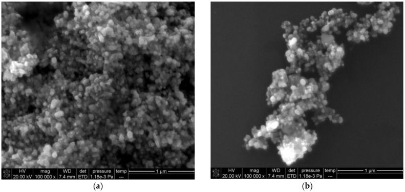
The AuNPb, AuNPc, and Ag NPs were in spherical shape in size of 5, 10, and 9 nm respectively (Figure S11). For the HAuCl4-H2O2 system, the reaction rate was slow in the absence of AuNPb and there were few gold nanoparticles in reaction solution. When nanocatalyst of AuNPb was added, a plenty of irregular gold nanoparticles were generated (Figure 5a). As Hg2+ was added, it inhibited the nanocatalytic reaction of AuNPb-H2O2-HAuCl4. The generated gold nanoparticles were reduced (Figure 5b).
Figure 5.
Scanning Electron Microscopy (SEM) images of the AuNPb catalytic system. (a) 0.67 mmol/L HCl + 4.48 µmol/L HAuCl4 + 3.33 mmol/L H2O2 + 19 ng/mL AuNPb; (b) a + 1 µmol/L Hg2+.
2.6. Optimization of Analytical Conditions
The effect of HCl concentration on the AuNPb-H2O2-HAuCl4 catalytic reaction was considered. The amount of HCl had great influences on the generated nanogold. When the concentration was 0.67 mmol/L, the ΔI value was the largest, and the color was pink with I370 nm of 3506, and the blank was colorless with I370 nm of 506. When HCl concentration continued to increase, a large number of hydrogen ions in the solution limited the redox of H2O2-HAuCl4. Therefore, the concentration of 0.67 mmol/L HCl was selected in this experiment (Figure S12). We also considered the effect of the HAuCl4 concentration on the catalytic reaction, and found that the ΔI value was the largest when the concentration was 4.48 µmol/L (Figure S13). When HAuCl4 concentration increased continuously the SERS value held constant due to forming the largest AuNPs. The effect of H2O2 concentration on ΔI was studied and the best concentration was 3.33 mmol/L (Figure S14). The effect of the temperature on the catalytic reaction was investigated. The ΔI value increased with a temperature increase in the range of 30–60 °C due to an increase in the number of formed AuNPs. When the temperature was 60 °C, the ΔI value was the largest due to formed largest AuNPs, and the blank formed small nanogolds with a pale pink color. When reaction temperature was higher than 60 °C, the ΔI value decreased due to the blank increasing significantly. Thus, 60 °C was chosen (Figure S15). We also investigated the heating time and 15 min was selected (Figure S16). We found that when the heating time exceeded 15 min, the blank generated nanogolds and the RRS intensity increased. Even when the heating time was 25 min, the blank solution presented a reddish color that meant a large amount of nanogold was produced. Moreover, after heating, the system was immediately cooled in an ice bath to stop the reaction. Effect of RhS, VBB, and safranine T concentration on the SERS intensity was investigated, and 7, 13.2, and 6.7 µmol/L were selected respectively (Figures S17–S19).
2.7. Effect of Foreign Substances
According to the procedure, the effect of foreign substances on the determination of 1.5 × 10⁻⁷ mol/L Hg2+ was tested, when the relative error was within ±10%. The results indicated that 8.3 × 10⁻⁵ mol/L Cu2+; 3.3 × 10⁻⁵ mol/L I− and Cd2+; 6.7 × 10⁻⁶ mol/L Zn2+ and Bi3+; 3.3 × 10⁻⁶ mol/L Pb2+; 2.0 × 10⁻⁶ mol/L S2−; Pd2+ and Pt2+; 1.7 × 10⁻⁶ mol/L Co2+; and Cr3+ and Ni2+ did not interfere with the determination. It indicated that this method had a good selectivity due to stronger intermolecular forces between Au and Hg than the other Au metals.
2.8. Working Curve
Under optimal conditions, the relationship between the nanocatalyst concentration of AuNPb, AuNPc, and AgNPs and their corresponding ΔI370 nm were obtained (Table 1, Figures S20–S22). The results showed that the AuNPb is the strongest catalyst with maximum slope, and it was chosen for use. For the AuNPb-HAuCl4-H2O2 nanocatalytic system, using RhS, VBB, and safranine T as molecular probes respectively, their SERS intensities were recorded. The experimental results indicated that VBB was the most sensitive SERS probe (Figures S23–S25). For the Hg2+ analytical system, the RRS, SERS, and Abs methods were studied (Figures S26–S28). From Table 1, we can see that the RRS is most sensitive and was chosen for detection of Hg. The RRS method was compared with the reported methods for determination of Hg2+ [34,35,36,37], this method is more simple, and the reagent is very easy to obtain, with high sensitivity and good selectivity.
Table 1.
Analytical features of the nanocatalytic analytical systems.
| Analyte | Method | Regression Equation | Linear Range (µmol/L) | Coefficient | Detection Limit (µmol/L) |
|---|---|---|---|---|---|
| AuNPb | RRS | ΔI370 nm = 131.3C + 300 | 0.025–25 | 0.9951 | 0.008 |
| AuNPc, | RRS | ΔI370 nm = 51.5C + 267 | 0.05–75 | 0.9941 | 0.02 |
| AgNP | RRS | ΔI370 nm = 23.4C + 73 | 0.5–50 | 0.9971 | 0.2 |
| AuNPb | SERS a | ΔI1645 cm−1 = 2.28C + 72 | 0.5–50 | 0.9786 | 0.2 |
| AuNPb | SERS b | ΔI1612 cm−1 = 5.94C + 86 | 0.2–50 | 0.9942 | 0.1 |
| AuNPb | SERS c | ΔI1372 cm−1 = 1.47C − 9.1 | 0.6–50 | 0.9879 | 0.3 |
| Hg2+ | RRS | ΔI370 nm = 3650C + 111 | 0.008–1.33 | 0.9958 | 0.003 |
| Hg2+ | SERS b | ΔI1612 cm−1 = 326C + 6.4 | 0.013–0.5 | 0.9932 | 0.03 |
| Hg2+ | Abs | ΔA600 nm = 0.083C + 0.0087 | 0.5–2.33 | 0.9876 | 0.2 |
a RhS; b VBB; c safranine T.
2.9. Analysis of Samples
Three water samples—including tap, river, and pond—were collected from Guangxi Normal University, and filtrated according to the reference [35]. The following operations were according to the procedure to detect Hg2+. The obtained results were accorded with the cold atomic absorption spectrometry (AAS) (Table S1). The average values (n = 5) for the water samples after adding a certain amount of Hg2+ were determined. The recovery was in the range of 97–102%, and the relative standard deviation (RSD) was in the range of 4.5–5.2%. This indicated the method is accuracy and reliable.
3. Materials and Methods
3.1. Apparatus
A DXR smart Raman spectrophotometer ( Thermo Fisher, Waltham, MA, USA) with a power of 2.5 mW, laser wavelength of 633 nm, and slit width of 50 mm; a Cary Eclipse fluorescence spectrophotometer (Varian, Santa Clara, CA, USA); a model of TU-1901 double-beam UV—visible spectrophotometer (Beijing Purkinje General Instrument Co., Ltd., Beijing, China); a C-MAG HS7 magnetic stirrer with heating (IKA, Staufen, Germany), and a constant temperature magnetic stirrer(Kewei Yongxing Instrument Co., Ltd., Beijing, China) were used. A FEI Quanta 200 FEG Field emission scanning electron microscope (FEI, Hillsboro, OR, USA) was recorded the SEM of nanoparticles, the sample preparation was as follows: put 1.5 mL of the reaction solution of the procedure into a 2 mL centrifuge tube and centrifuge for 20 min (150 × 100 r/min), discard the supernatant and add water to 1.5 mL, then dispers for 30 min with ultrasonication. After centrifuging again, add 1 mL water, and disperse for 30 min. Place 2 µL of the sample solution by pipette dripping on silicon slices and dry naturally for use.
3.2. Reagents
A 1% HAuCl4·4H2O (National Medicine Group Chemical Reagent Co., Ltd., Shanghai, China), 1% trisodium citrate, 10 mmol/L sodium borohydride, 0.01 mol/L HCl solution and 0.3% H2O2 (0.1 mol/L) were prepared. Victoria blue B solution: 0.025 g VBB was dissolved in 5.0 mL ethyl alcohol before diluting to 50.0 mL with water, and the concentration was 1.0 × 10⁻³ mol/L. It was stepwise diluted to 1.0 × 10⁻⁵ mol/L before use. A 5.23 × 10⁻⁵ mol/L RhS and 5.0 × 10⁻⁵ mol/L safranine T solution were prepared. Preparation of gold nano sol (AuNPb): At room temperature, 40 mL of water was added into a conical flask, 0.5 mL 1.0% HAuCl4 and 3.5 mL 1.0% trisodium citrate were added into the flask in order by stirring. Then, 4.0 mL 0.05% NaBH4 was added slowly. The mixture was stirred for 10 min, to obtain a concentration of 58 μg/mL 5 nm AuNPb . Preparation of AuNPc: 50 mL of water was added into a conical flask, heated to boiling. Then, 0.5 mL 1% HAuCl4 and 3.5 mL 1% trisodium citrate were added rapidly into the boiling water successively. After boiling for 10 min while stirring, the color went from colorless to wine red. The mixture was stirred continuously to room temperature, and then diluted to 50.0 mL to obtain about 10 nm of AuNPc at a concentration of 58 μg/mL. Preparation of silver nanosol (AgNPs): 40 mL of water was added into a conical flask, 385 μL 2.4 × 10⁻² mol/L AgNO3 and 3.5 mL 10 g/L trisodium citrate were added into the flask in order with stirring. Then, 4.0 mL 0.5 mg/mL NaBH4 was added slowly. The color turned from pale yellow to deep yellow. The mixture was stirred for 10 min, diluted to 50.0 mL to obtain a concentration of 20.0 μg/mL AgNPs about 9 nm in size, and stored at 4 °C. All reagents were of analytical grade and the water was doubly distilled.
3.3. Procedure
In a 5 mL marked test tube, 100 µL 0.57 µg/mL AuNPb, and a certain amount of Hg2+ were added and diluted to 200 µL, then left to rest for about 20 min. 80 µL 0.1% HAuCl4 (84 μmol/L), 100 μL 0.01 mol/L HCl, and 50 µL 0.3% (0.1 mol/L) H2O2 were added sequentially, diluted to 1.5 mL and mixed well. Then the mixture heated for 15 min in a 60 °C water bath and cooled with tap water. The RRS spectra were recorded by means of synchronous scanning excited wavelength λex and emission wavelength λem (λex − λem = Δλ = 0) on fluorescence spectrophotometer, with a photomultiplier tube (PMT) voltage of 400 v, and both excited and emission slit widths were 5 nm, emission filter = 1% T attenuator. The reaction solution RRS intensity at 370 nm I370 nm and the blank solution without Hg2+ (I370 nm)0 were recorded. The value of ΔI370 nm = I370 nm − (I370 nm)0 was calculated. For the SERS detection, a 200 μL 1.0 × 10⁻⁵ mol/L VBB was added in the reaction solution, mixed well, and transferred to a 1 cm quartz cell. Its SERS spectra were recorded by the Raman spectrophotometer. The SERS intensity at 1612cm−1 (I) and the blank value (I0) without Hg2+ were recorded. The value of ΔI = I − I0 was calculated.
4. Conclusions
The gold nanoreaction of HAuCl4-H2O2 is slow. Upon addition of nanocatalyst of AuNPb, the nanoreaction enhanced greatly to form gold nanoparticles with strong RRS, SERS and SPR absorption effects. When analyte of Hg2+ was added, the catalysis was greatly inhibited and the SPR effect decreased. Thus, two new RRS and SERS methods were developed for determination of Hg2+, with simplicity, high sensitivity, and good selectivity.
Acknowledgments
This work was supported by the National Natural Science Foundation of China (No. 21667006, 21367005, 21465006, 21477025, 21567001, 21567005), and the Natural Science Foundation of Guangxi (No. 2013GXNSFFA019003).
Supplementary Materials
The following are available online at http://www.mdpi.com/2079-4991/7/5/114/s1.
Author Contributions
Chongning Li and Huixiang Ouyang contributed equally to this article, and performed the experiments; Qingye Liu and Guiqing Wen analyzed the data; Aihui Liang and Zhiliang Jiang conceived and designed the experiments.
Conflicts of Interest
The authors declare no conflict of interest.
References
- 1.Luo W.J., Zhu C.F., Su S., Li D., He Y., Huang Q., Fan C.H. Self-catalyzed, self-limiting growth of glucose oxidase-mimicking gold nanoparticles. ACS Nano. 2010;4:7451–7458. doi: 10.1021/nn102592h. [DOI] [PubMed] [Google Scholar]
- 2.Rica R.D.L., Stevens M.M. Plasmonic ELISA for the ultrasensitive detection of disease biomarkers with the naked eye. Nat. Nanotechnol. 2012;7:821–824. doi: 10.1038/nnano.2012.186. [DOI] [PubMed] [Google Scholar]
- 3.Gao L.Z., Zhuang J., Nie L., Zhang J.B., Zhang Y., Gu N., Wang T.H., Feng J., Yang D.L., Perrett S., et al. Intrinsic peroxidase-like activity of ferromagnetic nanoparticles. Nat. Nanotechnol. 2007;2:577–583. doi: 10.1038/nnano.2007.260. [DOI] [PubMed] [Google Scholar]
- 4.Natalio F., Andre R., Hartog A.F., Stoll B., Jochum K.P., Wever R., Tremel W. Vanadium pentoxide nanoparticles mimic vanadium haloperoxidases and thwart biofilm formation. Nat Nanotechnol. 2012;7:530–535. doi: 10.1038/nnano.2012.91. [DOI] [PubMed] [Google Scholar]
- 5.Liu Y., Wu H., Li M., Yin J.J., Nie Z. pH dependent catalytic activities of platinum nanoparticles with respect to the decomposition of hydrogen peroxide and scavenging of superoxide and singlet oxygen. Nanoscale. 2014;6:11904–11910. doi: 10.1039/C4NR03848G. [DOI] [PubMed] [Google Scholar]
- 6.Li J.N., Liu W.Q., Wu X.C., Gao X.F. Mechanism of pH-switchable peroxidase and catalase-like activities of gold, silver, platinum and palladium. Biomaterials. 2015;48:37–44. doi: 10.1016/j.biomaterials.2015.01.012. [DOI] [PubMed] [Google Scholar]
- 7.Liu Y., Yuan M., Qiao L.L., Guo R. An efficient colorimetric biosensor for glucose based on peroxidase-like protein-Fe3O4 and glucose oxidase nanocomposites. Biosens. Bioelectron. 2014;52:391–396. doi: 10.1016/j.bios.2013.09.020. [DOI] [PubMed] [Google Scholar]
- 8.Wan L.J., Liu J.H., Huang X.J. Novel magnetic nickel telluride nanowires decorated with thorns: Synthesis and their intrinsic peroxidase-like activity for detection of glucose. Chem. Commun. 2014;50:13589–13591. doi: 10.1039/C4CC06684G. [DOI] [PubMed] [Google Scholar]
- 9.Shi W.B., Zhang X.D., He S.H., Li J., Huang Y.M. Fast screening nanoparticle mimenic enzyme by chemiluminescence. Sci. Sin. Chim. 2013;43:1591–1598. doi: 10.1360/032013-81. [DOI] [Google Scholar]
- 10.Shi W.B., Wang Q.L., Long Y.J., Cheng Z.L., Chen S.H., Zheng H.Z., Huang Y.M. Carbon nanodots as peroxidase mimetics and their applications to glucose detection. Chem. Commun. 2011;47:6695–6697. doi: 10.1039/c1cc11943e. [DOI] [PubMed] [Google Scholar]
- 11.Wang G.L., Jin L.Y., Dong Y.M., Wu X.M., Li Z.J. Intrinsic enzyme mimicking activity of gold nanoclusters upon visible light triggering and its application for colorimetric trypsin detection. Biosens. Bioelectron. 2015;64:523–529. doi: 10.1016/j.bios.2014.09.071. [DOI] [PubMed] [Google Scholar]
- 12.Ju Y., Li B.X., Cao R. Positively-charged gold nanoparticles as peroxidase mimic and their application in hydrogen peroxide and glucose detection. Chem. Commun. 2010;46:8017–8019. doi: 10.1039/c0cc02698k. [DOI] [PubMed] [Google Scholar]
- 13.Li Y., Ma Q., Liu Z.P., Wang X.Y., Su X.G. A novel enzyme-mimic nanosensor based on quantum dot-Au nanoparticle@silica mesoporous microsphere for the detection of glucose. Anal. Chim. Acta. 2014;840:68–74. doi: 10.1016/j.aca.2014.05.027. [DOI] [PubMed] [Google Scholar]
- 14.Romero V., Costas-Mora I., Lavilla I., Bendicho C. Cold vapor-solid phase microextraction using amalgamation in different Pd-based substrates combined with direct thermal desorption in a modified absorption cell for the determination of Hg by atomic absorption spectrometry. Spectrochem. Acta Part B. 2011;66:156–162. doi: 10.1016/j.sab.2011.01.005. [DOI] [Google Scholar]
- 15.Leopold K., Foulkes M., Worsfold P.J. Gold-coated silica as a preconcentration phase for the determination of total dissolved mercury in natural waters using atomic fluorescence spectrometry. Anal. Chem. 2009;81:3421–3428. doi: 10.1021/ac802685s. [DOI] [PubMed] [Google Scholar]
- 16.Chen J.G., Chen H.W., Jin C.H.T. Determination of ultra-trace amount methyl-, phenyl- and inorganic mercury in environmental and biological samples by liquid chromatography with inductively coupled plasma mass spectrometry after cloud point extraction preconcentration. Talanta. 2009;77:1281–1287. doi: 10.1016/j.talanta.2008.09.021. [DOI] [PubMed] [Google Scholar]
- 17.Liu C.W., Huang C.C., Chang H.T. Control over surface DNA density on gold nanoparticles allows selective and sensitive detection of mercury (II) Langmuir. 2008;24:8346–8350. doi: 10.1021/la800589m. [DOI] [PubMed] [Google Scholar]
- 18.Luo Y.H., Xu L.L., Liang A.H., Deng A.P., Jiang Z.L. A highly sensitive resonance Rayleigh scattering assay for detection of Hg(II) using immunonanogold as probe. RSC Adv. 2014;4:19234–19237. doi: 10.1039/c4ra02041c. [DOI] [Google Scholar]
- 19.Wen G.Q., Liang A.H., Jiang Z.L. Functional nucleic acid nanoparticle-based resonance scattering spectral probe. Plasmonics. 2013;8:899–911. doi: 10.1007/s11468-013-9489-y. [DOI] [Google Scholar]
- 20.Liu S.P., Liu Z.F., Luo H.Q. Resonance Rayleigh scattering method for the determination of trace amounts of cadmium with iodide-rhodamine dye systems. Anal. Chim. Acta. 2000;407:255–260. doi: 10.1016/S0003-2670(99)00816-8. [DOI] [PubMed] [Google Scholar]
- 21.Liang A.H., Liu Q.Y., Wen G.Q., Jiang Z.L. The surface-plasmon-resonance effect of nanogold/silver and its analytical applications. TrAC Trends Anal. Chem. 2012;37:32–47. doi: 10.1016/j.trac.2012.03.015. [DOI] [Google Scholar]
- 22.Shi Y., Luo H.Q., Li N.B. A highly sensitive resonance Rayleigh scattering method to discriminate a parallel-stranded G-quadruplex from DNA with other topologies and structures. Chem. Commun. 2013;49:6209–6211. doi: 10.1039/c3cc42140f. [DOI] [PubMed] [Google Scholar]
- 23.Liu Y., Huang C.Z. Screening sensitive nanosensors via the investigation of shape-dependent localized surface plasmon resonance of single Ag nanoparticles. Nanoscale. 2013;5:7458–7466. doi: 10.1039/c3nr01952g. [DOI] [PubMed] [Google Scholar]
- 24.Cheng Y.Q., Li Z.P., Su Y.Q., Fan Y.S. Ferric nanoparticle-based resonance light scattering determination of DNA at nanogram levels. Talanta. 2007;71:1757–1761. doi: 10.1016/j.talanta.2006.08.008. [DOI] [PubMed] [Google Scholar]
- 25.Yao D.M., Wen G.Q., Jiang Z.L. A highly sensitive and selective resonance Rayleigh scattering method for bisphenol a detection based on the aptamer-nanogold catalysis of the HAuCl4-vitamin C particle reaction. RSC Adv. 2013;3:13353–13356. doi: 10.1039/c3ra41845f. [DOI] [Google Scholar]
- 26.Dong J.C., Liang A.H., Jiang Z.L. A highly sensitive resonance Rayleigh scattering method for hemin based on the aptamer-nanogold probe catalysis of citrate-HAuCl4 particle reaction. RSC Adv. 2013;3:17703–17706. doi: 10.1039/c3ra43213k. [DOI] [Google Scholar]
- 27.Jiang Z.L., Zhang S.S., Liang A.H., Huang S.Y. Resonance scattering spectral detection of ultratrace IgG using immunonanogold-HAuCl4-NH2OH catalytic reaction. Sci. Chin. Chem. 2008;51:1–7. doi: 10.1007/s11426-008-0050-3. [DOI] [Google Scholar]
- 28.Liang A.H., Zou M.J., Jiang Z.L. Immunonanogold-catalytic resonance scattering spectral assay of trace human chorionic gonadotrophin. Talanta. 2008;75:1214–1220. doi: 10.1016/j.talanta.2008.01.015. [DOI] [PubMed] [Google Scholar]
- 29.Jiang Z.L., Zhang Y., Liang A.H., Chen C.Q., Tian J.N., Li T.S. Free-labeled nanogold catalytic detection of trace UO22+ based on the aptamer reaction and gold particle resonance scattering effect. Plasmonics. 2012;7:185–190. doi: 10.1007/s11468-011-9292-6. [DOI] [Google Scholar]
- 30.Song J., Huang Y.Q., Fan Y.X., Zhao Z.H., Yu W.S., Rasco B.A., Lai K.Q. Detection of prohibited fish drugs using silver nanowires as substrate for surface-enhanced Raman scattering. Nanomaterials. 2016;6:175. doi: 10.3390/nano6090175. [DOI] [PMC free article] [PubMed] [Google Scholar]
- 31.Zhang W.J., Cai Y., Qian R., Zhao B., Zhu P.Z. Synthesis of ball-Like Ag nanorod aggregates for surface-enhanced Raman scattering andcatalytic reduction. Nanomaterials. 2016;6:99. doi: 10.3390/nano6060099. [DOI] [PMC free article] [PubMed] [Google Scholar]
- 32.Li C.N., Ouyang H.X., Tang X.P., Wen G.Q., Liang A.H., Jiang Z.L. A surface enhanced Raman scattering quantitative analytical platform for detection of trace Cu coupled the catalytic reaction and gold nanoparticle aggregation with label-free Victoria blue B molecular probe. Biosens. Bioelectron. 2017;87:888–893. doi: 10.1016/j.bios.2016.09.053. [DOI] [PubMed] [Google Scholar]
- 33.Wen G.Q., Liang X.J., Liu Q.Y., Liang A.H., Jiang Z.L. A novel nanocatalytic SERS detection of trace human chorionic gonadotropin using labeled-free Victoria blue 4R as molecular probe. Biosens. Bioelectron. 2016;85:450–456. doi: 10.1016/j.bios.2016.05.024. [DOI] [PubMed] [Google Scholar]
- 34.Liu S.P., Liu Z.F., Zhou G.M. Resonant rayleigh scattering for the determination of trace smounts of mercury (II) with thiocyanate and basic triphenylmethane dyes. Anal. Lett. 1998;31:1247–1259. doi: 10.1080/00032719808002860. [DOI] [Google Scholar]
- 35.Wang G.Q., Lim C.S., Chen L.X., Chon H., Choo J., Hong J., deMello A.J. Surface-enhanced Raman scattering in nanoliter droplets: Towards high-sensitivity detection of mercury (II) ions. Anal. Bioanal. Chem. 2009;394:1827–1832. doi: 10.1007/s00216-009-2832-7. [DOI] [PubMed] [Google Scholar]
- 36.Lee C., II, Choo J. Selective trace analysis of mercury (II) Ions in aqueous media using SERS-based aptamer sensor. Bull. Korean Chem. Soc. 2011;32:2003–2007. doi: 10.5012/bkcs.2011.32.6.2003. [DOI] [Google Scholar]
- 37.Jiang Z.L., Fan Y.Y., Chen M.L., Liang A.H., Liao X.J., Wen G.Q., Shen X.C., He X.C., Pan H.C., Jiang H.S. Resonance scattering spectral detection of trace Hg(II) using aptamer modified nanogold as probe and nanocatalyst. Anal. Chem. 2009;81:5439–5445. doi: 10.1021/ac900590g. [DOI] [PubMed] [Google Scholar]
Associated Data
This section collects any data citations, data availability statements, or supplementary materials included in this article.


