Abstract
Background:
Identification of sex from skeletal remains is an important tool in forensic science. Mandibular ramus can be used for sex determination either on dry mandible or through orthopantomogram (OPG).
Aim:
To determine the sex from mandibular ramus using digital OPG.
Materials and Methods:
The morphometric analysis was conducted on mandibular ramus of 1000 digital OPG using Kodak Master View version 4.3 software. Statistical analysis was performed, and independent t-test and discriminant function were applied.
Results:
The participants’ age ranged from 21–60 years with an equal number of males and females. The mean dimensions of all parameters for ramus were higher in males and highly significant (P < 0.001). The total mean length of minimum and maximum ramus breadth was 27.44 ± 3.41 mm and 32.27 ± 3.40 mm, respectively. The maximum and projective ramus height was 71.78 ± 5.98 mm and 65.62 ± 6.19 mm, respectively. The coronoid height was 59.23 ± 6.08 mm. The correlation of gender with morphology of mandibular ramus was significant (P < 0.05). The overall accuracy for diagnosing sex was 69%, whereas for diagnosing male and female, the accuracy was 68% and 70%, respectively.
Conclusion:
Measurements of mandibular ramus using OPG are helpful in sex determination.
Keywords: Discriminant function analysis, mandibular ramus, orthopantomogram, sex determination, sexual dimorphism
Introduction
Identity is the set of physical characteristics, functional or psychic, normal, or pathological that defines a person.[1] Human identification is the most challenging issue faced by the forensic investigators, especially to determine age, sex, stature, ethnicity, etc. All humans have a unique identity in life, and the identification of living or deceased person using the unique traits and characteristics of teeth and jaws is of significance in forensic odontology.[2] Identification of age and sex determination poses certain difficulties and is based on the remains of skeletal structures and teeth, as these two structures survive long after the death of an individual.[1,3,4] If the entire adult skeleton is available for analysis, age and sex can be determined up to 100% accuracy, but in case of mass disasters where fragmented bones are available, the evaluation becomes difficult. Skull is the most dimorphic structure after pelvis for sex determination. From skull, mandible is the most reliable bone for sexual dimorphism.[5,6,7]
Several studies have been conducted using dry adult mandibles for sex determination. However, after going through various databases, till date, only one study is conducted for sex determination through mandibular ramus using digital orthopantomogram (OPG). Hence, the need was felt to design the present study to determine sex using five variables on mandibular ramus from digital OPG, with a larger sample size.
Materials and Methods
The present retrospective study was conducted with the permission of the Institutional Ethics Committee of Sumandeep Vidyapeeth University with protocol number SVIEC/ON/DENT/RP/1505 dated 07/07/2014. The total of 1000 digital OPG, which were available in the archives of department, were subjected to morphometric analysis of mandibular ramus. The parameters such as maximum ramus breadth, minimum ramus breadth, condylar height, projective height of ramus, and coronoid height were considered on the ramus [Figure 1], and the measurements were performed using Kodak Master View software version 4.3. [Figures 2 and 3]. The parameters were defined as under:[8]
Figure 1.
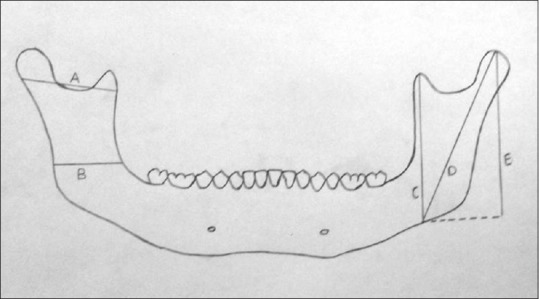
Schematic diagram with five parameters on mandibular ramus where A: Maximum ramus breadth, B: Minimum ramus breadth, C: Coronoid height, D: Condylar/maximum ramus height, E: Projective ramus height
Figure 2.
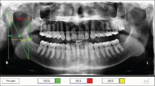
Digital orthopantomogram of male participant showing measurements
Figure 3.
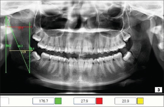
Digital orthopantomogram of female participant showing measurements
Maximum ramus breadth: The distance between the most anterior point on the mandibular ramus and a line connecting the most posterior point on the condyle and the angle of jaw
Minimum ramus breadth: Smallest anterior–posterior diameter of the ramus
Coronoid height: Projective distance between coronion and lower wall of the bone
Condylar/maximum ramus height: Height of the ramus from the most superior point on the mandibular condyle to the tubercle or most protruding portion of the inferior border of the ramus
Projective ramus height: Projective height of ramus between the highest point of the mandibular condyle and lower margin of the bone.
The collected data were analyzed using SPSS software version 16 (Chicago, SPSS Inc.), and the tests applied were independent t-test and discriminant function analysis.
Results and Observations
The participants in the present study ranged from 21 to 60 years with an equal number of males (n = 500) and females (n = 500). The maximum number of participants was in the age group of 31–40 years, in which 149 (14.9%) were males and 152 (15.2%) were females [Graph 1]. The total mean of minimum and maximum ramus breadth was 27.44 ± 3.41 mm and 32.27 ± 3.40 mm, respectively. The maximum and projective ramus height was 71.78 ± 5.98 mm and 65.62 ± 6.19 mm, respectively, and the coronoid height was 59.23 ± 6.08 mm [Table 1].
Graph 1.
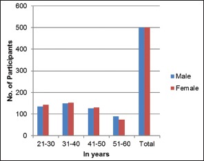
Distribution of participants correlating age and sex
Table 1.
Discriminant analysis of different parameters of mandibular ramus
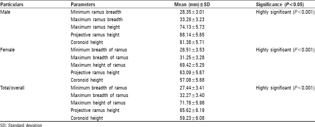
In our study, it was distinctly observed that the mean of minimum ramus breadth (m = 28.35 ± 3.01 mm, f = 26.51 ± 3.53 mm), maximum ramus breadth (m = 33.29 ± 3.23 mm, f = 31.25 ± 3.28 mm), maximum ramus height (m = 74.13 ± 5.73 mm, f = 69.42 ± 5.25 mm), projective ramus height (m = 68.14 ± 5.65 mm f = 63.09 ± 5.67 mm), and coronoid height (m = 61.38 ± 5.71 mm, f = 57.08 ± 5.68 mm) was noted and suggested that males had higher values as compared to females, and this was highly significant (P < 0.001) after applying independent t-test [Table 1].
The Box's M statistics was applied to verify the applicability of mandibular ramus in determining sex. Our study confirmed that male and female sex can be differentiated and this was highly significant (P < 0.001) [Table 2].
Table 2.
Box's M statistics for sex verification

The accuracy for sex determination was obtained using canonical discriminant function coefficient and constant value from the dimensions of mandibular ramus [Table 3]. The estimated sex was calculated using the following equations:
Table 3.
Canonical discriminant function for male and female
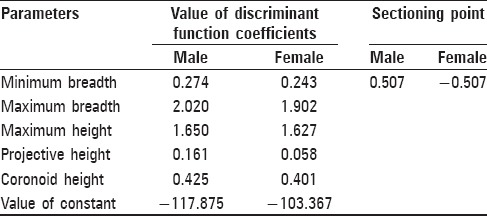
For males = −117.875 + (0.274 × minimum ramus breadth) + (2.020 × maximum ramus breadth) + (1.650 × maximum ramus height) + (0.161 × projective ramus height) + (0.425 × coronoid height)
For females = −103.367+ (0.243 × minimum ramus breadth) + (1.902 × maximum ramus breadth) + (1.627 × maximum ramus height) + (0.058 × projective ramus height) + (0.401 × coronoid height).
The sectioning point was found to be 0.507. The discriminant function value if near to 0.507, then the person is probably male, whereas if it is near to −0.507, then the person is probably female [Table 3].
By this in our study, out of total 500 males, 340 (68.0%) were correctly predicted as males, whereas out of total 500 females, 350 (70.0%) were correctly predicted as females. Thus, it was observed that the overall accuracy for diagnosing sex from mandibular ramus was 69.0% [Table 4].
Table 4.
Prediction accuracy

Discussion
Mandible is the strongest structure of skull because of dense layer of compact bone. It has a vital role because of its sexual dimorphism and radiomorphometric features although it undergoes morphological changes in size and remodeling during growth up to certain age. Dimorphism in mandible is due to size and shape, and male bones are usually large and strong than female bones. Mandible is the last skull bone to cease growth and is sensitive to the adolescent growth spurt. The stages of mandibular development, growth rates, and its duration vary in males and females and hence useful in differentiating sexes. Various structures of mandible are used for sex determination, out of which ramus is the most sexually dimorphic structure.[2,5,7]
In the present study, the dimensions of different parameters on mandibular ramus when correlated with sex; it was distinctly observed that the mean values were significantly higher in males when compared to females and were statistically highly significant (P < 0.001). This finding matched with the study of Indira et al.[8]
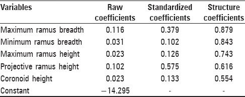
The utility of mandible for sex determination is proved by various studies.[5,9,10,11,12,13,14] In our study, the ramus measurements were subjected to discriminant function analysis. Each of the five variables measured on mandibular ramus showed statistically significant (P < 0.05). Sex differences suggesting that the mandibular ramus has strong sexual dimorphism. The ramus presented highest univariate sexual dimorphism in relation to maximum ramus breadth and projective ramus height [Table 5], which did not simulate with the results of Indira et al.[8] and studies cited in Humphrey et al.[9] In our study, Box's M statistics was applied to verify the applicability of mandibular ramus in determining sex which was proved to be highly significant (P < 0.001). The sites on mandible, where bone deposition or resorption or remodeling occurs, have a high potential for becoming sexually dimorphic. Ramus and condyle are usually the most sexually dimorphic because of consistent morphological changes in size and remodeling during the growth.[9]
Table 5.
Descriptive analysis
We could successfully diagnose 68% of males and 70% of females accurately, and with an overall accuracy of 69%, from the morphological analysis of mandibular ramus. Our these findings did not match with the findings of Indira et al.,[8] wherein the overall accuracy for diagnosing sex was 76%. The reasons for this may be the larger sample size (n = 1000) of our study.
Various researchers have conducted studies[5,10,11] on dry adult mandibles of known sex using anthropometric measurements and found an accuracy of 81%–85% and reported that ramus height and breadth were highly significant parameters.
Conclusion
The morphometric analysis of mandibular ramus using digital OPG serves as an important and valuable aid for sex identification up to certain extent although social and environmental factor does influence the development and structure of mandible. The skeletal characteristic varies geographically. The limitations of this study are a failure to consistently designate sex in the subadult range and to assess the gender in edentulous cases. Further studies are recommended on varied population, larger sample size, other imaging modalities, and the measurements shall be carried out by more than two observers as it may result in comparatively better discrimination.
Financial support and sponsorship
Nil.
Conflicts of interest
There are no conflicts of interest.
References
- 1.Sweet D. Why a dentist for identification? Dent Clin North Am. 2001;45:237–51. [PubMed] [Google Scholar]
- 2.Phillip E. Introduction to forensic science. Dent Clin North Am. 2001;45:217–27. [PubMed] [Google Scholar]
- 3.Patel D, Kumar S, Sheikh M. Age estimation in 70 cases by applying Gustafson's method on incisors and canines. J Forensic Med Toxicol. 2007;24:32–5. [Google Scholar]
- 4.Valenzuela A, Martin-De Las Heras S, Mandojana JM, De Dios Luna J, Valenzuela M, Villanueva E. Multiple regression models for age estimation by assessment of morphologic dental changes according to teeth source. Am J Forensic Med Pathol. 2002;23:386–9. doi: 10.1097/00000433-200212000-00018. [DOI] [PubMed] [Google Scholar]
- 5.Saini V, Srivastava R, Rai RK, Shamal SN, Singh TB, Tripathi SK. Mandibular ramus: An indicator for sex in fragmentary mandible. J Forensic Sci. 2011;56(Suppl 1):S13–6. doi: 10.1111/j.1556-4029.2010.01599.x. [DOI] [PubMed] [Google Scholar]
- 6.Scheuer L. Application of osteology to forensic medicine. Clin Anat. 2002;15:297–312. doi: 10.1002/ca.10028. [DOI] [PubMed] [Google Scholar]
- 7.Carvalho SP, Brito LM, Paiva LA, Bicudo LA, Crosato EM, Oliveira RN. Validation of a physical anthropology methodology using mandibles for gender estimation in a Brazilian population. J Appl Oral Sci. 2013;21:358–62. doi: 10.1590/1679-775720130022. [DOI] [PMC free article] [PubMed] [Google Scholar]
- 8.Indira AP, Markande A, David MP. Mandibular ramus: An indicator for sex determination – A digital radiographic study. J Forensic Dent Sci. 2012;4:58–62. doi: 10.4103/0975-1475.109885. [DOI] [PMC free article] [PubMed] [Google Scholar]
- 9.Humphrey LT, Dean MC, Stringer CB. Morphological variation in great ape and modern human mandibles. J Anat. 1999;195(Pt 4):491–513. doi: 10.1046/j.1469-7580.1999.19540491.x. [DOI] [PMC free article] [PubMed] [Google Scholar]
- 10.Giles E. Sex determination by discriminant function analysis of the mandible. Am J Phys Anthropol. 1964;22:129–35. doi: 10.1002/ajpa.1330220212. [DOI] [PubMed] [Google Scholar]
- 11.Steyn M, Iscan MY. Sexual dimorphism in the crania and mandibles of South African whites. Forensic Sci Int. 1998;98:9–16. doi: 10.1016/s0379-0738(98)00120-0. [DOI] [PubMed] [Google Scholar]
- 12.Duric M, Rakocevic Z, Donic D. The reliability of sex determination of skeletons from forensic context in the Balkans. Forensic Sci Int. 2005;147:159–64. doi: 10.1016/j.forsciint.2004.09.111. [DOI] [PubMed] [Google Scholar]
- 13.Hu KS, Koh KS, Han SH, Shin KJ, Kim HJ. Sex determination using nonmetric characteristics of the mandible in Koreans. J Forensic Sci. 2006;51:1376–82. doi: 10.1111/j.1556-4029.2006.00270.x. [DOI] [PubMed] [Google Scholar]
- 14.Franklin D, O’Higgins P, Oxnard CE, Dadour I. Discriminant function sexing of the mandible of indigenous South Africans. Forensic Sci Int. 2008;179:84.e1–5. doi: 10.1016/j.forsciint.2008.03.014. [DOI] [PubMed] [Google Scholar]


