Abstract
Background:
Age and sex determinations are important tools in forensic odontology which help in the identification of an individual. Radiographic method of sex and age estimation is a noninvasive simple technique. Measurements of the maxillary sinuses can be used for the estimation of age and gender when other methods are inconclusive. Maxillary sinus dimensions were used as an important tool in the identification of unknown.
Aim:
This study aims to estimate age and sex using the dimensions and volume of the maxillary sinus in magnetic resonance imaging (MRI).
Materials and Methods:
This study included sixty patients visiting Department of Radiology Mamata General Hospital, Khammam requiring MRI of the brain and paranasal sinuses. Maxillary sinus dimensions were measured using Siemens software, and statistical analysis was done.
Results:
The volume and dimensions of the maxillary sinus were more in males when compared to the females with a statistically significant difference. The highest percentage of sexual dimorphism was seen in the volume of left maxillary sinus. Age estimated using the volume of maxillary sinus showed no statistically significant difference from the actual age of the subjects.
Conclusion:
The dimensions and volume of the maxillary sinuses were larger in males than in females, in addition to that they tend to be less with the older age. MRI measurements of maxillary sinuses may be useful to support gender and age estimation in forensic radiology.
Keywords: Age determination by skeleton, forensic dentistry, magnetic resonance imaging, maxillary sinus, sex determination analysis
Introduction
Measurements of the maxillary sinuses in magnetic resonance imaging (MRI) can be used for the age and gender estimation.[1] Radiology can assist in giving accurate dimensions for which certain formulae can be applied to estimate gender. Age estimation is one of the several indicators employed to establish identity in forensic cases. Such estimations of living individuals are made for refugees or other persons who arrived in a country without acceptable identification papers and may require a verification of age, to be entitled to civil rights and/or social benefits in a modern society.[1] Maxillary sinuses remain intact even in explosions, warfare, and other mass disasters such as aircraft crashes, the skull, and other bones are badly disfigured.[2] Hence, the dimensions of the maxillary sinuses are reliable indicators in forensics.
The extent of pneumatization of the maxillary sinus varies from person to person, and its volume is influenced by age.[3] The maxillary sinus is a triangular pyramid in the body of the maxilla which presents three recesses: An alveolar recess pointed inferiorly, bounded by the alveolar process of the maxilla; a zygomatic recess pointed laterally, delimited by the zygomatic bone, and an infraorbital recess pointed superiorly, bounded by the inferior orbital surface of the maxilla.[4]
The size of sinuses varies in different skulls, and even on the two sides of the same skull. Measurement of the internal dimension of maxillary sinus has proven to be a challenge for researchers. Considering the complex structure of maxillary sinuses, MRI and computed tomography (CT) are the gold standard methods to depict the true anatomy of the Highmore's antrum. Nevertheless, their use is limited by high dose, cost, or restricted accessibility.[4] For the last 10 years, CT and MRI techniques have replaced the imaging of paranasal sinuses by traditional X-ray. These two technological modalities can be used in the determination of the more detailed anatomic structure of the diseased region. The axial, sagittal, and coronal sections obtained by CT and MRI enable better evaluation of these structures.[5]
Hence, the aim of this study was to know whether the width, the depth, and the height of maxillary sinuses can be used for the estimation of age and gender.
Materials and Methods
The present study was carried out in the Department of Radiology, Mamata General Hospital Campus, Khammam after required approval from the Institutional Ethical Committee. A prospective study comprising 60 subjects (30 males and 30 females) ranged from 21 to 73 years age group over a period of 7 months (January to July 2015) was selected for the purpose of this study. Healthy subjects were taken randomly attending the outpatient department were included, and informed consent was obtained. Subjects with the history of facial trauma, fracture of maxillary sinus, congenital developmental abnormalities, sinusitis, and pathology associated with maxillary sinus were excluded from the study.
All subjects were examined on MRI. The width, height, and depth measurements were made where the maxillary sinus was in its widest position with the help of the measurement equipment on SIEMENS AVANTO TIM DOT (NUMARIS/4 Software, Syngo MR D13 Version, Germany).
The three dimensions (height, width, and depth) were measured on the axial and coronal views. The width and depth were measured on the axial views while the height was measured on coronal view. The following measurements were performed:
The right and left side height of the maxillary sinus (maximal craniocaudal diameter)
The right and left side width of the maxillary sinus (maximal width)
The right and left side depth of the maxillary sinus (anteroposterior diameter).[2]
The depth and width of maxillary sinus were measured above the most apical level of the maxillary sinus floor. The width was defined as the longest distance perpendicular from the medial wall of the sinus to the most lateral wall of the lateral process of the maxillary sinus in the axial view. The depth was defined as the longest distance from the most anterior point to the most posterior point of the medial wall in the axial wall. The height was measured away from the inner surface of the anterior border of the maxillary sinus. The height of the maxillary sinus was measured as the longest distance from the lowest point of the sinus floor to the highest point of the sinus roof in the coronal view [Figures 1 and 2].[1] All the three dimensions of the maxillary sinus were measured by single observer and the volume of each maxillary sinus was also calculated using the following equation:
Figure 1.
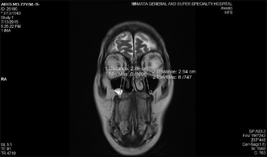
Magnetic resonance imaging coronal view revealing height of maxillary sinus
Figure 2.
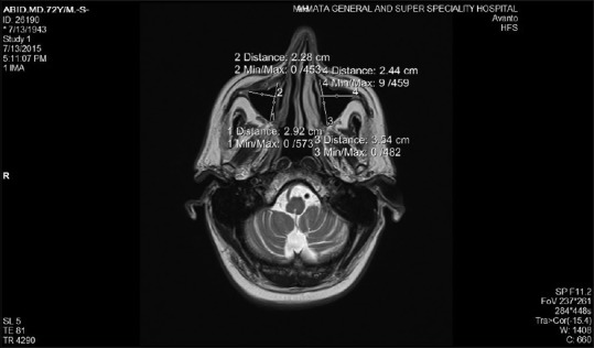
Magnetic resonance imaging axial view depicting width and depth of maxillary sinus
Volume = (height × depth × width × 0.5)
A multiple linear regression model was then developed to estimate the age of the subjects using the dimensions and volume of the maxillary sinuses. The formula derived was:
Age = 51.84 + 135.43 (height) − 146.03 (width) + 69.93 (depth) − 6.78 (total maxillary sinus volume)
Example: Sample 1: Chronological age – 66 years.
Height = 3.73, width = 3.94, depth = 4.22.
Volume = (height × width × depth) × 0.5 = (3.73 × 3.94 × 4.22) × 0.5 = 31
Estimated age = 51.84 + 135.43 (3.73) − 146.03 (3.94) + 69.93 (4.22) − 6.78 (31) = 66.6.
Statistical analysis
After obtaining the results, statistical analysis was performed using Mann–Whitney U-test and Student's t-test.
Results
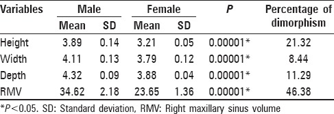
The mean values of the right side maxillary sinus height, width, depth, and volume in males were 3.89, 4.11, 4.32, and 34.62, respectively, whereas in case of females it was 3.21, 3.79, 3.88, and 23.65, respectively. There was a statistically significant difference between the mean dimension of height, width, depth, and volume of the right maxillary sinus with P values of 0.00001, 0.00001, 0.00001, and 0.00001, respectively [Table 1].
Table 1.
Comparison of volume of the right maxillary sinus between males and females
The mean values of the left side maxillary sinus height, width, depth, and volume for males were 3.88, 4.09, 4.27, and 33.91, respectively while in case of females it was 3.18, 3.74, 3.82, and 22.70, respectively. There was a statistically significant difference between the mean dimension of height, width, depth and volume of the left maxillary sinus with P values of 0.00001, 0.00001, 0.00001, and 0.00001, respectively [Table 2].
Table 2.
Comparison of volume of the left maxillary sinus between males and females
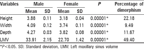
The percentage of sexual dimorphism among the selected subjects in right maxillary sinus height, width, depth and volume were 21.32%, 8.44%, 11.29, and 46.38%, respectively. The percentage of sexual dimorphism among the selected subjects in the left maxillary sinus height, width, depth, and volume were 22.18%, 9.49%, 11.67, and 49.40%, respectively.
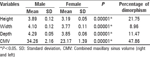
The percentage of sexual dimorphism on combining the height, width, depth, and volume on the right and left side maxillary sinuses were 21.75, 8.96, 11.47, and 47.86, respectively. The highest percentage of sexual dimorphism was shown by the volume of the maxillary sinuses on the left side [Table 3].
Table 3.
Comparison of the right and left maxillary sinus volume between males and females
On comparison of the estimated age and the actual age of the selected subjects, there was no statistically significant difference between the estimated age and the actual age of the patients (P > 0.05). This indicates that the dimensions and volume of the maxillary sinuses can be used in the estimation of the age [Table 4 and Figure 3].
Table 4.
Comparison of actual and estimated age in total, male and female samples
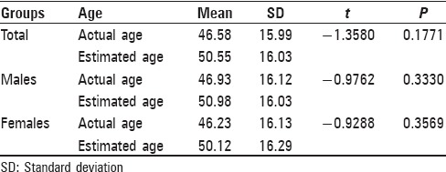
Figure 3.
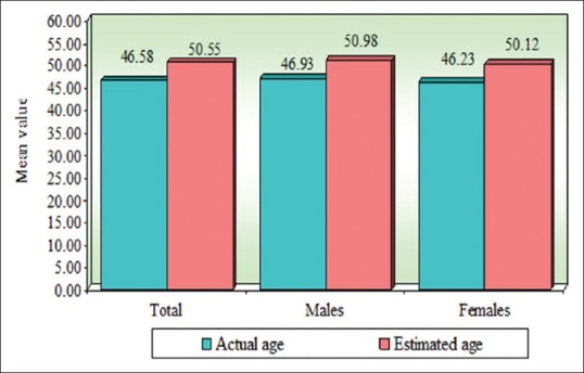
Comparison of actual and estimated age in total, male and female samples
Discussion
The maxillary sinus is the largest and the first paranasal sinus to develop.[6] It is located in the maxillary bones and consists of two spaces, which is air-filled cavity lined with pseudostratified ciliated columnar epithelium. It serves many functions such as, to decrease the weight of the skull, increases voice resonance, protects against blows to the face, insulation of the eyes and roots of the teeth against temperature fluctuations, humidification of inhaled air, and contributes to the maxillary growth.[7]
Identification on skeletal and decomposing human remains is one of the most difficult skills in forensic medicine. Sex determination is also an important problem in the identification. When the skeleton exists completely, sex can be determined with 100% accuracy. This estimation rate is 98% in the existence of pelvis and cranium, 95% with only pelvis and 80%–90% with long bones. Next to the pelvis, the skull is the most easily sexed portion of the skeleton, but the determination of sex from the skull is not reliable until after puberty. Sex estimation can be accomplished using either morphological or metric methodologies.[8]
Identification from the remnants of human skeletons is a significant forensic procedure. Determination of age and gender is an essential part of identification. Gender determination is certainly significant for identification.[6] Maxillary sinus dimensions and volume show a wide range in different studies that may reflect the influential effects such as human variability and triggering of pneumatization.[1]
Regarding age grouping, the four variables (volume, width, depth, and height) showed a significant difference, where all of them found to decrease with the age. It is found that the maxillary sinuses in males were larger in volume and wider in width than that of females, as well as the depth and height are higher in males than that of females.[1] Previous studies found that there was a significant difference of the maxillary sinus volume between males and females, mainly because males exhibit higher and wider maxillary sinuses than females and they also found no significant difference between the left and right maxillary sinus volume that is consistent with this study.[1]
In literature, the effects of gender on maxillary sinus volume were evaluated. Some of these studies reported no significant differences while the others concluded that males had larger maxillary sinus volumes than the females. On the other hand, Park et al. and Kim et al. did not consider gender discrimination in the evaluation of maxillary sinus volume.[9,10]
Some authors found that the volume decrease with the age and this might be related to skeletal size and physique.[1] The maxillary sinus has been shown to increase in size in childhood and adolescence.[11] In this study, the estimated age and the actual age of the subjects were closely related, indicating that the dimensions and volume of the maxillary sinuses can be used in the estimation of the age.
The paranasal sinus anatomy varies from person to person. The volume of the paranasal sinuses tends to decrease with increasing age.[3] The main characteristics of these structures are pneumatic. Genetic diseases, environmental conditions, and past infections can affect the process of pneumatization of paranasal sinuses.[12]
Uthman et al. studied maxillary sinus dimensions in 88 patients between the age group of 20–49 years by CT scan. The width, length, and height of the maxillary sinuses in addition to the total distance across both sinuses were measured. Data were subjected to discriminant analysis for gender using multiple regression analysis. Maxillary sinus height was the best parameter that could be used to study sexual dimorphism with an overall accuracy of 71.6% using multivariate analysis 74.4% of male sinus and 73.3% of female sinus were sexed correctly.[13]
On the other hand, Teke et al. reported lower accuracy rates for the same measurements, which was 69.3% for men and 69.4% for women with an overall accuracy of 69.3%.[14] Further, Deshmuk and Deversh tested maxillary sinus measurements for gender assessment, and they found that the average accuracy reached 80%–87%.[15]
Conclusion
The result of the present work showed that the maxillary sinus exhibits anatomic variability between genders and can be used in the sexing of a population. The dimensions and volume of the maxillary sinuses can be used in estimating the age of the subjects. The MRI images could provide adequate measurements for maxillary sinuses that cannot be approached by other means. Hence, morphometric analysis of maxillary sinus can assist in gender and age determination.
Financial support and sponsorship
Nil.
Conflicts of interest
There are no conflicts of interest.
References
- 1.Jasim HH, Al-Taei JA. Computed tomographic measurement of maxillary sinus volume and dimension in correlation to the age and gender (comparative study among individuals with dentate and edentulous maxilla) J Bagh Coll Dent. 2013;25:87–93. [Google Scholar]
- 2.Kanthem RK, Guttikonda VR, Yeluri S, Kumari G. Sex determination using maxillary sinus. J Forensic Dent Sci. 2015;7:163–7. doi: 10.4103/0975-1475.154595. [DOI] [PMC free article] [PubMed] [Google Scholar]
- 3.Abdulhameed A, Zagga AD, Maaji SM, Bello A, Bello SS, Usman JD, et al. Three dimensional volumetric analysis of the maxillary sinus using computed tomography from Usmanu Danfodiyo University Teaching Hospital, Sokoto, Nigeria. Int J Health Med Inf. 2013;2:1–9. [Google Scholar]
- 4.Saccucci M, Cipriani F, Carderi S, Di Carlo G, D’Attilio M, Rodolfino D, et al. Gender assessment through three-dimensional analysis of maxillary sinuses by means of cone beam computed tomography. Eur Rev Med Pharmacol Sci. 2015;19:185–93. [PubMed] [Google Scholar]
- 5.Ozkadif S, Eken E. Three-dimensional reconstruction of multi detector computed tomography images of paranasal sinuses of New Zealand rabbits. Turk J Vet Anim Sci. 2013;37:675–81. [Google Scholar]
- 6.Kiruba LN, Gupta C, Kumar S, D’Souza AS. A study of morphometric evaluation of the maxillary sinuses in normal subjects using computer tomography images. Arch Med Health Sci. 2014;2:12–5. [Google Scholar]
- 7.Masri AA, Yusof1 A, Hassan R. A three dimensional computed tomography (3D-CT): A study of maxillary sinus in Malays. Can J Basic Appl Sci. 2013;01:125–34. [Google Scholar]
- 8.Sidhu R, Chandra S, Devi P, Taneja N, Sah K, Kaur N. Forensic importance of maxillary sinus in gender determination: A morphometric analysis from Western Uttar Pradesh, India. Eur J Gen Dent. 2014;3:53–6. [Google Scholar]
- 9.Park IH, Song JS, Choi H, Kim TH, Hoon S, Lee SH, et al. Volumetric study in the development of paranasal sinuses by CT imaging in Asian: A pilot study. Int J Pediatr Otorhinolaryngol. 2010;74:1347–50. doi: 10.1016/j.ijporl.2010.08.018. [DOI] [PubMed] [Google Scholar]
- 10.Kim HY, Kim MB, Dhong HJ, Jung YG, Min JY, Chung SK, et al. Changes of maxillary sinus volume and bony thickness of the paranasal sinuses in longstanding pediatric chronic rhinosinusitis. Int J Pediatr Otorhinolaryngol. 2008;72:103–8. doi: 10.1016/j.ijporl.2007.09.018. [DOI] [PubMed] [Google Scholar]
- 11.Ariji Y, Kuroki T, Moriguchi S, Ariji E, Kanda S. Age changes in the volume of the human maxillary sinus: A study using computed tomography. Dentomaxillofac Radiol. 1994;23:163–8. doi: 10.1259/dmfr.23.3.7835518. [DOI] [PubMed] [Google Scholar]
- 12.Baweja S, Dixit A, Baweja S. Study of age related changes of maxillary air sinus from its anteroposterior, transverse and vertical dimensions using computerized tomographic (CT) scan. Int J Biomed Res. 2013;04:21–5. [Google Scholar]
- 13.Uthman AT, Al-Rawi NH, Al- Naaimi AS, Al- Timimi JF. Evaluation of maxillary sinus dimensions in gender determination using helical CT scanning. Journal of Forensic Sciences. 2011;56:403–8. doi: 10.1111/j.1556-4029.2010.01642.x. [DOI] [PubMed] [Google Scholar]
- 14.Teke HY, Duran S, Canturk N, Canturk G. Determination ofgender by measuring the size of themaxillary sinuses in computed tomographyscans. Surg Radiol Anat. 2007;29:9–13. doi: 10.1007/s00276-006-0157-1. [DOI] [PubMed] [Google Scholar]
- 15.Deshmukh AG, Deversh DB. Comparison of cranial sex determination by univariate and multivariateanalysis. J Anat Soc India. 2006;55:1–5. [Google Scholar]


