Abstract
Efforts to implement family cord blood banking have been developed in the past decades for siblings requiring stem cell transplantation for conditions such as sickle cell disease. However, public banks are faced with challenging decisions about the units to be stored, discarded, or used for other endeavors. We report here 20 years of experience in family cord blood banking for sickle cell disease in two dedicated public banks. Participants were pregnant women who had a previous child diagnosed with homozygous sickle cell disease. Participation was voluntary and free of charge. All mothers underwent mandatory serological screening. Cord blood units were collected in different hospitals, but processed and stored in two public banks. A total of 338 units were stored for 302 families. Median recipient age was six years (11 months-15 years). Median collected volume and total nucleated cell count were 91 mL (range 23–230) and 8.6×10⁸ (range 0.7–75×108), respectively. Microbial contamination was observed in 3.5% (n=12), positive hepatitis B serology in 25% (n=84), and homozygous sickle cell disease in 11% (n=37) of the collections. Forty-four units were HLA-identical to the intended recipient, and 28 units were released for transplantation either alone (n=23) or in combination with the bone marrow from the same donor (n=5), reflecting a utilization rate of 8%. Engraftment rate was 96% with 100% survival. Family cord blood banking yields good quality units for sibling transplantation. More comprehensive banking based on close collaboration among banks, clinical and transplant teams is recommended to optimize the use of these units.
Introduction
Sickle cell disease (SCD) is the most common inherited hemoglobin disorder. Particularly frequent in people of African descent, the disease is associated with numerous complications and early mortality.1,2 Major progress has been made in the management of SCD and this has improved patients’ survival without curing the disease;3 this includes implementation of antenatal counseling and neonatal screening programs,3 screening and prevention of neurological complications,4 prevention of pneumococcal infections,5,6 and the use of hydroxycarbamide5,7 and chronic transfusion therapy.8
However, hematopoietic stem cell transplantation (HSCT) remains, to date, the only known curative therapy for SCD,9–12 offering a cure rate exceeding 90%.9–17 Walters et al., in a recent review of HSCT in SCD children after HLA-identical sibling HSCT reported overall survival and event-free survival rates of 95% and 92%, respectively.17 Similar results were reported in retrospective registry-based surveys from the Center for International Blood and Marrow Transplant Research18 and the European Society for Blood and Marrow Transplantation9,10,13 with overall survival rates of over 91%.
Despite a cure rate exceeding 90%, regardless of the stem cell source, HSCT is still under-used for patients with SCD.9,10 Limiting factors include the availability of suitable donors and, for the majority of patients for whom a matched sibling donor has been identified, the reluctance of families and physicians to consider transplantation because of possible transplant-related toxicity.
Umbilical cord blood transplantation (CBT) from a related family member has proved to be an effective alternative for patients with SCD, resulting in survival rates similar or superior to adult donor transplant9,10,13,19,20 and lower probability of acute and chronic graft-versus-host diseases (GvHD).9,10 More recently, a promising, but still experimental, approach based on gene and cellular therapy methods has been proposed aiming to correct the sickle gene defect in the patient’s own stem cells.21,22
With the emergence of the gene-therapy biotechnology to treat genetic disorders22 and the improved outcomes of CBT, many directed public or private cord blood (CB) banking programs have been established for affected siblings who would benefit from a related HSCT. Consequently, cord blood banks (CBB) are now confronted with major challenges regarding storage and disposal of the units.
We report here our 20-year experience in two public family-directed cord blood banks for SCD.
Methods
Participants were pregnant women who had a child homozygous for SCD. Recruitment was carried out through referral addressed to the CBB by the clinician treating the affected child. All mothers, including those who refused prenatal diagnosis, were offered sibling CB banking. Participation was voluntary. Informed consent was obtained from the mother before delivery, in accordance with local ethical requirements. Collections were organized by two public banks, free of charge for the families. Both banks had a wide experience in unrelated CB banking and were affiliated to the French network of CBB accredited by the “Agence Nationale de Biomedecine”.
The CBUs were reserved for family use and were shipped to the transplant center once the decision to proceed with transplantation was made. All mothers underwent a panel of mandatory serology testing prior to banking, which included hepatitis B and C viruses, HIV 1–2, HTLV I–II and syphilis.
The CBU were collected in the health care institution selected by the mother for child delivery. Twenty-seven institutions were involved and were contacted by the CBB. Informed consents were collected by midwives or designated medical personnel locally before delivery. Detailed instructions and training to local staff were provided by the CBB team. CBU collection kits, including a collection pack and materials necessary for harvesting, were sent to the contact team in the delivery hospital along with the standard regulatory forms. All CBUs were transported to the designated CBB to be processed within 24 hours of collection.
The CBBs policy was to process and store all the collected CBUs, independent of the unit volume, cell counts and HLA compatibility. Cell counts, cell viability, sterility tests, progenitor cell quantification and functional assays were performed for all CBUs. Due to related cost and absence of immediate patient indication for transplantation, histocompatibility testing was not conducted routinely, unless requested by the referring clinician. There was no volume reduction before cryopreservation. The CBUs were cryop-reserved using a controlled-rate freezer, then transferred to the vapor phase of a liquid nitrogen storage tank and maintained at less than −150°C.
The CBUs and aliquots collected from mothers having positive infectious markers or awaiting results were stored in quarantine tanks. Abnormal results that could affect donor suitability were considered individually at the time a CBU was requested for transplantation.
Hemoglobin genotyping was not performed on CBU samples. Information about donor SCD status was available through the national neonatal screening database for genetic diseases. CBUs collected from infants found to have SS genotype, were not discarded unless a written request was formulated by the referring physician.
Once the decision to proceed with transplantation was made, confirmatory HLA typing was conducted. CBUs were thawed and shipped to the transplant unit for infusion. Total nucleated cells and CD34+ cell counts, viability and sterility tests were performed after thawing.
Data were collected from CBB databases, Eurocord registry and patient records. Neutrophil engraftment was defined as the first of three consecutive days with neutrophils 0.5×10⁹/L or more, and platelet engraftment as the first of three consecutive days with platelets 20×109/L or more with no platelet transfusions for at least seven days before reconstitution.
Results
From 1995 to 2014, a total of 338 CBUs were collected with a sustained increase in the number of referrals for family CB banking for SCD over the years. These units were stored for 302 families, including 32 mothers recruited more than once. All collections were for an existing homozygous sibling. Fifteen families (5%) had more than one affected child. Figure 1 details the distribution of the CBUs collected in each CBB.
Figure 1.
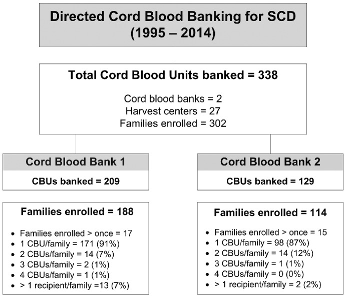
Family cord blood banking program. Flow diagram. SCD: sickle cell disease; CBU: cord blood units.
Characteristics of banked CBUs
Median values for specific quality parameters of the collected CBUs are shown in Table 1. Sixty-one percent (n=207) of the collections exceeded 80 mL (Figure 2) and 40% (n=135) exceeded 10.0×10⁸ total nucleated cell count (TNC).
Table 1.
Characteristics of collected cord blood units.
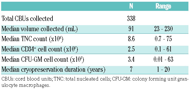
Figure 2.
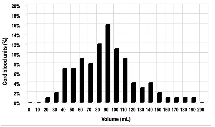
Volume distribution (%) of collected cord blood units.
Sixty-five percent (n=218) of the units had a minimal TNC of 5×10⁸ (based on 20 kg recipient, a target dose of ≥2.5×10⁷ nucleated cells/kg), considered suitable for pediatric transplantation according to the US FDA23 criteria for banking CBUs and 44% of the units (n=148) exceeded the minimal pre-freezing thresholds for cell counts and volume adopted by many unrelated CBBs24–27 (i.e. volume >40 mL and TNC >9×10⁸).
A microbial contamination rate of 3.5% was observed in our cohort as 12 CBUs failed sterility testing; these units were stored because antimicrobial sensitivity results did not preclude their use for transplantation with the provision of appropriate antibiotics. Eighty-four CBUs (25%) had positive hepatitis B serological markers: 76% (n=64) of these were due to positive anti-HBs and/or anti-HBc antibodies with negative HBs antigen. Two units tested positive for hepatitis C (HCV) antibody, but HCV RNA was not detected by PCR, despite maternal active HCV infection. All CBUs were negative for HIV.
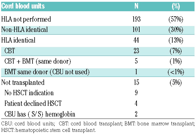
HLA-typing was carried out for 145 donor-recipient pairs (43% of the collected CBU) upon request of the referring physicians (Table 2). Forty-four units had a full HLA antigen match with the intended recipient: these represented 30% of the typed units and 13% of all collected units.
Table 2.
HLA typing and utilization of cord blood units.
Eleven percent (n=37) of the collections had homozygous SCD and 45% (n=152) a carrier disease status (Figure 3). Of the 145 HLA typed CBU, 8% (n=11) were homozygous for SCD, and 26% (n=38) were both HLA identical to the recipient and had normal hemoglobin or sickle cell carrier status, thus considered as “potential graft source” for the intended sibling.
Figure 3.
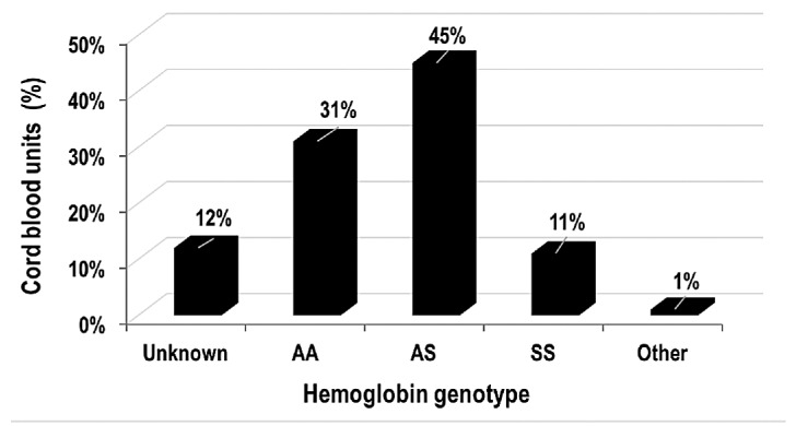
Hemoglobin genotype of collected cord blood units. AA: normal hemoglobin genotype; AS: heterozygous βS; SS: homozyous βS/βS.
To date, 28 units were released for transplantation either alone (CBT, n=23) or in combination with a bone marrow (BM) graft from the same donor (CBT+BMT, n=5), reflecting a utilization rate of 8% for the whole cohort over 20 years, and 19% transplant rate for the typed units. Another 13 HLA-identical CBUs with adequate cell dose to perform transplantation remained in storage for future use, as their intended recipient was not an immediate indication for transplantation (n=9) or decided not to undergo the procedure (n=4) because of transplant-related risks. Three HLA identical CBUs were deemed unsuitable for transplantation due to their biological features: 2 units were homozygous for HbS and one unit had low cell counts (2.7×10⁷ TNC/kg and 0.2×10⁶ CD34+ cell/kg) (Table 2).
Of further interest, 41% (n=137) of the collected CBUs were referred from a single hematologic center.9,11 Histocompatibility testing was requested and performed for 87% (n=119) of these CBUs and 30% (n=35) were HLA identical to the affected sibling. To date, 25 of these collections were used for CBT. This represents an 18% (25 of 137) utilization rate for collections referred from this center, 21% (25 of 119) transplant rate for typed units, and a 71% (25 of 35) transplant rate when a matched sibling is available. These are noteworthy rates and reflect a 2.3-fold increase in the utilization rate when the collections are requested by clinicians willing to use CBUs to transplant the affected child once transplantation is indicated. When compared to other referrals that were not affiliated to a transplant center (n=201), only 29 CBUs (14%) were typed for HLA, of which 3 units were used for transplantation; this reflects a 1.5% (3 of 201) utilization rate for the units referred from these centers, 10% (3 of 29) of the typed units, and 33% (3 of 9) of the HLA-identical units.
Transplant characteristics
Over a period of 20 years, 28 units were released for transplantation to 28 patients. Characteristics of transplanted patients are shown in Table 3. The median age of recipients at transplantation was 6.8 (3.2–13.6) years. All patients for whom data were available (n=25) received a conditioning regimen including busulfan, cyclophosphamide and anti-thymocyte globulin (ATG). All CBUs were HLA identical to the intended recipient, and 54% of the donors were sickle cell carriers. The CBUs were transplanted after a median storage time of 2.2 years (range 0.4–8.1) (Table 4).
Table 3.
Transplant characteristics.
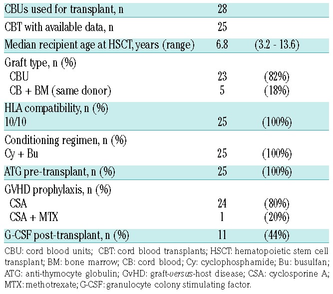
Table 4.
Characteristics of transplanted cord blood units.

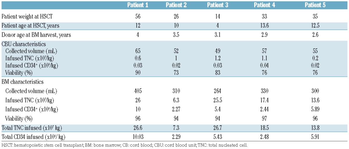
The median cell dose collected was 4.0×10⁷/kg (0.5–13.1×10⁷) TNC and 1.6×10⁵/kg (0.2–14.8×10⁵) CD34+ cells, and the median infused cell dose was 3.1×10⁷/kg (0.2–7.6×10⁷) TNC and 1.4×10⁵/kg (0.2–11.8×10⁵) CD34+ cells, with a median recipient body weight of 22 kg (14–56) for the 28 patients transplanted (Table 4). Five CBUs with a collected TNC dose less than 2×10⁷/kg (0.5–1.7×10⁷/kg) were combined with the BM collected from the same donor (Table 5). After adding the BM, the median infused TNC and CD34+ cell doses were 18.5×10⁷/kg (7.3–26.7×10⁷) and 5.4×10⁶/kg (2.3–10×106), respectively, of which a median dose of 1×10⁷/kg TNC (0.2–1.2×10⁶) and 0.03×10⁶/kg CD34+ (0.02–0.04 ×10⁶) were provided by the CBUs.
Table 5.
Characteristics of the combined cord blood + bone marrow grafts.
The median time to engraftment was 28 days (range 17–60) for neutrophils, and 54 days (range 12–91) for platelets (n=18). All but one patient engrafted for neutrophils before day 60, with a median time of 33 days after single CBT and 26 days after BMT+CBT. Chimerism at day 100 for those patients with available data (n=22) was 100% donor for 9 patients (41%) and 60–80% donor for 12 patients (55%). At last follow up, donor chimerism exceeded 80% for all patients with available data.
Notably, one patient transplanted in 1996 with an adequate cell dose (4×10⁷ TNC/kg) had primary graft failure; this patient also failed to engraft after a second transplant using the BM collected from the same sibling donor. At last follow up, the patient was alive with autologous reconstitution and remained anemic but free of vasoocclusive events. This is due to sustained increase in fetal hemoglobin production, as previously reported after autologous reconstitution in patients affected by SCD11,28 and β-hemoglobin disorders.29
Five patients who received single CBT experienced grade I–II acute GvHD that required steroids in 4 cases. One recipient of combined CB+BM transplant developed limited liver chronic GvHD, which was the only chronic GvHD event reported for the entire cohort.
All 28 patients are alive and free of SCD symptoms to date (except the sole case of persistent anemia after primary graft failure), with a median follow up of 4.3 years (4 months-14 years).
Discussion
The aim of this study was to investigate whether sibling CBU collections had sufficient cellular quantity and quality to support allogeneic CBT of the intended recipient and to assess the utilization pattern of these collections.
Family CB banking differs in many important aspects from public CBU donations. Besides the cost related to this stem cell source, when less expensive alternatives using the sibling BM are possible, there are major challenges related to cellular characteristics of the collections, such as the long-term storage and low utilization rate noted by many teams13,30–32 and confirmed in our cohort.
The cellular and biological characteristics of our banked units were comparable to those reported by other directed CBB programs13,26,33 with a utilization rate reaching 8% of the 338 CBUs collected over 20 years, and 19% of the typed units. Similar low utilization rates of banked sibling CBUs for hematologic disorders have been reported by many authors.30–33
Both of our CBBs had previously published their experience13 in banking sibling CBUs for malignant and nonmalignant disorders, with more than 400 directed CBUs collected in one bank and 111 units (80% for SCD affected siblings) in the second, a utilization rate for HLA identical sibling transplants of 3% and 9%, respectively, and 5-year overall survival of 83% and 100%, respectively, in the subgroup of patients with hemoglobin disorders.11,13 Most of the patients in our cohort were included in these studies.
Adequate volume and TNC dose are critical factors in achieving a successful CB transplantation; therefore, the use of procedures that might increase collection volume or decrease cell loss during processing is especially important in family CB banking. While 65% of our CBUs had cellular characteristics (minimal collected TNC count of 5×10⁸) adequate to support allogeneic CBT in the pediatric setting,26,34 only 13% (n=45) of our collections provided the TNC dose and volume recommended for unrelated CBU banking (initial volume >80 mL and TNC >18×108). However, the low cellular content should not be a limitation for the use of these CBU,31,33 particularly when there is an opportunity to increase the cell dose by combining CB with BM collected from the same sibling donor, an approach that proved to be effective with excellent overall survival and low incidence of GvHD, as noted by many authors.13,30,31,35
Indeed, all but one of the patients transplanted in our cohort engrafted within less than 60 days. Adding the same donor’s BM to the CBUs with low cell counts probably improved the engraftment rate of our patients.
The 3.5% bacterial contamination rate observed in our cohort was comparable to those reported in other studies;33,34 bacterial contaminations could be addressed at the time of transplantation with prophylactic antibiotics if necessary.
Homozygous SCD was detected in 11% of the collections, a lower rate than might be expected in autosomal recessive genetic disorders. This might be explained by the quite frequent antenatal diagnosis testing performed in this high-risk population.
Many important questions require further investigation, including the cost related to long-term storage of this graft source and its low utilization rate.
Ideally, the CBUs reported in our cohort should have been collected from families at risk of transmitting homozygous SCD, rather than those with an affected child. Storage-related cost remains a major limitation to such an ambitious program, when faced with the low utilization rate of these units.
In our cohort, 43% of the units were typed for HLA upon the request of the referring physicians, and 30% of the typed units were HLA-identical to the intended recipient; an expected proportion that reflects the 25% probability of finding a matched sibling donor. To limit the unnecessary long-term storage and its associated costs, HLA testing and identification of SCD status for all collections could be implemented, so a better-informed decision could be made about keeping or disposing of those units with a low usage rate, such as non-HLA-identical units or those with HbS homozygous status. However, the decision-making process must be managed with caution; non-HLA-identical CBUs could be kept to transplant homozygous identical siblings resulting from future pregnancies. Moreover, alternative experimental treatments might emerge in the future to support haploidentical-related allogeneic CBT36 or autologous CBT in the setting of gene therapy.37–42
Finally, enhancing the utilization rate of the banked unit will also achieve a significant reduction in storage needs and related costs. The transplantation experience reported here with a single center being involved in most of the transplants, strongly suggests that close collaboration with clinical and transplant teams willing to support CBT of the affected sibling is likely to increase the proportion of transplants using directed CBUs. An increase in the awareness of the excellent outcomes of CBT in this setting is also crucial to overcome the reluctance of physicians and families to consider transplant.
However, despite the increase in number of homozygous cases of SCD in the developed countries and the excellent transplant outcomes, referrals for transplantation remain limited, as reported by our group in a recent review of sibling transplants.10
The current study demonstrates that, despite the challenges associated with public directed CBB programs, sibling CBU represents a useful stem cell resource for families with specific medical indications, such as a child with sickle cell anemia or other genetic conditions that could benefit from a related transplant. These families should be encouraged to bank the CBUs from siblings. Family banking may yield good quality HLA-identical CBUs, with post-transplant outcomes and survival similar or superior to other stem cell sources.
The potential scope of medically indicated sibling cord blood banking is considerable, including hemoglobin disorders, as well as genetic and metabolic diseases. For this reason, banks should adopt a more comprehensive approach for the identification and collection of CBUs for family-directed uses, and work closely with the clinical and transplant teams to optimize the use of these units.
Supplementary Material
Acknowledgments
The authors thank all participating families, transplant centers and delivery teams for the valuable contributions to the study and the organization “Cordon de vie” in Monaco, for continuous support.
Footnotes
Check the online version for the most updated information on this article, online supplements, and information on authorship & disclosures: www.haematologica.org/content/102/6/976
References
- 1.Modell B, Darlison M. Global epidemiology of haemoglobin disorders and derived service indicators. Bull World Health Organ. 2008;86(6):480–487. [DOI] [PMC free article] [PubMed] [Google Scholar]
- 2.Weatherall DJ. Hemoglobinopathies worldwide: present and future. Curr Mol Med. 2008;8(7):592–599. [DOI] [PubMed] [Google Scholar]
- 3.Evidence-Based Management of Sickle Cell Disease: Expert Panel Report - National Heart, Lung, and Blood Institute; 2014. https://www.nhlbi.nih.gov/sites/www.nhlbi.nih.gov/files/sickle-cell-disease-report.pdf. Last accessed 14 Dec 2016.
- 4.Adams RJ, McKie VC, Hsu L, et al. Prevention of a first stroke by transfusions in children with sickle cell anemia and abnormal results on transcranial Doppler ultrasonography. N Engl J Med. 1998; 339(1):5–11. [DOI] [PubMed] [Google Scholar]
- 5.Quinn CT, Rogers ZR, McCavit TL, Buchanan GR. Improved survival of children and adolescents with sickle cell disease. Blood. 2010;115(17):3447–3452. [DOI] [PMC free article] [PubMed] [Google Scholar]
- 6.Gaston MH, Verter JI, Woods G, et al. Prophylaxis with oral penicillin in children with sickle cell anemia. A randomized trial. N Engl J Med. 1986;314(25):1593–1599. [DOI] [PubMed] [Google Scholar]
- 7.Wang WC, Ware RE, Miller ST, et al. Hydroxycarbamide in very young children with sickle-cell anaemia: a multicentre, randomised, controlled trial (BABY HUG). Lancet. 2011;377(9778):1663–1672. [DOI] [PMC free article] [PubMed] [Google Scholar]
- 8.Yawn BP, Buchanan GR, Afenyi-Annan AN, et al. Management of sickle cell disease: summary of the 2014 evidence-based report by expert panel members. JAMA. 2014;312(10):1033–1048. [DOI] [PubMed] [Google Scholar]
- 9.Gluckman E. Allogeneic transplantation strategies including haploidentical transplantation in sickle cell disease. Am Soc Hematol Educ Program Hematol. 2013; 2013:370–376. [DOI] [PubMed] [Google Scholar]
- 10.Gluckman E, Cappelli B, Bernaudin F, et al. Sickle cell disease: an international survey of results of HLA-identical sibling hematopoietic stem cell transplantation. Blood. 2017;129(11):1548–1556. [DOI] [PMC free article] [PubMed] [Google Scholar]
- 11.Bernaudin F, Socie G, Kuentz M, et al. Long-term results of related myeloablative stem-cell transplantation to cure sickle cell disease. Blood. 2007;110(7):2749–2756. [DOI] [PubMed] [Google Scholar]
- 12.Hsieh MM, Fitzhugh CD, Tisdale JF. Allogeneic hematopoietic stem cell transplantation for sickle cell disease: the time is now. Blood. 2011;118(5):1197–1207. [DOI] [PMC free article] [PubMed] [Google Scholar]
- 13.Gluckman E, Ruggeri A, Rocha V, et al. Family-directed umbilical cord blood banking. Haematologica. 2011;96(11):1700–1707. [DOI] [PMC free article] [PubMed] [Google Scholar]
- 14.Arnold SD, Bhatia M, Horan J, Krishnamurti L. Haematopoietic stem cell transplantation for sickle cell disease - current practice and new approaches. Br J Haematol. 2016;174(4):515–525. [DOI] [PubMed] [Google Scholar]
- 15.Angelucci E, Matthes-Martin S, Baronciani D, et al. Hematopoietic stem cell transplantation in thalassemia major and sickle cell disease: indications and management recommendations from an international expert panel. Haematologica. 2014; 99(5):811–820. [DOI] [PMC free article] [PubMed] [Google Scholar]
- 16.Hsieh MM, Fitzhugh CD, Weitzel RP, et al. Nonmyeloablative HLA-matched sibling allogeneic hematopoietic stem cell transplantation for severe sickle cell phenotype. JAMA. 2014;312(1):48–56. [DOI] [PMC free article] [PubMed] [Google Scholar]
- 17.Walters MC, De Castro LM, Sullivan KM, et al. Indications and Results of HLA-Identical Sibling Hematopoietic Cell Transplantation for Sickle Cell Disease. Biol Blood Marrow Transplant. 2016;22(2):207–211. [DOI] [PMC free article] [PubMed] [Google Scholar]
- 18.Panepinto JA, Walters MC, Carreras J, et al. Matched-related donor transplantation for sickle cell disease: report from the Center for International Blood and Transplant Research. Br J Haematol. 2007;137(5):479–485. [DOI] [PubMed] [Google Scholar]
- 19.Locatelli F, Rocha V, Reed W, et al. Related umbilical cord blood transplantation in patients with thalassemia and sickle cell disease. Blood. 2003;101(6):2137–2143. [DOI] [PubMed] [Google Scholar]
- 20.Locatelli F, Kabbara N, Ruggeri A, et al. Outcome of patients with hemoglobinopathies given either cord blood or bone marrow transplantation from an HLA-identical sibling. Blood. 2013;122(6):1072–1078. [DOI] [PubMed] [Google Scholar]
- 21.Kumar SR, Markusic DM, Biswas M, High KA, Herzog RW. Clinical development of gene therapy: results and lessons from recent successes. Mol Ther Methods Clin Dev. 2016;3:16034. [DOI] [PMC free article] [PubMed] [Google Scholar]
- 22.Hoban MD, Cost GJ, Mendel MC, et al. Correction of the sickle cell disease mutation in human hematopoietic stem/progenitor cells. Blood. 2015;125(17):2597–2604. [DOI] [PMC free article] [PubMed] [Google Scholar]
- 23.Cellular & Gene Therapy Guidances-Guidance for Industry and FDA Staff: IND Applications for Minimally Manipulated, Unrelated Allogeneic Placental/Umbilical Cord Blood Intended for Hematopoietic and Immunologic Reconstitution in Patients with Disorders Affecting the Hematopoietic System. http://www.fda.gov/biologicsbloodvaccines/guidancecomplianceregulatoryinformation/guidances/cellularandgenetherapy/ucm38821.htm. Last accessed 11 Dec 2016.
- 24.Rocha V, Gluckman E, Eurocord-Netcord registry and European Blood and Marrow Transplant group. Improving outcomes of cord blood transplantation: HLA matching, cell dose and other graft- and transplantation-related factors. Br J Haematol. 2009; 147(2):262–274. [DOI] [PubMed] [Google Scholar]
- 25.Saccardi R, Tucunduva L, Ruggeri A, et al. Impact of cord blood banking technologies on clinical outcome: a Eurocord/Cord Blood Committee (CTIWP), European Society for Blood and Marrow Transplantation and NetCord retrospective analysis. Transfusion. 2016;56(8):2021–2029. [DOI] [PubMed] [Google Scholar]
- 26.Querol S, Mufti GJ, Marsh SGE, et al. Cord blood stem cells for hematopoietic stem cell transplantation in the UK: how big should the bank be? Haematologica. 2009; 94(4):536–541. [DOI] [PMC free article] [PubMed] [Google Scholar]
- 27.Kurtzberg J, Cairo MS, Fraser JK, et al. Results of the cord blood transplantation (COBLT) study unrelated donor banking program. Transfusion. 2005;45(6):842–855. [DOI] [PubMed] [Google Scholar]
- 28.Ferster A, Corazza F, Vertongen F, et al. Transplanted sickle-cell disease patients with autologous bone marrow recovery after graft failure develop increased levels of fetal haemoglobin which corrects disease severity. Br J Haematol. 1995; 90(4):804–808. [DOI] [PubMed] [Google Scholar]
- 29.Paciaroni K, Gallucci C, De Angelis G, et al. Sustained and full fetal hemoglobin production after failure of bone marrow transplant in a patient homozygous for beta 0-thalassemia: a clinical remission despite genetic disease and transplant rejection. Am J Hematol. 2009;84(6):372–373. [DOI] [PubMed] [Google Scholar]
- 30.Goussetis E, Peristeri I, Kitra V, et al. Low usage rate of banked sibling cord blood units in hematopoietic stem cell transplantation for children with hematological malignancies: implications for directed cord blood banking policies. Blood Cells Mol Dis. 2011;46(2):177–181. [DOI] [PubMed] [Google Scholar]
- 31.Smythe J, Armitage S, McDonald D, et al. Directed sibling cord blood banking for transplantation: the 10-year experience in the national blood service in England. Stem Cells. 2007;25(8):2087–2093. [DOI] [PubMed] [Google Scholar]
- 32.Reed W, Smith R, Dekovic F, et al. Comprehensive banking of sibling donor cord blood for children with malignant and nonmalignant disease. Blood. 2003; 101(1):351–357. [DOI] [PubMed] [Google Scholar]
- 33.Screnci M, Murgi E, Valle V, et al. Sibling cord blood donor program for hematopoietic cell transplantation: the 20-year experience in the Rome Cord Blood Bank. Blood Cells Mol Dis. 2016;57:71–73. [DOI] [PubMed] [Google Scholar]
- 34.Allan D, Petraszko T, Elmoazzen H, Smith S. A review of factors influencing the banking of collected umbilical cord blood units. Stem Cells. 2013;2013:463031. [DOI] [PMC free article] [PubMed] [Google Scholar]
- 35.Tucunduva L, Volt F, Cunha R, et al. Combined cord blood and bone marrow transplantation from the same human leucocyte antigen-identical sibling donor for children with malignant and non-malignant diseases. Br J Haematol. 2015;169(1):103–110. [DOI] [PubMed] [Google Scholar]
- 36.Bolaños-Meade J, Fuchs EJ, Luznik L, et al. HLA-haploidentical bone marrow transplantation with post transplant cyclophosphamide expands the donor pool for patients with sickle cell disease. Blood. 2012;120(22):4285–4291. [DOI] [PMC free article] [PubMed] [Google Scholar]
- 37.Levasseur DN, Ryan TM, Pawlik KM, Townes TM. Correction of a mouse model of sickle cell disease: lentiviral/antisickling beta-globin gene transduction of unmobilized, purified hematopoietic stem cells. Blood. 2003;102(13):4312–4319. [DOI] [PubMed] [Google Scholar]
- 38.Chou Y-C, Chen R-L, Lai Z-S, et al. Pharmacological Induction of Human Fetal Globin Gene in Hydroxyurea-Resistant Primary Adult Erythroid Cells. Mol Cell Biol. 2015;35(14):2541–2553. [DOI] [PMC free article] [PubMed] [Google Scholar]
- 39.Xu J, Peng C, Sankaran VG, et al. Correction of sickle cell disease in adult mice by interference with fetal hemoglobin silencing. Science. 2011;334(6058):993–996. [DOI] [PMC free article] [PubMed] [Google Scholar]
- 40.Cavazzana M, Ribeil JA, Payen E, et al. Outcomes of Gene Therapy for Severe Sickle Disease and Beta-Thalassemia Major Via Transplantation of Autologous Hematopoietic Stem Cells Transduced Ex Vivo with a Lentiviral Beta AT87Q-Globin Vector. Blood. 2015;126(23):202. [Google Scholar]
- 41.Malik P. Gene therapy for hemoglobinopathies: tremendous successes and remaining caveats. Mol Ther. 2016; 24(4):668–670. [DOI] [PMC free article] [PubMed] [Google Scholar]
- 42.Kanter J, Walters MC, Hsieh MM, et al. Interim Results from a Phase 1/2 Clinical Study of Lentiglobin Gene Therapy for Severe Sickle Cell Disease. Blood. 2016; 128(22):1176–1176. [Google Scholar]
Associated Data
This section collects any data citations, data availability statements, or supplementary materials included in this article.


