Abstract
211At is a most promising radionuclide for targeted alpha therapy. However, its limited availability and poorly known basic chemistry hamper its use. Based on the analogy with iodine, labelling is performed via astatobenzoate conjugates, but in vivo deastatination occurs, particularly when the conjugates are internalized in cells. Actually, the chemical or biological mechanism responsible for deastatination is unknown. In this work, we show that the C−At “organometalloid” bond can be cleaved by oxidative dehalogenation induced by oxidants such as permanganates, peroxides or hydroxyl radicals. Quantum mechanical calculations demonstrate that astatobenzoates are more sensitive to oxidation than iodobenzoates, and the oxidative deastatination rate is estimated to be about 6 × 10⁶ faster at 37 °C than the oxidative deiodination one. Therefore, we attribute the “internal” deastatination mechanism to oxidative dehalogenation in biological compartments, in particular lysosomes.
Introduction
Halogens are usually named according to ancient Greek words denoting one of their characteristics. The heaviest of these elements befell the name astatine, as a reference to its instability1. While this name was proposed from a physical perspective (i.e. the absence of any stable isotope), one might tend to consider that it is also suitable from a chemical point of view, since most bonds involving this atom are less stable than the ones involving its closest analogue, iodine. This affects the potential application of 211At to medicine. Nevertheless, 211At is considered as one of the most promising radionuclides for targeted alpha therapy, due to its favourable physical properties (notably its half-life time of 7.2 h and its α-particle emission yield of 100%)2, 3.
Clinical trials using either monoclonal antibodies (mAbs) or antibody fragments labelled by astatobenzoate conjugates afforded encouraging results against both recurrent brain tumours and recurrent ovarian cancers2, 4–6. However, labelling with astatobenzoate conjugates suffers from in vivo dehalogenation, which diminishes the tumour uptake and leads to the release of free astatine and its accumulation in stomach and thyroid. Even if stomach and thyroid uptake can be mitigated7–9, a more stable labelling is needed for systemic administration8. It is particularly interesting to note that when mAbs are labelled with astatobenzoates, deastatination is limited10–14, while when antibody fragments are used, considerable dehalogenation occurs10, 11, 15. Unfortunately, the slow mAbs pharmacokinetics are not well-suited to be combined with the 211At half-life time7, 16, and astatine-labelled antibodies have been so far limited to locoregional treatments. Moreover, this behaviour is remarkable, as it echoes the one of proteins, iodinated through direct labelling or using the Bolton-Hunter reagent17. In these cases the dehalogenation mechanism has been elucidated: the 2-iodophenol moiety of the catabolites released through carrier metabolization undergoes dehalogenation catalysed by deiodinases. Thus, the deastatination mechanism is most probably initiated through internalization into cells and lysosomal degradation. To overcome deiodination, reagents such as the N-succinimidyliodobenzoate (SIB) have been synthesized by electrophilic radioiodination of N-succinimidylaryltrialkylstannane derivatives18, 19. Indeed, these reagents lack the phenolic hydroxyl group required in the catalytic mechanism of mammal deiodinases18–21.
Therefore, an analogous astatination reagent, the N-succinimidylastatobenzoate (SAB), has been developed10. However, the aforementioned studies of astatobenzoate-labelled proteins showed that such labelling with 211At is unstable, leading to dehalogenation, contrary to the iodine case. Hence, it is clear that carrier catabolism favours astatobenzoate deastatination via mechanisms that remain unknown. It should also be mentioned that besides proteins, the injection of small organic compounds such as astatobenzoate-labelled biotin derivatives22 or simply 3-astatobenzoate itself11 leads to a radioactivity biodistribution similar to that of astatide, and very dissimilar to the iodobenzoate one, denoting a fast deastatination. Alternatively, labelling with astatodecaborates instead of astatobenzoate conjugates was envisaged, leading to stable labelling even with small molecules and very encouraging preclinical results23–25. However, this approach seems hindered by high uptake in kidneys and liver7, 26. Note that the study of such compounds is beyond the scope of the present work, which aims at revealing the mechanism(s) responsible for astatobenzoate dehalogenation.
Two explanations have been proposed so far to justify the astatobenzoate dehalogenation, (i) the action of unidentified enzymes that would catalyse the C−At bond cleavage (similarly to what happens to radioiodinated proteins by direct labelling)27, and (ii) the relative weakness of the C−At bonds compared to the C−I ones. One may argue that since astatine is absent from the biosphere (it is the rarest naturally occurring element on Earth)28, 29, no At-specific enzyme that catalyses C−At bond breakages is likely to exist. However, some proteins such as the sodium-iodide symporter recognize both astatide and iodide30, 31, demonstrating that the presence of iodine-processing enzymes may also affect astatine compounds. Nevertheless, C−At bond cleavages induced by promiscuous enzymes are unlikely to happen since the analogous C−I bonds are not cleaved. The second and most often quoted justification, that the C−At bonds are weaker than the corresponding C−I ones, although true32, is not sufficient to explain why astatobenzoate-labelled proteins are seemingly stable in blood and not when internalized inside living cells.
In order to explain the biodistribution observations from the literature, the sought in vivo deastatination mechanism(s) should satisfy the following criteria, (i) the analogous C−I bonds must remain stable under conditions that are sufficient for allowing C−At bond cleavages, (ii) these conditions could not be met in blood, but rather in other biological compartments such as the ones the carrier and its catabolites enter during the catabolism process, and (iii) the C−At bond breakage, should occur in the absence of any enzyme. This work aims at providing a satisfactory explanation that fully meets these criteria. One of the most striking differences between I and At lies in the astatine metalloid properties33, 34: its Pourbaix (E−pH) diagram displays cationic species35, 36, contrarily to the iodine one37, and the ionization potential of its free atom28 is lower than the one of iodine by more than 1 eV. Therefore, one may hypothesize that astatinated compounds are more sensitive to oxidation than iodinated ones, which seems particularly relevant since carrier catabolism (which favours deastatination) exposes astatobenzoate moieties to critical changes in redox conditions. Indeed, internalization in cells will lead labelled carriers into lysosomes, where myriads of strong oxidants such as the so-called reactive oxygen species (ROS), a family of compounds including peroxides and oxygen radicals, are present.
Here, we report the stability of an astatobenzoate conjugate in the presence of various oxidants to assess if its oxidation is possible, and if it eventually leads to deastatination. Indeed, an extensive metabolic study has been made on a 125I-iodobenzoate-labelled antibody fragment38, showing the presence of iodobenzoate and of its lysine and glycine conjugates as main catabolites, free iodine being absent. One could thus assume that the astatobenzoate catabolites would be similar to the iodinated ones. Oxidants such as permanganates, peroxides or hydroxyl radicals have been tested. Also, we show that the Fenton reaction, which happens in vivo in lysosomes, leads to deastatination within seconds. Since astatine is only produced in minute quantities, no spectroscopic tool can be used to probe the nature of its chemical species. Therefore, we also present relativistic density functional theory (DFT) calculations to elucidate or at least get insight on the “microscopic” mechanism that eventually leads to deastatination. It is indeed of great interest to combine quantum calculations and experiments to obtain information on astatine species at the molecular scale35, 39–41. Finally, we conclude by attributing the deastatination mechanism to oxidative dehalogenation.
Results
Oxidative dehalogenation of astatobenzoate conjugates
To probe the oxidative dehalogenation hypothesis, we have selected ethyl 3-astatobenzoate (1a) as a model compound for performing stability studies in presence of oxidants such as permanganates (Fig. 1a) or peroxides (Fig. 1b) by measuring the proportion of “intact” astatobenzoate conjugate by reverse-phase high-performance liquid chromatography (HPLC). The influence of acidity (up to pH = 1) and strong reductants was also briefly investigated, but did not result in any noticeable deastatination.
Figure 1.

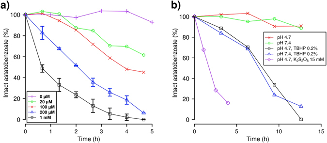
Influence of oxidants on the deastatination of ethyl 3-astatobenzoates. The proportion of intact ethyl 3-astatobenzoate is assessed by reverse-phase HPLC coupled to a dual-flow cell gamma detection system42. (a) Concentration-dependent deastatination promoted by permanganate. The NaMnO4 concentration is varied between 0 and 1 mM while the pH value is fixed at 4.7 with a phosphate-acetate buffer (50 mM). (b) Effect of peroxodisulfate (purple) and tert-butyl hydroperoxide (TBHP, black and blue) on the ethyl 3-astatobenzoate stability (see text).
Figure 1a clearly evidences that, at pH = 4.7 (an average pH value for lysosomes), the presence of ions leads to the deastatination of astatobenzoates, in a concentration-dependent way. This demonstrates for the first time that astatobenzoate conjugates can be altered by oxidative dehalogenation, contrarily to what was previously thought43. By contrast, no discernible deiodination is observed after 12 h incubation of ethyl 3-iodobenzoate (1b) in the presence of 2 mM NaMnO4 (not shown). Figure 1b also shows that some peroxides (tert-butyl hydroperoxide, referred to as “TBHP” and peroxodisulfate) are also able to induce deastatination at the same pH value. This is particularly interesting since the most notorious oxidants occurring in vivo are the ROS, among which peroxides can be found. Also, one should note that oxidative dehalogenation induced by TBHP occurs at physiological pH, i.e. 7.4, with similar kinetics as observed at pH = 4.7.
Oxidative dehalogenation of astatobenzoate conjugates induced by Fenton and Fenton-like conditions
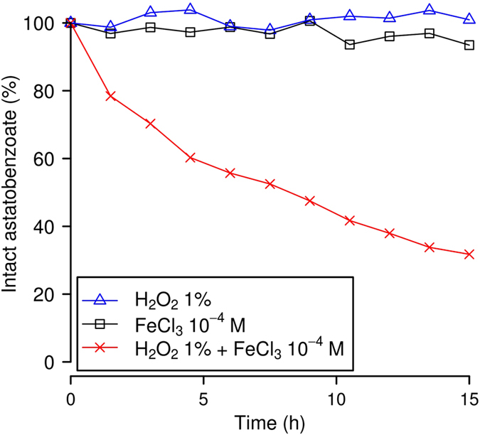
The most common in vivo ROS, namely hydrogen peroxide (H2O2), does not promote any noticeable deastatination (see Fig. 2). By contrast, the combination of catalytic amount of ferrous iron with hydrogen peroxide (and other peroxides as well) was proven to be a powerful oxidant more than 120 years ago44. While radicals were not known at that time, it is now well-established that this combination produces hydroxyl radicals, which are highly reactive species45. When astatobenzoate conjugates are incubated under Fenton conditions (50 mM of the phosphate-acetate buffer − pH ≈ 3, 10−4 M of Fe2+ ions, and 1% H2O2), they undergo an extremely fast deastatination: within the few tens of seconds needed for the HPLC injection, most of the astatobenzoate moieties were dehalogenated, and the majority of the activity was found in a peak having a retention time that appears to match the one of an oxygen adduct of astatine (in particular an At (III) species).
Figure 2.

Influence of Fenton-like conditions on the deastatination of the 1a astatobenzoate. Amounts of intact ethyl 3-astatobenzoate are assessed by reverse-phase HPLC coupled to a dual-flow cell gamma detection system42.
Catalytic amounts of ferric iron (i.e. trivalent iron instead of divalent) coupled with hydrogen peroxide are also known to produce hydroxyl radicals, but at a much slower rate45. These conditions are often referred to as “Fenton-like”. The dehalogenation kinetics of ethyl 3-astatobenzoate (1a) incubated under Fenton-like conditions is displayed on Fig. 2. Note that, for illustration purposes, the radiochromatograms obtained after 3 h with 1% H2O2 and 1% H2O2 plus 10−4 M of Fe3+ ions are displayed in Fig. S1. These experimental results prove that astatobenzoate conjugates are sensitive to oxidation via the Fenton reaction, that is actually at play in lysosomes46, 47. They also indicate that the released astatine species should be an oxygen adduct, but they do not indicate how and why oxidation leads to deastatination. One should note that this part of the study has been performed at pH = 3 to favour the efficiency of the Fenton reaction in order to probe the effect of the presence of hydroxyl radicals (which are produced in lysosomes) on the dehalogenation mechanism(s). Furthermore, one should note that no discernible deiodination of ethyl 3-iodobenzoate (1b) is observed after 12 h incubation under Fenton-like conditions. In order to gain more insight on the oxidative dehalogenation mechanism(s) and on the differences between the C−At and C−I bonds of interest, a quantum mechanical study was performed.
Accuracy of the computational approach
As no spectroscopic information can be obtained at ultratrace concentrations, it is necessary to consider quantum mechanical calculations to get any “microscopic” information on the deastatination mechanism(s). It is of particular importance to treat spin-orbit coupling (SOC), since this relativistic interaction has a strong influence on the geometries and properties of At compounds40, 48. The B3LYP hybrid exchange-correlation functional was selected, owing to its “safe choice” label for investigating astatine species48. Before studying the bond energies of interest, it was worth checking the validity of the used level of theory on well-known systems for which experimental data are available49. In the case of astatobenzene and iodobenzene, the correct dissociation limit is the homolytic one (i.e. radical fission, A − B → A· + B·), and test calculations confirmed that this dissociation limit is found to be favoured by more than a hundred kcal.mol−1 compared to ionic limits at the considered level of theory.
A good agreement between the experimental dissociation energies of astatobenzene and iodobenzene (44.9 ± 5.1 and 61.1 ± 4.7 kcal.mol−1, respectively)49 and the computed ones (44.7 and 59.6 kcal.mol−1, respectively) was obtained. Furthermore, first ionisation potentials (IP 1s) were computed as a model descriptor for the oxidation propensity of halobenzoates. We obtained an IP 1 value of 195.5 kcal.mol−1 for iodobenzene, which fits well with the experimental value of 201.3 ± 0.7 kcal.mol−1 50. All these results provide a firm ground to the used level of theory prior to starting the study of the oxidation of halobenzoate conjugates. Note that for the interested reader, additional calculations to illustrate the importance of SOC on the computed quantities are reported in Tables S1 and S2. Since reliable values can only be obtained when SOC is accounted for, we only report in the main text results that do include this relativistic interaction.
Probing the sensitivity to oxidation of halobenzoates: first ionisation potentials
To probe the relative feasibility of the oxidation of 1a and 1b, we first computed their IP 1s. At first, we checked that these IP 1s were indeed related to electron removal at the halogen moiety of halobenzoates by computing the condensed-to-atom Fukui index, a quantity that resides in the realm of conceptual DFT. This index is computed through a finite difference approximation to the so-called Fukui function; for an electrophilic attack, it has the f k − = q k(N) − q k(N − 1) form, where k is an atom, q k(N) is the electron population of the k atom in the neutral system (N electrons) and q k(N − 1) is the electron population of the k atom in the ionized system (N − 1 electrons). Note that the electron populations are obtained in the present work from natural population analyses. We found that the condensed-to-atom Fukui index, f − 51, is not only maximum for At in 1a, and for I in 1b, but is also at least four times larger for At or I, respectively, than for any other atom of the system. The corresponding f − values are 0.7 and 0.5, meaning that the removed electron is more than 50% localized on the halogen moiety in each case.
It appears that the iodobenzoate compound 1b presents a higher IP 1 (196.2 kcal.mol−1) than the one computed for astatobenzoate 1a (185.8 kcal.mol−1). The important difference, 10.4 kcal.mol−1, makes 1a actually significantly easier to oxidize than its iodine counterpart. Indeed, the previous difference is greater than the one between iodobenzene and bromobenzene, and even the one between iodobenzene and chlorobenzene (5.8 and 7.8 kcal.mol−1 according to the experimental IP 1 s)50. Thus, our calculations support the fact that astatobenzoates should be more prone to oxidation than their iodinated counterparts, since the first conceptual step of any potential oxidation, i.e. the withdrawal of one electron, is more favourable in the X = At case. Therefore, since we do not aim at fully elucidating the oxidation mechanism(s), we continue by directly studying the consequences of oxidation on the C−X bonds of interest.
The effect of oxidation on the C−X bond dissociation energies
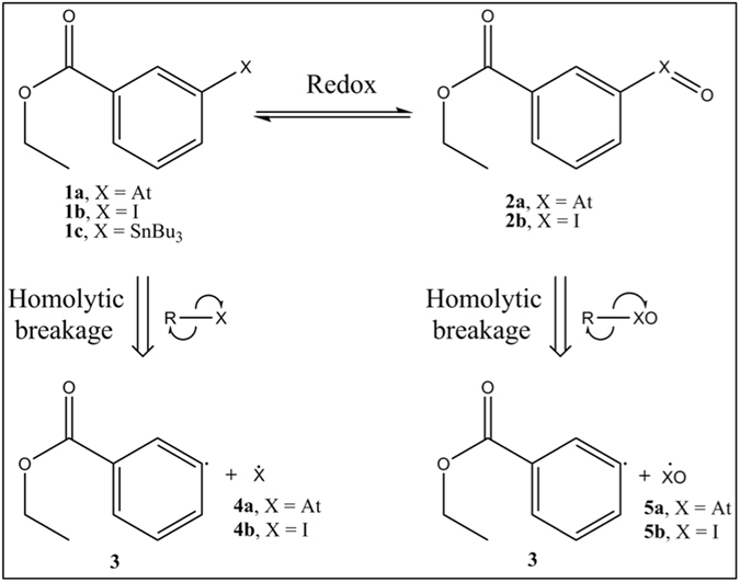
As the immediate product of deastatination resulting from the Fenton reaction is attributed to an oxygen adduct of astatine, and since the 2b species is known to exist, it seems reasonable to study the dissociation energies of the 2a and 2b compounds (see Fig. 3), where the halogen atoms formally bear a +III oxidation state, and to compare them with the ones of 1a and 1b. The bond dissociation energies are calculated considering the most favourable process, i.e a homolytic cleavage, by subtracting the energies of the two radical products (see Fig. 3), from the energy of the whole molecule. The obtained numerical results are displayed in Table 1.
Figure 3.

Scheme to assess the effect of oxidation on the C−X bond dissociation energies of halobenzoate compounds (X = At, I).
Table 1.
C−X bond dissociation energies (kcal.mol−1) of halobenzoates (X = At, I).
| Compound | DFT |
|---|---|
| 1a | 44.6 |
| 2a | 28.2 |
| 1b | 59.4 |
| 2b | 37.8 |
By comparing the results obtained for 1a and 1b with the ones of the corresponding halobenzenes (44.7 and 59.6 kcal.mol−1, respectively), one can first observe that the C−At and C−I bond energies are almost unchanged. Much more enthralling is the comparison between halobenzoates and their oxidized counterparts, as the bond dissociation energies drop in 2a and 2b by more than a third compared to 1a and 1b, respectively, e.g. diminishing the C−At bond energy from 44.6 to 28.2 kcal.mol−1. While these energy decreases are of course noticeable, their significance may be further assessed with transition state theory in these undoubtedly kinetically controlled systems. As test calculations showed no energy barrier on the potential energy surfaces corresponding to the homolytic dissociations of the C−X bonds in 2a and 2b, as well as in 1a and 1b, these bond breakages are kinetically controlled by the corresponding bond dissociation energies. According to the Eyring equation52, the ratio between the bond breakages rates in 2a and 2b is affected by the following temperature-dependent factor:
| 1 |
where Ed(2b) and Ed(2a) are the bond dissociation energies of 2b and 2a, respectively, R is the universal gas constant and T is the temperature. Hence, the 9.6 kcal.mol−1 difference between Ed(2b) and Ed(2a) leads to a dissociation rate in 2a larger by a factor of roughly 6 × 10⁶ at 37 °C (human body temperature) than in 2b, which could explain the iodobenzoate relative stabilities towards oxidizing conditions compared to the astatobenzoate ones. Indeed, the iodosobenzoates will not only be produced much less efficiently than their astatinated counterparts, but also the astatosobenzoates are more prone to homolytic dehalogenation, unlike the iodosobenzoates for which the halogen will be more efficiently reduced back while proteins are oxidized53. On the other hand, the 16.4 kcal.mol−1 difference between Ed(1a) and Ed(2a) leads at 37 °C to an impressive relative increase of the dissociation rate for 2a, by a factor of roughly 4 × 10¹¹ with respect to 1a. This difference is compatible with stable astatobenzoates in blood, given its antioxidant protections54, and efficient dehalogenation of astatosobenzoates after the oxidation of astatine to its +II oxidation state.
Discussion
We report the first experiments of astatobenzoate dehalogenations, and shed light on the probable in vivo mechanism by which these therapeutically relevant compounds are catabolized. We propose that the in vivo C−At bond cleavage occurs through oxidative dehalogenation of the astatobenzoate moiety. This would explain the stability of the labelled carriers in blood, where the conjugates are protected from oxidative dehalogenation, notably by the thiolates of the human serum albumin (≈43 g.L−1) in its mercaptalbumin form, and by the strong antioxidants properties of erythrocytes54. Also, no strong oxidant is known to be present in blood. On the other hand, after internalisation in cells, the halobenzoates moieties would no longer be shielded from encountering strong oxidants.
We propose reactions with ROS in lysosomes as the most likely path to oxidation. Indeed, lysosomes are the organelles that are responsible for protein degradation, and will be met in the first step of the carrier catabolism. They are known to be acidic (with average pH values ranging from 4.5 to 5)46 and more oxidizing than other subcellular organelles55, 56. However, the overall oxidation level is not the only parameter at play. Indeed, strong oxidants and reductants coexist in the lysosomes microdomains56, 57. Similarly, the pH value experiences strong local variations, notably in the strict vicinity of proton pumps. Thus, when catabolized, the astatobenzoate conjugates will be exposed to strong oxidants, typically under acidic conditions in lysosomes. We have shown that the most common in vivo ROS, i.e. hydrogen peroxide (H2O2), does not intrinsically promote any noticeable deastatination. When coupled with ferrous (Fenton conditions) or even of ferric (“Fenton-like” conditions) ions, it promotes a fast cleavage of the C−At bond. It is thus clear that astatobenzoates undergo an extremely fast deastatination in the presence of hydroxyl radicals, ROS known to exist in lysosomes as products of the Fenton reaction. Indeed, due to the degradation of iron-containing macromolecules, many lysosomes are rich in redox-active iron compounds, which results in Fenton-type reactions in these organelles46, 47. The ubiquity of lysosomes in mammalian cells is also consistent with the observed deastatination after cell internalisation regardless of the nature of the cells.
Alternatively, another possible oxidation path for halobenzoates is worth mentioning. Indeed, P-450 cytochromes (CYPs) catalyse the oxidation of iodobenzene into iodosobenzene53, akin to the conversion of 1b into 2b. Moreover, it has also been shown, within the halobenzene series, that, the heavier the halogen, the easier it is for CYPs to oxidize it ref. 58. Therefore, it seems possible that CYPs catalyses the oxidation of astatobenzoates as well. Note that even though iodosobenzene is formed in vivo, it could not be abundant, as it is reduced while it oxidizes proteins53, and that no important dehalogenation of the iodosobenzene has been reported. It is therefore possible that following carrier metabolization, the astatobenzoates conjugates are released in the blood circulation, then captured by the liver, and later undergo oxidation catalysed by CYPs, which ultimately yields to deastatination.
The oxidative dehalogenation hypothesis nicely meets the criteria we proposed: deastatination by oxidation has been proven to be easily doable in the absence of any enzyme; it explains why the astatobenzoate conjugate stabilities must be very different in blood, where they are protected from oxidation, and within lysosomes where they are exposed to Fenton conditions (or alternatively after catabolization of the carrier and oxidation by CYPs in the liver). It is also consistent with the observed stability of the corresponding iodobenzoates under the same in vivo conditions: they are stable under conditions that are sufficient to provoke deastatination.
Besides experimental evidences and a proposed in vivo mechanism, we also aimed at giving an insight of the oxidative deastatination process at the molecular level, especially in regard to iodinated analogues of astatobenzoates. We hypothesized that the difference in in vivo stability between iodobenzoate- and astatobenzoate-labelled proteins with respect to dehalogenation is due to (i) the different sensitivities of the At and I atoms toward oxidation and (ii) the difference in the C−X bond strengths in the oxidized compounds. A plausible scenario is oxidative dehalogenation in which the At atom is oxidized to its +III oxidation state, which weakens enough the C−At bond and eventually leads to its breakage. Our DFT calculations show that it is much easier to start oxidizing astatobenzoates than their iodinated counterparts (by 10.4 kcal.mol−1 according to the calculated first ionization potentials). We also show that this oxidation results in a vast decrease of the C−X bond dissociation energies, illustrated by a drop of the C−At bond energy in the astatobenzoate from 44.6 to 28.2 kcal.mol−1. Finally, to link this quantity to a more in vivo relevant one, we provide a rough estimate of the kinetic enhancement of the homolytic cleavage rate, showing that the reaction should be accelerated by a factor of about 4 × 10¹¹ (a difference greater than the one between decades and milliseconds).
Finally, we deem that our results could be of interest to the conception of innovative 211At-labelling agents, particularly in stimulating new ideas concerning their design and screening. Indeed, just as knowledge of the deiodinase involvement was a necessary step prior to designing SIB, our research demonstrates which types of mechanism should be inhibited for obtaining stable in vivo labelling with 211At. Also, to speed up the quest for new compounds, as the in vivo experiments are costly, labour intensive, and time-consuming, we suggest that prior in silico screenings based on relativistic DFT calculations must be undertaken given both the accuracy and the cost-effective favour of this approach.
Methods
Radiolabelling
The synthesis of 1a was done following the previously described methods for the synthesis of SAB59, 60. Briefly, to 10 µL of acetic acid were added 25 µL of 2 mg/mL of N-chlorosuccinimide in methanol and 1,1 mg of ethyl 3-(tri-n-butylstannyl) benzoate (1c) in 25 µL of methanol in an HPLC vial. Then 50 µL of 211At in chloroform were added (roughly corresponding to 5–10 MBq of activity). After 20 min incubation, 1a was purified by HPLC using a Dionex Ultimate3000 HPLC device with an Interchrom C18 column piloted by the Chromeleon 6.80 software (ThermoFisher Scientific Inc.). It was coupled with a dual-flow cell gamma detection system42 using a γ-ray detector (raytest GABI Star) piloted by the Gina software (raytest Isotopenmeßgeräte GmbH).
Kinetics of deastatination
1.4 mL samples of 1a (≃2–4 MBq of activity) were incubated in various media at 20 °C directly in the HPLC apparatus (same as described above), and 50 µL samples were injected onto column for analysis. The HPLC sequence was the following: 20 s of 100% acetonitrile (ACN) at 1.05 mL.min−1, a 5 s gradient decrease from 100 to 0% ACN, 65 s of 0% ACN, a 90 s gradient from 0 to 60% ACN, a 720 s gradient from 60 to 72% ACN during which the flow is increased from 1.05 mL.min−1 to 1.3 mL.min−1 during 90 s after the first 6o s of the gradient, a 60 s gradient from 72 to 100% ACN, 300 s of 100% ACN, a 60 s gradient from 100 to 0% ACN during which the flow is decreased from 1.3 mL.min−1 to 1.05 mL.min−1 and 150 s of 0% CAN for a total run duration of 24.5 min. Two subsequent injections of 50 µL of Na2S2O3 (50 mM) and of NaMnO4 (2 mM) were run in a “short run mode” between two kinetic points to wash out any potential residual activity. The short run program consisted of 1 min of 100% H2O, 1 min of 100% H2O to 100% of CH3CN (gradient) and 2 min of 100% CH3CN. The sums of the counts in the “intact” astatobenzoate peak were corrected by the intrinsic decay of 211At (considering its 7.21 h half-life time).
Computational methods
All the calculations have been performed in gas phase. The two-component (2c) DFT methods61 relying on relativistic effective core potentials (namely ECP28MDF and ECP60MDF for I62 and At63, respectively) and implemented in the NWChem64 and Turbomole65 program packages were used. The hybrid B3LYP exchange-correlation functional66 was selected, according to results of a recent benchmark study led on At compounds48. For treating the 25 valence electrons on both heavy atoms, we have selected triple zeta basis sets supplemented with 2c extensions, referred to as aug-cc-pVTZ-PP-2c 62, 63, 67. The aug-cc-pVTZ basis sets68, 69 have been used for the remaining atoms (C, H and O).
Electronic supplementary material
Acknowledgements
D.T. is thankful to Michel Chérel for helpful discussions. This work has been supported by grants funded by the French National Agency for Research with “Investissements d’Avenir” (ANR-11-EQPX-0004, ANR-11-LABX-0018). This work was performed using HPC resources from CCIPL (“Centre de Calcul Intensif des Pays de la Loire”).
Author Contributions
D.T. and G.M. conceived and performed the experimental study. D.T., D.-C.S., N.G. and R.M. conceived and performed the computational studies. All authors jointly discussed the results and their interpretations and participated in writing the manuscript.
Competing Interests
The authors declare that they have no competing interests.
Footnotes
Electronic supplementary material
Supplementary information accompanies this paper at doi:10.1038/s41598-017-02614-2
Publisher's note: Springer Nature remains neutral with regard to jurisdictional claims in published maps and institutional affiliations.
Contributor Information
David Teze, Email: david.teze@gmail.com.
Rémi Maurice, Email: remi.maurice@subatech.in2p3.fr.
References
- 1.Corson DR, Mackenzie KR, Segre E. Astatine: the element of Atomic Number 85. Nature. 1947;159:24–24. doi: 10.1038/159024b0. [DOI] [Google Scholar]
- 2.Zalutsky MR, Reardon DA, Pozzi OR, Vaidyanathan G, Bigner DD. Targeted alpha-particle radiotherapy with 211At-labeled monoclonal antibodies. Nucl. Med. Biol. 2007;34:779–85. doi: 10.1016/j.nucmedbio.2007.03.007. [DOI] [PMC free article] [PubMed] [Google Scholar]
- 3.Vaidyanathan G, Zalutsky M. Applications of 211At and 223Ra in Targeted Alpha-Particle Radiotherapy. Curr Radiopharm. 2011;4:283–294. doi: 10.2174/1874471011104040283. [DOI] [PMC free article] [PubMed] [Google Scholar]
- 4.Zalutsky MR, et al. Clinical experience with alpha-particle emitting 211At: treatment of recurrent brain tumor patients with 211At-labeled chimeric antitenascin monoclonal antibody 81C6. J. Nucl. Med. 2008;49:30–8. doi: 10.2967/jnumed.107.046938. [DOI] [PMC free article] [PubMed] [Google Scholar]
- 5.Andersson H, et al. Intraperitoneal alpha-particle radioimmunotherapy of ovarian cancer patients: pharmacokinetics and dosimetry of 211At-MX35 F(ab’)2-a phase I study. J. Nucl. Med. 2009;50:1153–60. doi: 10.2967/jnumed.109.062604. [DOI] [PubMed] [Google Scholar]
- 6.Aneheim E, et al. Automated astatination of biomolecules–a stepping stone towards multicenter clinical trials. Sci. Rep. 2015;5:1–11. doi: 10.1038/srep12025. [DOI] [PMC free article] [PubMed] [Google Scholar]
- 7.Steffen A-C, et al. Biodistribution of 211At labeled HER-2 binding affibody molecules in mice. Oncol. Rep. 2007;17:1141–1147. [PubMed] [Google Scholar]
- 8.Vaidyanathan G, Zalutsky MR. Astatine Radiopharmaceuticals: Prospects and Problems. Curr Radiopharm. 2008;1:1–42. doi: 10.2174/1874471010801010001. [DOI] [PMC free article] [PubMed] [Google Scholar]
- 9.Larsen RH, Slade S, Zalutsky MR. Blocking [211At]Astatide Accumulation in Normal Tissues: Preliminary Evaluation of Seven Potential Compounds. Nucl. Med. Biol. 1998;25:351–357. doi: 10.1016/S0969-8051(97)00230-8. [DOI] [PubMed] [Google Scholar]
- 10.Hadley SW, Wilbur SD, Gray MA, Atcher RW. Astatine-211 Labeling of an Antimelanoma Antibody and Its Fab Fragment Using N-Succinimidyl p-Astatobenzoate: Comparisons in Vivo with the p-[125I]Iodobenzoyl Conjugate. Bioconjugate Chem. 1991;2:171–179. doi: 10.1021/bc00009a006. [DOI] [PubMed] [Google Scholar]
- 11.Garg PK, Harrison CL, Zalutsky MR. Comparative Tissue Distribution in Mice of the α-Emitter 211At and 131I as Labels of a Monoclonal Antibody and F(ab′)2 Fragment. Cancer Res. 1990;50:3514–3520. [PubMed] [Google Scholar]
- 12.Persson, M. I. et al. Astatinated trastuzumab, a putative agent for radionuclide immunotherapy of ErbB2-expressing tumours. Oncol. Rep. 15, 673–80, doi:10.3892/or.15.3.673 (2006). [DOI] [PubMed]
- 13.Gustafsson AME, et al. Comparison of therapeutic efficacy and biodistribution of 213Bi- and 211At-labeled monoclonal antibody MX35 in an ovarian cancer model. Nucl. Med. Biol. 2012;39:15–22. doi: 10.1016/j.nucmedbio.2011.07.003. [DOI] [PubMed] [Google Scholar]
- 14.Zalutsky MR, Stabin MG, Larsen RH, Bigner DD. Tissue distribution and radiation dosimetry of astatine-211-labeled chimeric 81C6, an alpha-particle-emitting immunoconjugate. Nucl. Med. Biol. 1997;24:255–61. doi: 10.1016/S0969-8051(97)00060-7. [DOI] [PubMed] [Google Scholar]
- 15.Wilbur DS, et al. Reagents for Astatination of Biomolecules. 2. Conjugation of Anionic Boron Cage Pendant Groups to a Protein Provides a Method for Direct Labeling that is Stable to in Vivo Deastatination. Bioconjugate Chem. 2007;18:1226–1240. doi: 10.1021/bc060345s. [DOI] [PubMed] [Google Scholar]
- 16.Orlova A, Wållberg H, Stone-Elander S, Tolmachev V. On the selection of a tracer for PET imaging of HER2-expressing tumors: direct comparison of a 124I-labeled affibody molecule and trastuzumab in a murine xenograft model. J. nucl. med. 2009;50:417–25. doi: 10.2967/jnumed.108.057919. [DOI] [PubMed] [Google Scholar]
- 17.Bolton AE, Hunter WM. The labelling of proteins to high specific radioactivities by conjugation to a 125I-containing acylating agent. Biochem. J. 1973;133:529–39. doi: 10.1042/bj1330529. [DOI] [PMC free article] [PubMed] [Google Scholar]
- 18.Zalutsky R, Narula AS. A Method for the Radiohalogenation of Proteins Resulting in Decreased Thyroid Uptake of Radioiodine. Appl. Radiat. Isot. 1987;38:1051–1055. doi: 10.1016/0883-2889(87)90069-4. [DOI] [PubMed] [Google Scholar]
- 19.Vaidyanathan G, Zalutsky MR. Preparation of N -succinimidyl 3-[*I]iodobenzoate: an agent for the indirect radioiodination of proteins. Nat. Protoc. 2006;1:707–713. doi: 10.1038/nprot.2006.99. [DOI] [PubMed] [Google Scholar]
- 20.Friedman JE, Watson JA, Rokita SE. Iodotyrosine Deiodinase Is the First Mammalian Member of the NADH Oxidase/Flavin Reductase Superfamily. J. Biol. Chem. 2006;281:2812–2819. doi: 10.1074/jbc.M510365200. [DOI] [PubMed] [Google Scholar]
- 21.Thomas SR, Mctamney PM, Adler JM, Laronde-leblanc N, Rokita SE. Crystal Structure of Iodotyrosine Deiodinase, a Novel Flavoprotein Responsible for Iodide Salvage in Thyroid Glands. J. Biol. Chem. 2009;284:19659–19667. doi: 10.1074/jbc.M109.013458. [DOI] [PMC free article] [PubMed] [Google Scholar]
- 22.Wilbur DS, et al. Biotin reagents in antibody pretargeting. 6. Synthesis and in vivo evaluation of astatinated and radioiodinated aryl- and nido-carboranyl-biotin derivatives. Bioconjug. Chem. 2004;15:601–616. doi: 10.1021/bc034229q. [DOI] [PubMed] [Google Scholar]
- 23.Chen Y, et al. Durable donor engraftment after radioimmunotherapy using alpha- emitter astatine-211 – labeled anti-CD45 antibody for conditioning in allogeneic hematopoietic cell transplantation. Blood. 2012;119:1130–1138. doi: 10.1182/blood-2011-09-380436. [DOI] [PMC free article] [PubMed] [Google Scholar]
- 24.Orozco JJ, et al. Anti-CD45 radioimmunotherapy using the alpha-emitting radionuclide 211At combined with bone marrow transplantation prolongs survival in a disseminated murine leukemia model. Blood. 2013;120:3759–3768. doi: 10.1182/blood-2012-11-467035. [DOI] [PMC free article] [PubMed] [Google Scholar]
- 25.Green DJ, et al. Astatine-211 conjugated to an anti-CD20 monoclonal antibody eradicates disseminated B-cell lymphoma in a mouse model. Blood. 2015;125:2111–2119. doi: 10.1182/blood-2014-11-612770. [DOI] [PMC free article] [PubMed] [Google Scholar]
- 26.Wilbur DS, Chyan M, Hamlin DK, Nguyen H, Vessella RL. Reagents for Astatination of Biomolecules. 5. Evaluation of Hydrazone Linkers in 211At- and 125I-Labeled closo-Decaborate(2-) Conjugates of Fab as a Means of Decreasing Kidney Retention. Bioconjugate Chem. 2011;22:1089–1102. doi: 10.1021/bc1005625. [DOI] [PMC free article] [PubMed] [Google Scholar]
- 27.Orlova A, et al. Targeting Against Epidermal Growth Factor Receptors. Cellular Processing of Astatinated EGF After Binding to Cultured Carcinoma Cells. Anticancer Res. 2004;24:4035–4041. [PubMed] [Google Scholar]
- 28.Rothe S, et al. Measurement of the first ionization potential of astatine by laser ionization spectroscopy. Nat. Commun. 2013;4:1–6. doi: 10.1038/ncomms2819. [DOI] [PMC free article] [PubMed] [Google Scholar]
- 29.Wilbur DS. Enigmatic astatine. Nat. Chem. 2013;5:246–246. doi: 10.1038/nchem.1580. [DOI] [PubMed] [Google Scholar]
- 30.Carlin S, Mairs RJ, Welsh P, Zalutsky MR. Sodium-iodide symporter (NIS)-mediated accumulation of [211At]astatide in NIS-transfected human cancer cells. Nucl. Med. Biol. 2002;29:729–739. doi: 10.1016/S0969-8051(02)00332-3. [DOI] [PubMed] [Google Scholar]
- 31.Carlin S, Akabani G, Zalutsky MR. In Vitro Cytotoxicity of 211At-Astatide and 131I-Iodide to Glioma Tumor Cells Expressing the Sodium/Iodide Symporter. J. Nucl. Med. 2003;44:1827–1838. [PubMed] [Google Scholar]
- 32.Ayed T, et al. 211At-labeled agents for alpha-immunotherapy: On the in vivo stability of astatine-agent bonds. Eur. J. Med. Chem. 2016;116:156–164. doi: 10.1016/j.ejmech.2016.03.082. [DOI] [PubMed] [Google Scholar]
- 33.Vernon RE. Which Elements Are Metalloids? J. Chem. Educ. 2013;90:1703–1707. doi: 10.1021/ed3008457. [DOI] [Google Scholar]
- 34.Hermann A, Hoffmann R, Ashcroft NW. Condensed astatine: Monatomic and metallic. Phys. Rev. Lett. 2013;111:1–5. doi: 10.1103/PhysRevLett.111.116404. [DOI] [PubMed] [Google Scholar]
- 35.Sergentu D-C, et al. Advances on the Determination of the Astatine Pourbaix Diagram: Predomination of AtO(OH)2− over At− in Basic Conditions. Chem. Eur. J. 2016;22:2964–2971. doi: 10.1002/chem.201504403. [DOI] [PubMed] [Google Scholar]
- 36.Champion J, et al. Astatine Standard Redox Potentials and Speciation in Acidic Medium. J. Phys. Chem. A. 2010;3:576–582. doi: 10.1021/jp9077008. [DOI] [PubMed] [Google Scholar]
- 37.Tigeras A, Bachet M, Catalette H, Simoni E. PWR iodine speciation and behaviour under normal primary coolant conditions: An analysis of thermodynamic calculations, sensibility evaluations and NPP feedback. Prog. Nucl. Energy. 2011;53:504–515. doi: 10.1016/j.pnucene.2011.02.002. [DOI] [Google Scholar]
- 38.Garg PK, Alston KL, Zalutsky MR. Catabolism of Radioiodinated Murine Monoclonal Antibody F(ab’)2 Fragment Labeled Using N-Succinimidyl 3-Iodobenzoate and Iodogen Methods. Bioconjugate Chem. 1995;6:493–501. doi: 10.1021/bc00034a020. [DOI] [PubMed] [Google Scholar]
- 39.Champion J, et al. Investigation of Astatine(III) Hydrolyzed Species: Experiments and Relativistic Calculations. J. Phys. Chem. A. 2013;117:1983–1990. doi: 10.1021/jp3099413. [DOI] [PubMed] [Google Scholar]
- 40.Champion J, et al. Assessment of an effective quasirelativistic methodology designed to study astatine chemistry in aqueous solution. Phys. Chem. Chem. Phys. 2011;13:14984–14992. doi: 10.1039/c1cp20512a. [DOI] [PubMed] [Google Scholar]
- 41.Guo N, et al. The Heaviest Possible Ternary Trihalogen Species, IAtBr −, Evidenced in Aqueous Solution: An Experimental Performance Driven by Computations. Angew. Chemie Int. Ed. 2016;55:15369–15372. doi: 10.1002/anie.201608746. [DOI] [PubMed] [Google Scholar]
- 42.Lindegren S, Jensen H, Jacobsson L. A radio-high-performance liquid chromatography dual-flow cell gamma-detection system for on-line radiochemical purity and labeling efficiency determination. J. Chromatogr. A. 2014;1337:128–132. doi: 10.1016/j.chroma.2014.02.043. [DOI] [PubMed] [Google Scholar]
- 43.Milius RA, et al. Organoastatine Chemistry. Astatination via Electrophilic Destannylation. Appl. Rad. Isot. 1986;37:799–802. doi: 10.1016/0883-2889(86)90274-1. [DOI] [PubMed] [Google Scholar]
- 44.Fenton HJH. Oxidation of tartaric acid in presence of iron. J. Chem. Soc., Trans. 1894;65:899–910. doi: 10.1039/CT8946500899. [DOI] [Google Scholar]
- 45.Walling C. Fenton’s reagent revisited. Acc. Chem. Res. 1975;8:125–131. doi: 10.1021/ar50088a003. [DOI] [Google Scholar]
- 46.Kurz T, Terman A, Gustafsson B, Brunk UT. Lysosomes in iron metabolism, ageing and apoptosis. Histochem. Cell Biol. 2008;129:389–406. doi: 10.1007/s00418-008-0394-y. [DOI] [PMC free article] [PubMed] [Google Scholar]
- 47.Lin Y, Epstein DL, Liton PB. Intralysosomal iron induces lysosomal membrane permeabilization and cathepsin D-mediated cell death in trabecular meshwork cells exposed to oxidative stress. Investig. Ophthalmol. Vis. Sci. 2010;51:6483–6495. doi: 10.1167/iovs.10-5410. [DOI] [PMC free article] [PubMed] [Google Scholar]
- 48.Sergentu D, David G, Montavon G, Maurice R, Galland N. Scrutinizing ‘invisible’ astatine: a challenge for modern density functionals. J. Comput. Chem. 2016;37:1–16. doi: 10.1002/jcc.24326. [DOI] [PubMed] [Google Scholar]
- 49.Вашарош Л, Норсеев ЮВ, Халкин ВА. ОПРЕДЕЛЕНИЕ ЭНЕРГИИ РАЗРЫВА ХИМИЧЕСКОЙ СВЯЗИ УГЛЕРОД-АСТАТ. Dokl Akad Nauk SSSR. 1981;263:119–123. [Google Scholar]
- 50.Watanabe K. Ionization Potentials of Some Molecules. J. Chem. Phys. 1957;26:542–547. doi: 10.1063/1.1743340. [DOI] [Google Scholar]
- 51.Chattaraj PK, Sarkar U, Roy DR. Electrophilicity Index. Chem. Rev. 2006;106:2065–2091. doi: 10.1021/cr040109f. [DOI] [PubMed] [Google Scholar]
- 52.Eyring H. The Activated Complex in Chemical Reactions. J. Chem. Phys. 1935;3:107–115. doi: 10.1063/1.1749604. [DOI] [Google Scholar]
- 53.Burka LT, Thorsen A, Guengerich FP. Enzymatic Monooxygenation of Halogen Atoms: Cytochrome P-450 Catalyzed Oxidation of Iodobenzene by Iodosobenzene. J. Am. Chem. Soc. 1980;102:7615–7616. doi: 10.1021/ja00545a062. [DOI] [Google Scholar]
- 54.Turell L, Radi R, Alvarez B. The thiol pool in human plasma: The central contribution of albumin to redox processes. Free Radic. Biol. Med. 2013;65:244–253. doi: 10.1016/j.freeradbiomed.2013.05.050. [DOI] [PMC free article] [PubMed] [Google Scholar]
- 55.Go YM, Jones DP. Redox compartmentalization in eukaryotic cells. Biochim. Biophys. Acta. 2008;1780:1273–1290. doi: 10.1016/j.bbagen.2008.01.011. [DOI] [PMC free article] [PubMed] [Google Scholar]
- 56.Kaludercic N, Deshwal S, Di Lisa F. Reactive oxygen species and redox compartmentalization. Front. Physiol. 2014;5:1–15. doi: 10.3389/fphys.2014.00285. [DOI] [PMC free article] [PubMed] [Google Scholar]
- 57.Kemp M, Go Y, Jones D. Nonequilibrium thermodynamics of thiol/disulfide redox systems: a perspective on redox systems biology. Free Radic. Biol. Med. 2008;44:921–937. doi: 10.1016/j.freeradbiomed.2007.11.008. [DOI] [PMC free article] [PubMed] [Google Scholar]
- 58.Burka LT, Plucinskit T, Macdonaldt TL. Mechanisms of hydroxylation by cytochrome P-450: Metabolism of monohalobenzenes by phenobarbital-induced microsomes. Proc. Natl. Acad. Sci. 1983;80:6680–6684. doi: 10.1073/pnas.80.21.6680. [DOI] [PMC free article] [PubMed] [Google Scholar]
- 59.Vaidyanathan G, Zalutsky MR. Preparation of N-succinimidyl 3-[*I]iodobenzoate: an agent for the indirect radioiodination of proteins. Nat. Protoc. 2006;1:707–13. doi: 10.1038/nprot.2006.99. [DOI] [PubMed] [Google Scholar]
- 60.Pozzi OR, Zalutsky MR. Radiopharmaceutical chemistry of targeted radiotherapeutics, Part 3: alpha-particle-induced radiolytic effects on the chemical behavior of 211At. J. Nucl. Med. 2007;48:1190–6. doi: 10.2967/jnumed.106.038505. [DOI] [PubMed] [Google Scholar]
- 61.Armbruster MK, Weigend F, van Wüllen C, Klopper W. Self-consistent treatment of spin-orbit interactions with efficient Hartree-Fock and density functional methods. Phys. Chem. Chem. Phys. 2008;10:1748–56. doi: 10.1039/b717719d. [DOI] [PubMed] [Google Scholar]
- 62.Peterson KA, Shepler BC, Figgen D, Stoll H. On the spectroscopic and thermochemical properties of ClO, BrO, IO, and their anions. J. Phys. Chem. A. 2006;110:13877–13883. doi: 10.1021/jp065887l. [DOI] [PubMed] [Google Scholar]
- 63.Peterson KA, Figgen D, Goll E, Stoll H, Dolg M. Systematically convergent basis sets with relativistic pseudopotentials. I. Correlation consistent basis sets for the post-d group 13–15 elements. J. Chem. Phys. 2003;119:11099–11112. doi: 10.1063/1.1622923. [DOI] [Google Scholar]
- 64.Valiev M, et al. NWChem: A comprehensive and scalable open-source solution for large scale molecular simulations. Comput. Phys. Commun. 2010;181:1477–1489. doi: 10.1016/j.cpc.2010.04.018. [DOI] [Google Scholar]
- 65.R. Ahlrichs et al. 6.6, a development of University of Karlsruhe and Forschungszentrum Karlsruhe GmbH, TURBOMOLE GmbH (2014).
- 66.Stephen PJ, Devlin FJ, Chabalowski CF, Frisch MJ. Ab Initio Calculation of Vibrational Absorption. J. Phys. Chem. 1994;98:11623–11627. doi: 10.1021/j100096a001. [DOI] [Google Scholar]
- 67.Armbruster MK, Klopper W, Weigend F. Basis-set extensions for two-component spin-orbit treatments of heavy elements. Phys. Chem. Chem. Phys. 2006;8:4862–5. doi: 10.1039/B610211E. [DOI] [PubMed] [Google Scholar]
- 68.Kendall Ra, Dunning TH, Jr., Harrison R. J. Electron affinities of the first-row atoms revisited. Systematic basis sets and wave functions. J. Chem. Phys. 1992;96:6796–6806. doi: 10.1063/1.462569. [DOI] [Google Scholar]
- 69.Dunning TH., Jr. Gaussian basis sets for use in correlated molecular calculations. I. The atoms boron through neon and hydrogen. J. Chem. Phys. 1989;90:1007–1023. doi: 10.1063/1.456153. [DOI] [Google Scholar]
Associated Data
This section collects any data citations, data availability statements, or supplementary materials included in this article.


