

Abstract
Fibroadenoma of the breast is the most common benign neoplasm in young women who present with a palpable, movable mass. Malignancy inside fibroadenomas is rare, with reported rates ranging from 0.002% to 0.125%. Carcinoma in situ inside a fibroadenoma is usually found incidentally when tumours are excised. Whether fibroadenoma is a risk factor for breast cancer remains controversial. Due to the rarity of carcinomas inside fibroadenomas, medical institutes have little experience with this phenomenon. We report an unusual case in which progression occurred from benign fibroadenoma to ductal carcinoma in situ, infiltrating ductal carcinoma and lymph node metastasis. A nipple-areolar complex-preserving mastectomy with immediate breast reconstruction with a gel implant and contralateral augmentation was performed. No local recurrence or metastasis was found during 5 years of follow-up.
INTRODUCTION
Most breast lumps in young female patients are found to be fibroadenomas [1–3], which are usually benign and self-limiting. Asymptomatic fibroadenomas can be conservatively managed [2]. Excision may be needed if the mass enlarges or if other atypical symptoms occur. Cancer within fibroadenoma is usually found incidentally during pathologic examination, with reported incidences ranging from 0.002% to 0.125% [4–6].
Malignant findings inside fibroadenomas are usually carcinoma in situ; <15% are invasive breast cancers [7]. Whether fibroadenoma is a risk factor for breast cancer remains controversial [5, 8–10]. Here, we present the case report of a patient with a long history of fibroadenoma that then progressed during follow-up to ductal carcinoma in situ (DCIS), invasive ductal carcinoma (IDC) and lymph node metastasis. The treatment details are reported along with a literature review.
CASE REPORT
A 31-year-old woman had had a tumour in her left breast for 7 years. A sonogram at the time of initial diagnosis revealed a hypoechoic mass ~1.6 cm in diameter. Fine-needle aspiration revealed no malignant cells. Benign fibroadenoma was diagnosed based on the imaging and cytology results, and the patient received regular follow-up at a local clinic. The breast mass enlarged gradually, and the patient decided to have the tumour excised. She visited Changhua Christian Hospital for treatment. A breast sonogram revealed one hypoechoic tumour with increased cellularity (Fig. 1a). A core needle biopsy (CNB) was performed, and pathologic analysis revealed low-grade DCIS in solid and cribriform patterns in a background of complex fibroadenoma (Fig. 2a).
Figure 1:

(a) Sonogram of the left breast reveals a 3.5-cm hypoechoic lesion with increased vascularity in the 2 o'clock/1-cm region. (b) Mammogram (mediolateral-oblique view) of the left breast shows a well-defined sharply circumscribed mass. (c) A maximal intensity projection magnetic resonance imaging image of the left breast revealed a 4.0-cm well-defined tumour with an engorged drainage vein beneath the nipple areolar complex region.
Figure 2:

(a) Preoperative CNB (low-power field). The upper left tissue shows a complex glandular growth pattern in the fibro-myxomatous stroma. The lower right tissue shows DCIS in solid and cribriform patterns. (b) Postoperative final pathology (high power field). Grade II infiltrating ductal carcinoma characterized by nesting to glandular structure with intermediate-sized nuclei and rare mitotic figures.
Preoperative mammography revealed one well-defined, sharply circumscribed lesion (Fig. 1b). Magnetic resonance imaging (MRI) showed one 4-cm circumscribed mass beneath the nipple-areolar complex (NAC) region of the left breast, with focal haemorrhage, nipple retraction, skin thickening, dilated ducts and lymphadenopathy (Fig. 1c).
Breast conservative surgery was not suggested due to the relatively large tumour in a small- to medium-sized breast and the tumour's location just beneath the NAC. The patient had a strong desire to preserve the NAC and her body image. The results of a sub-nipple frozen biopsy were negative for malignancy. Axillary lymph node dissection was performed because sentinel lymph node biopsy revealed metastatic carcinoma. Endoscopy-assisted nipple-sparing mastectomy was performed with immediate breast reconstruction using a gel implant combined with contralateral augmentation (Fig. 3).
Figure 3:
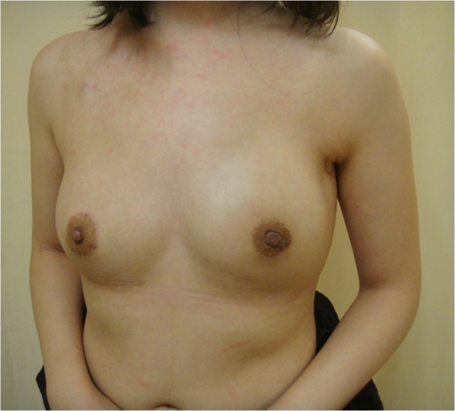
Postoperative view 3 months after nipple-sparing mastectomy with immediate breast reconstruction with a gel implant combined with contralateral augmentation.
The postoperative pathology report revealed IDC with DCIS arising from a complex background of fibroadenoma (Fig. 2b). The stage classification was pT1a (0.5 cm) N1(1/18)M0, Stage IIA. The hormone receptor status was oestrogen receptor–positive and progesterone receptor–positive. The HER-2 receptor was not overexpressed, and the Ki-67 level was 10%. In consideration of the patient's young age (31 years) and lymph node metastasis, she underwent adjuvant chemotherapy with 5-fluorouracil (500 mg/m2), epirubicin (90 mg/m2) and cyclophosphamide (500 mg/m2) (four cycles) followed by docetaxel (75 mg/m2) (four cycles). She is currently undergoing adjuvant endocrine therapy with tamoxifen (20 mg/per day) and regular follow-up at our outpatient clinic. There has been no disease recurrence or distant metastasis.
DISCUSSION
We present the rare case of a patient with a diagnosis of CNB with DCIS arising from fibroadenoma. When the tumour was excised, IDC with lymph node metastasis was found. This unusual case demonstrates the rare transition of benign fibroadenoma to malignant DCIS, invasive carcinoma and then lymph node metastasis.
Whether fibroadenoma is a risk factor for breast cancer remains uncertain, and several reports have discussed this phenomenon [5, 10–13]. Dupont et al. [5] reported that the risk of invasive breast cancer was 2.17 times higher in patients with fibroadenoma and increased to 3.10 times higher in complex fibroadenoma. The risk of malignant change persisted even 20 years after the diagnosis of fibroadenoma. Other studies also provide strong evidence that fibroadenoma with hyperplasia is associated with an increased risk of breast cancer [11, 12]. However, Bonardi et al. [10] reported that the observed transformation of fibroadenoma to breast cancer was due to selection bias. Levi et al. [13] claimed that the evidence was too weak to support the connection of fibroadenoma to subsequent breast cancer.
Imaging studies may suggest characteristics of carcinomas arising from fibroadenomas [14–16]. In sonographic examination of fibroadenomas, irregular shape and contour, extensive hypo-echogenicity, shadowing, echogenic halo and distortion of surrounding tissue are considered to be hints of suspicious malignancy [15]. On mammography, irregular shape, ill-defined spiculated margins or pleomorphic/linear micro-calcifications should also be considered signs of malignancy [14]. With dynamic MRI, benign fibroadenoma and DCIS/IDC can be differentiated according to differences in vascularity [16]. Parameter colour maps can also demonstrate the extent of DCIS within a fibroadenoma [16]. However, in the case described here, sonography, mammography and MRI revealed a well-defined, sharply circumscribed hypoechoic lesion with increased vascularity (Fig. 1a–c). Preoperative imaging to differentiate benign from malignant fibroadenoma was not easy, and CNB was therefore performed to obtain a tissue diagnosis.
The surgical treatment of benign fibroadenoma is to excise the mass. Tumourectomy or lumpectomy may be sufficient if the surgical margin is free of cancer or if only lobular carcinoma in situ is found inside the fibroadenoma [17, 18]. However, if the resection margin is involved or close, further wide excision may be needed [5]. In the current case, because the tumour was large in a relatively small breast, a nipple-sparing mastectomy was performed instead of breast conservative surgery. Due to the usually well-defined capsule-like nature of fibroadenomas [3, 19], malignancy inside a fibroadenoma is often regarded as more benign than other types of breast cancer. Intraoperative sub-nipple biopsy was performed to decide whether the NAC could be preserved [20].
This unusual case involved the transition of benign fibroadenoma to malignant DCIS, IDC and lymph node metastasis. Although rare, the risk of malignancy inside a fibroadenoma should be kept in mind. Adequate action (CNB or excision) should be performed when an enlarging mass or atypical presentation of fibroadenoma is found.
FUNDING
This study was supported by Changhua Christian Hospital Breast Cancer Research Program (105-CCH-IRP-025).
CONFLICT OF INTEREST STATEMENT
None declared.
ACKNOWLEDGEMENTS
The authors would like to thank Ya-Ling Lin for her assistance in this study.
REFERENCES
- 1. Goehring C, Morabia A. Epidemiology of benign breast disease, with special attention to histologic types. Epidemiol Rev 1997;19:310–27. [DOI] [PubMed] [Google Scholar]
- 2. Cant PJ, Madden MV, Coleman MG, Dent DM. Non-operative management of breast masses diagnosed as fibroadenoma. Br J Surg 1995;82:792–4. [DOI] [PubMed] [Google Scholar]
- 3. Foster ME, Garrahan N, Williams S. Fibroadenoma of the breast: a clinical and pathological study. J R Coll Surg Edinb 1988;33:16–9. [PubMed] [Google Scholar]
- 4. Feschenes L, Jacob S, Fabia J, Christen A. Beware of breast fibroadenomas in middle-aged women. Can J Surg 1985;28:372–4. [PubMed] [Google Scholar]
- 5. Dupont WD, Page DL, Parl FF, Vnencak-Jones CL, Plummer WD Jr, Rados MS, et al. Long-term risk of breast cancer in women with fibroadenoma. N Engl J Med 1994;331:10–5. [DOI] [PubMed] [Google Scholar]
- 6. Buzanowski-Konakry K, Harrison e.g. Jr, Payne WS. Lobular carcinoma arising in fibroadenoma of the breast. Cancer 1975;35:450–6. [DOI] [PubMed] [Google Scholar]
- 7. Fukuda M, Nagao K, Nishimura R, Matsuda M, Baba K, Ueno Y, et al. Carcinoma arising in fibroadenoma of the breast: A case report and review of the literature. Jpn J Surg 1989;19:593–6. [DOI] [PubMed] [Google Scholar]
- 8. Worsham MJ, Raju U, Lu M, Kapke A, Botttrell A, Cheng J, et al. Risk factors for breast cancer from benign breast disease in a diverse population. Breast Cancer Res Treat 2009;118:1–7. [DOI] [PMC free article] [PubMed] [Google Scholar]
- 9. Chun J, Pocock B, Joseph KA, El-Tamer M, Klein L, Schnabel F. Breast cancer risk factors in younger and older women. Ann Surg Oncol 2009;16:96–9. [DOI] [PubMed] [Google Scholar]
- 10. Ciatto S, Bonardi R, Zappa M, Giorgi D. Risk of breast cancer subsequent to histological or clinical diagnosis of fibroadenoma: retrospective longitudinal study of 3938 cases. Ann Oncol 1997;8:297–300. [DOI] [PubMed] [Google Scholar]
- 11. McDivitt RW, Stevens JA, Lee NC, Wingo PA, Rubin GL, Gersell D. Histologic types of benign breast disease and the risk for breast cancer. The Cancer and Steroid Hormone Study Group. Cancer. 1992;69:1408–14. [DOI] [PubMed] [Google Scholar]
- 12. Hutchinson WB, Thomas DB, Hamlin WB, Roth GJ, Peterson AV, Williams B. Risk of breast cancer in women with benign breast disease. J Natl Cancer Inst 1980;65:13–20. [PubMed] [Google Scholar]
- 13. Levi F, Randimbison L, Te VC, La Vecchia C. Incidence of breast cancer in women with fibroadenoma. Int J Cancer 1994;57:681–3. [DOI] [PubMed] [Google Scholar]
- 14. Borecky N, Rickard M. Preoperative diagnosis of carcinoma within fibroadenoma on screening mammograms. J Med Imaging Radiat Oncol 2008;52:64–7. [DOI] [PubMed] [Google Scholar]
- 15. Skaane P, Engedal K. Analysis of sonographic features in the differentiation of fibroadenoma and invasive ductal carcinoma. AJR Am J Roentgenol 1998;170:109–14. [DOI] [PubMed] [Google Scholar]
- 16. Kato F, Omatsu T, Matsumura W, Takahashi M, Hosoda M, Takahashi H, et al. Dynamic MR findings of ductal carcinoma in situ within a fibroadenoma. Magn Reson Med Sci 2011;10:129–32. [DOI] [PubMed] [Google Scholar]
- 17. Sarela AI, Madvanur AA, Soonawala ZF, Shah HK, Pandit AA, Samsi AB. Carcinoma in a fibroadenoma. J Postgrad Med 1995;41:19–20. [PubMed] [Google Scholar]
- 18. Rosen PP, Kosloff C, Lieberman PH, Adair F, Braun DW Jr. Lobular carcinoma in situ of the breast: detailed analysis of 99 patients with average follow-up of 24 years. Am J Surg Pathol 1978;2:225–51. [DOI] [PubMed] [Google Scholar]
- 19. Greenberg R, Skornick Y, Kaplan O. Management of breast fibroadenomas. J Gen Intern Med 1998;13:640–5. [DOI] [PMC free article] [PubMed] [Google Scholar]
- 20. Kissin MW, Kark AE. Nipple preservation during mastectomy. Br J Surg 1987;74:58–61. [DOI] [PubMed] [Google Scholar]