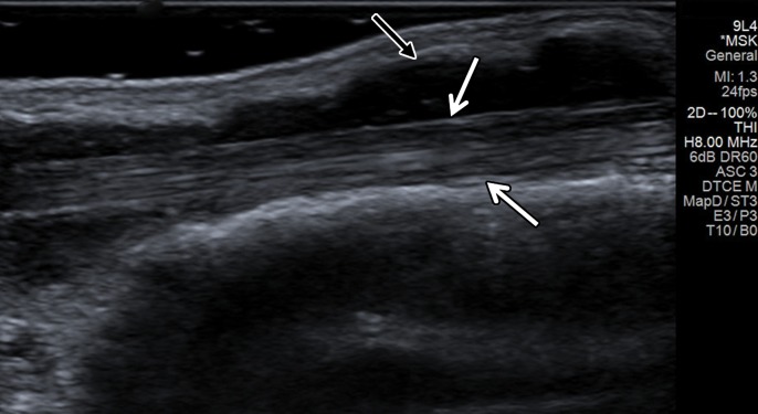
Figure 7a.
Tenosynovitis of the first extensor compartment tendons of the wrist in a 59-year-old woman with a history of rheumatoid arthritis. (a) Long-axis gray-scale US image shows mild hypoechogenicity of the extensor pollicis brevis (EPB) tendon fibers (white arrows) and a mildly heterogeneous synovial-fluid complex in the massively distended tendon sheath (black arrow). (b) Color elastogram of the same region shows predominantly intermediate shear-wave velocities (arrows) in the EPB tendon (mean, 8.65 m/sec) and low shear-wave velocities (mean, 3.04 m/sec) in the region of synovitis (black arrow) within the tendon sheath. SWE data were collected using an Acuson S3000 US scanner with an L9–4-MHz linear transducer. The right side of the images is proximal.

