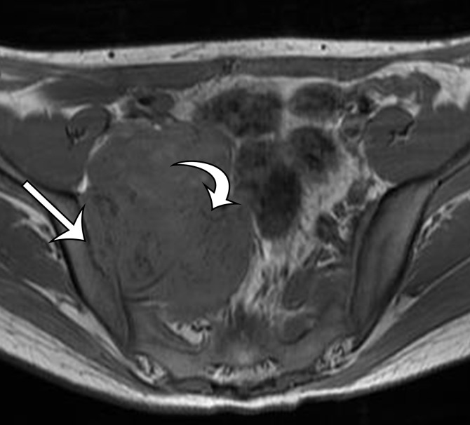
Figure 7c.
Chondrosarcoma in a 21-year-old woman with right lower quadrant pain. (a, b) Axial CT images obtained with soft-tissue window (a) and bone window (b) show a sharply marginated soft-tissue–attenuation mass closely approximating the right iliac wing (arrow on a), with chondroid-type calcifications in rings and arcs (arrows on b). (c, d) Axial T1-weighted (c) and T2-weighted (d) MR images better show the well-defined margins with scalloping of the iliac wing, but without medullary infiltration (straight arrow). Note the hypointense foci of chondroid calcifications (curved arrow).

