Abstract
Melorheostosis is a rare, non-hereditary, benign, sclerotic bone dysplasia with no sex predilection, typically occurring in late childhood or early adulthood which can lead to substantial functional morbidity depending on the sites of involvement. We report on a patient with extensive melorheostotsis in the axial and appendicular skeleton as well as in the soft-tissues, who was evaluated with whole-body 18F-NaF PET/CT scan. All melorheostotic lesions of the skeleton and of the ossified soft-tissue masses, demonstrated intensely increased 18F-NaF activity suggesting the application of this modality in assessing and monitoring the disease activity.
Melorheostosis is a rare, non-heridatary, benign, sclerotic bone-dysplasia of unknown etiology which typically occurs in late childhood or early adulthood [1, 2]. The characteristic radiographic appearance of melorheostosis consists of cortical and medullary hyperostosis of the affected bones, giving the impression of “dripping candle wax” [3, 4]. Melorheostosic lesions in the axial skeleton and soft-tissue ossified masses like in the case presented herein, are rare features of this uncommon disorder [4–6]. The presented data indicates 18F-NaF-avidity of all melorheostotic lesions both in the skeleton and in sites of extra-osseous ossifications, making 18F-NaF the optimal bone-seeking PET-radiopharmaceutical [7–9] to assess and monitor the disease activity.
Figure 1.
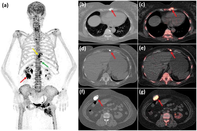
A 67-year-old woman with established diagnosis of melorheostosis since the age of 22 was referred to our institute. Although independent in self-care activities, the patient has been suffering from chronic pain secondary to the melorheostosis with three to five episodes of pain flare-ups per year. The patient developed lymphedema of the left lower extremity and leg length discrepancy with the left leg being longer. In order to assess the disease activity the patient underwent whole body 18F-NaF PET/CT scan, which showed multiple 18F-NaF avid lesions corresponding to the hyperdense bone abnormalities and the soft-tissue ossifications. A prominent lesion with intensely elevated 18F-NaF activity (SUVmax: 45,4) resided in the left lower sternum (Fig. 1a: Maximum Intensity Projection (MIP) PET image of the torso, yellow arrow; Fig 1b & 1c: axial CT and axial fused PET/CT images of the chest, red arrows). Also, two sites of extra-osseous bone formation were observed in the anterior abdominal wall (Fig. 1a: green arrow; Fig 1d & 1e: axial CT and axial fused PET/CT images of the upper abdomen, red arrows) and in the right anterolateral abdominal wall (Fig. 1a: red arrow; Fig 1f & 1g: axial CT and axial fused PET/CT images of the abdomen, red arrows) which also showed markedly elevated 18F-NaFuptake (SUVmax: 30,3 & 42,6 respectively).
Figure 2.
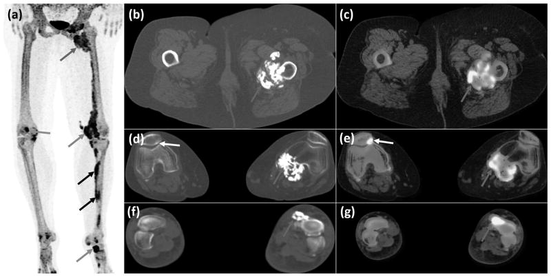
In the lower extremities, abnormal left femoral osteosclerosis with associated bulky soft-tissue ossifications was seen between the left proximal femur and the pelvis (Fig 2a: Maximum Intensity Projection (MIP) PET image of the lower extremities, red arrow; Fig 2b & 2c: axial CT and fused PET/CT images of the femurs, red arrows), with intensely increased 18F-NaF activity (SUVmax: 52,2). Linear elevated 18F-NaF activity was seen along the left femoral shaft as well as in an extra-osseous ossified mass adjacent to the medial femoral condyle (Fig 2a: green arrow; Fig 2d & 2e: axial CT and fused PET/CT images of the knees, red arrows,) with high uptake as well (SUVmax: 51,7). Focal intense uptake (SUVmax: 55,3) in the right patella corresponded to an osteosclerotic lesion (Fig 2a: blue arrow; Fig 2d & 2e: white arrows). Sites of intense 18F-NaF avidity (SUVmax: 52,7) were seen in the medial left tibia (Fig 2a: black arrows) and in the left talor-navicular joint (SUVmax: 56,2) corresponding to melorheostotic lesion observed on CT (Fig 2a: orange arrow; Fig 2f & 2g: axial CT and fused PET/CT images of the feet, red arrows).
Footnotes
Disclosure: All authors have nothing to disclose
References
- 1.Jain VK, Arya RK, Bharadwaj M, et al. Melorheostosis: clinicopathological features, diagnosis, and management. Orthopedics. 2009;32:512. doi: 10.3928/01477447-20090527-20. [DOI] [PubMed] [Google Scholar]
- 2.Faruqi T, Dhawan N, Bahl J, et al. Molecular, phenotypic aspects and therapeutic horizons of rare genetic bone disorders. Biomed Res Int. 2014;2014:670842. doi: 10.1155/2014/670842. [DOI] [PMC free article] [PubMed] [Google Scholar]
- 3.Ihde LL, Forrester DM, Gottsegen CJ, et al. Sclerosing bone dysplasias: review and differentiation from other causes of osteosclerosis. Radiographics. 2011;31:1865–1882. doi: 10.1148/rg.317115093. [DOI] [PubMed] [Google Scholar]
- 4.Suresh S, Muthukumar T, Saifuddin A. Classical and unusual imaging appearances of melorheostosis. Clin Radiol. 2010;65:593–600. doi: 10.1016/j.crad.2010.02.004. [DOI] [PubMed] [Google Scholar]
- 5.Goldman AB, Schneider R, Huvos AS, et al. Case report 778. Melorheostosis presenting as two soft-tissue masses with osseous changes limited to the axial skeleton. Skeletal Radiol. 1993;22:206–210. doi: 10.1007/BF00206157. [DOI] [PubMed] [Google Scholar]
- 6.Yu JS, Resnick D, Vaughan LM, et al. Melorheostosis with an ossified soft tissue mass: MR features. Skeletal Radiol. 1995;24:367–370. doi: 10.1007/BF00197070. [DOI] [PubMed] [Google Scholar]
- 7.Bastawrous S, Bhargava P, Behnia F, et al. Newer PET application with an old tracer: role of 18F-NaF skeletal PET/CT in oncologic practice. Radiographics. 2014;34:1295–1316. doi: 10.1148/rg.345130061. [DOI] [PubMed] [Google Scholar]
- 8.Papadakis GZ, Millo C, Bagci U, et al. 18F-NaF and 18F-FDG PET/CT in Gorham-Stout Disease. Clin. Nucl. Med. 2016;41:884–885. doi: 10.1097/RLU.0000000000001369. [DOI] [PMC free article] [PubMed] [Google Scholar]
- 9.Agrawal A, Agrawal R, Purandare N, et al. Extensive melorheostosis of the ribs demonstrated on 18F-Fluoride PET/CT. Eur J Nucl Med Mol Imaging. 2015;42:533–534. doi: 10.1007/s00259-014-2947-8. [DOI] [PubMed] [Google Scholar]


