Abstract
Background
Training intensity is an important variable in strength training and above 80% of one repetition maximum is recommended for promoting strength for athletes. Four dynamic and two isometric on-field exercises are included in the Hölmich groin-injury prevention study that initially failed to show a reduction in groin injuries in soccer players. It has been speculated that exercise-intensity in this groin-injury prevention program was too low to induce the strength gains necessary to protect against groin-related injuries.
Purpose
To estimate the intensity of the six exercises from the Hölmich program using electromyography (EMG) and possibly categorize them as strength-training exercises.
Study Design
Cross-sectional study.
Methods
21 adult male soccer players training >5 hours weekly were included. Surface-EMG was recorded from adductor longus, gluteus medius, rectus abdominis and external obliques during isometric adduction against a football placed between the ankles (IBA), isometric adduction against a football placed between the knees (IBK), folding knife (FK), cross-country skiing on one leg (CCS), adduction partner (ADP) and abduction partner (ABP). The EMG-signals were normalized (nEMG) to an isometric maximal voluntary contraction for each tested muscle.
Results
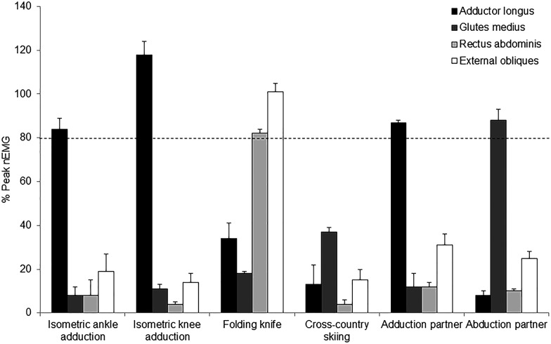
Adductor longus activity during IBA was 84% nEMG (95% CI: 70-98) and during IBK it was 118% nEMG (95% CI 106-130). For the dynamic exercises, ADP evoked 87% nEMG (95% CI 69-105) in adductor longus, ABP evoked 88% nEMG (95% CI 76-100) in gluteus medius, FK evoked 82% nEMG (95% CI 68-96) rectus abdominis, and 101% nEMG (95% CI 85-118) in external obliques. During CSS < 37% nEMG was evoked from all muscles.
Conclusion
These data suggest that exercise-intensity of all the six investigated exercises in the Hölmich groin injury prevention program, except cross-county skiing, is sufficient to be considered strength-training for specific muscle groups in and around the groin region.
Level of Evidence
3
Keywords: Abdominals, adductor longus, electromyography, gluteus medius, soccer
INTRODUCTION
Groin injuries are one of the most common types of injuries in soccer, accounting for 8-18% of all injuries.1-3 Groin injuries in soccer are primarily related to the hip adductors, accounting for approximately 60% of all acute and long-standing groin injuries, but can also be related to varied anatomical locations/tissues, such as the hip flexors, the abdominal muscles, and the inguinal canal.3-5
The latest systematic reviews on risk factors for groin injuries concluded that there is relatively consistent Level 1 and 2 evidence to suggest that hip adductor and abductor strength is associated with increased risk of groin injuy.6-7 A randomized controlled trial by Hölmich et. al.8 concerning injury prevention of groin injuries in soccer included exercises targeting these risk factors by performing a comprehensive exercise program focusing on strength and stability. The trial included nearly 1000 players and was aimed at reducing groin injury rates by 50%, however, only a 30% non-significant difference in injury rates was found.8 One of the possible reasons for the lack of an intended effect (≥50% reduction) on the prevention of groin injuries in soccer has been suggested to be related to a lack of adequate intensity of the included exercise.8
To promote strength gains in athletes, it is recommended to recruit the entire pool of motor units, based on the size principle.9-11 The threshold for high-threshold motor units varies for different muscles, with small muscles, such as muscles of the fingers, requiring as little as 30% of maximum effort to reach total recruitment,12 and larger muscles, such as the tibialis anterior and biceps brachialis, having thresholds at 80% and 90%, respectively.12,13 In that respect, intensity is recommended to be at least 80% of one repetition maximum (RM) for a stimulus that can induce strength gains in athletes.9 As opposed to joint-torque or external load of a given exercise, EMG (electromyography) is often used to quantify exercise intensity of specific muscles by measuring the recruitment and firing characteristics during exercise.14-19 In that respect, longitudinal strength gains have been repeatedly found when utilizing exercises at above 80% EMG intensity,19-24 as a quasi-linear relationship between EMG and force output has been described.14-16,25-29 Thus, EMG normalized (nEMG) to a maximal voluntary isometric contraction30 (MVC) with correct electrode placement14,15 and in a non-fatigued state15,16,29 is suggested as a valid option when estimating exercise intensity of specific muscles,14-19 with 80% of maximum as the rough cut-off required to promote strength gains in athletes.9
Six exercises were included in the Hölmich prevention program of which the two isometric exercises were shown in a previous investigation to produce high levels of muscle activity (nEMG values > 80%) in the adductor longus.19 However, resistance training is recommended to include dynamic muscle contractions9,31,32 and no information exists on the specific intensity of the dynamic exercises in the prevention program introduced by Hölmich et al.8 Considering that the dynamic exercises in the program require no use of additional equipment for resistance, compared to weight training machines, free weights, elastic bands or other forms of exercise equipment that help quantify intensity, it may be that these exercises do not induce sufficient intensity to promote strength gains in soccer players.
The purpose of this study was therefore to estimate the intensity of the six exercises from the Hölmich program using electromyography (EMG) and possibly categorize them as strength-training exercises.
METHODS
Study design
The present study used a cross-sectional design in which participants were investigated once having been familiarized with the trial procedures on a previous occasion. During the first session, they performed a 10-min. standardized warm-up of running drills and mobility exercises and were subsequently familiarized with all the exercises. The second session was conducted at least four days after to avoid delayed onset of muscle soreness. After the same standardized warm-up, the subjects performed MVCs to be used as reference contractions followed by two trials of the six exercises in a random order with minimum 60 s between, while muscle activity was recorded using surface EMG. Participants rated the level of perceived exertion during all exercises on the BorgCR10-scale, a categorical numeric rating scale with verbal anchors ranging from 0 ∼ ‘Nothing at all’ to 10 ∼ ‘Extremely strong’33 to provide additional data on subjective intensity. No exercise or training was allowed the day before testing and both sessions took place at Hvidovre Hospital, Copenhagen, Denmark. The reporting of the study follows the STROBE (Strengthening the Reporting of Observational Studies in Epidemiology) guidelines, using the checklist for cross-sectional studies34 and was approved by the Danish National Committee on Health Research Ethics (H-3-2011-145.). All participants gave written consent according to the Helsinki Declaration before testing was initiated. The study is not pre-registered.
Participants
Through convenience sampling, participants were recruited on a voluntary basis through contacts from local rugby and soccer clubs in Copenhagen, Denmark, from March to June 2012. Players were eligible for inclusion if they did not have injuries which could be presumed to influence the execution of the exercises, and had no or only minimal self-reported hip or groin pain, indicated by a score of above 80 out of 100 on the Hip and Groin Outcome Score (Sports subscale).35
The six exercises from the Hölmich groin-injury prevention program
The present study included two isometric and four dynamic exercises introduced by Hölmich et al.8 Three repetitions of each dynamic exercise were used for analysis of the EMG signal in order to reduce the accumulation of fatigue. The isometric exercises were performed for 10 s, as described by Hölmich et al.8
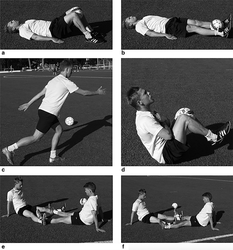
1: Isometric adduction against a soccer ball placed between the ankles (IBA): when lying supine, the thigh in neutral position, applying pressure against the ball as hard as possible (Figure 1a).8
2: Isometric adduction against a soccer ball placed between the knees (IBK): when lying supine with flexed hips and knees and feet flat on the surface, the thigh in neutral position, applying pressure against the ball as hard as possible (Figure 1b).8
3: ‘Folding knife” (FK): a combined abdominal sit-up and hip flexion. Starting from the supine position, with a football between flexed knees, repetitions are performed in a slow pace by flexing the hip and the lower back, bringing knees and chest together (Figure 1c).8
-
4: Standing one-leg coordination exercise called “cross-country skiing on one leg” (CSS): flexing and extending the knee and swinging the arms in the same rhythm, repetitions are performed on the dominant leg (defined as the preferred kicking leg) as the standing leg (Figure 1d).8
Exercises
5 and 6 are partner exercises where two players perform both exercises together simultaneously:
5: Hip adduction against a partner's hip abduction called “adduction partner” (ADP): in the sitting position, supported by the hands placed on the ground behind the trunk, the tested player places his legs straight and wide apart with the feet and lower shin on the outside of the partners feet and lower shin. He adducts while the partner abducts eccentrically and slowly presses his feet together (Figure 1e).8
-
6: Hip abduction against a partner's hip adduction called “abduction partner” (ABP): from the reversed starting position as “adduction partner” with feet and lower shins now placed medially on his partner's feet and lower shin, the player abducts concentrically (“abduction partner”) while the partner adducts eccentrically, and is slowly brought into abduction (Figure 1f).8
Both partner-exercises were performed for 6 seconds per repetition with a 3 s concentric and a 3 s eccentric contraction, with as much effort as possible, still allowing the partner to follow the tempo for the duration of the exercise.8
Figure 1.
The six included exercises: a = ”isometric adduction against a football placed between the ankles”; b = ”isometric adduction against a football placed between the knees”; c = “folding knife”; d = “crosscountry skiing on one leg”; e = “adduction partner” performed by the player to the left; f = “abduction partner” performed by the player to the left.
Figure 1.
Mean peak of median nEMG with standard error for adductor longus, gluteus medius, external obliques and rectus abdominis during the exercises. nEMG = Normalized electromyography.
ELECTROMYOGRAPHY (EMG)
Rectangular (20 x 30 mm) non-disposable differential surface-electrodes with 1 cm inter-electrode distance (DE-2.1, Delsys, Boston, MA, USA) were placed unilaterally to collect data from adductor longus, gluteus medius, rectus abdominis and the external oblique muscles after standard skin preparation. Using electrode gel and medical grade adhesive (Delsys electrode interface, Delsys, Boston, MA, USA), the electrodes were aligned parallel with the direction of the muscle fibres. Verification of EMG signal quality was conducted by visual inspection of the raw EMG signal while the subjects performed movements similar to the six evaluated exercises and isometric contractions specific to each muscle of interest after initial electrode placement and again after the warm-up routine. The electrodes were attached to 150 cm shielded wires, connected directly to small built-in preamplifiers and further to a main amplifier unit (Bagnoli-16, Delsys, Boston, MA, USA) with a band-pass of 15–450 Hz and a common-mode rejection ratio of 92 dB. Sampling was done at 1 kHz using a 16-bit A/D converter (6036E, National Instruments, Austin, TX, USA). Data analysis was performed on a personal computer (EMGworks acquisition 3.1, Delsys, Boston, MA, USA).
Electrode placements
Adductor longus; medially on the thigh equivalent to the proximal third of the distance from the pubic tubercle to the insertion on femur.17,36,37 Gluteus medius; half the distance between the iliac crest and the greater trochanter antero-superiorly from the gluteus maximus.36,37 Rectus abdominis; half the distance between the umbilicus and the pubic symphysis38 and centered on the muscle belly, i.e. 2-4 cm laterally from the umbilicus, depending on the size of the participant. External obliques; directly below the most inferior point of the ribs in direction towards the opposite pubic tubercle.39 Reference electrode; on the contralateral patella. A previous study of dynamic exercises for adductor longus has shown a high intra session reliability of EMG-activity in both legs.19 It was therefore decided only to measure unilaterally on the dominant side, defined as the side of the preferred kicking leg.
Reference contractions
The participants performed two MVCs in three different test positions in a randomized order to estimate the maximal muscle activity. The highest value from any of the contractions was used as the reference contraction, i.e. 100%, to normalize the EMG recordings from the exercises. MVCs lasted 5 s each and participants were given at least 30 s rest between each contraction.40 A standard verbal encouragement of “three, two, one, start, push, push, push, push, stop” was used. Adductor longus MVC; supine with straight hips and knees, bilateral isometric hip adduction was performed against a ball placed between the knees. Gluteus medius MVC; side-lying with hip and knees straight, the tested leg is held in approximately 25 degrees of hip abduction by the therapist.37,41An isometric hip abduction was performed against a belt fixed around the lateral femoral epicondyle, as the use of belts has been shown to be less demanding on the therapist, preferred by participants and increases the reliability of maximal hip abduction torque measurements.42 Rectus abdominis and external obliques; a lumbar spine flexion with isometric abdominal contraction in a supine position against a fixed training belt strapped around the chest.
Data reduction
The raw EMG signals were visually inspected and contractions with signal artifacts were discarded. All raw EMG signals were digitally filtered using a Butterworth 4th order high-pass filter (10 Hz cutoff frequency), and subsequently smoothed and filtered using a moving root-mean-square (RMS) filter of 500 ms.43 Peak EMG of each muscle within each contraction was identified as the maximum value of the smoothed RMS EMG signal and normalized to the maximal RMS EMG obtained during the MVC.
Statistical methods
Based on a mean of 108% nEMG and a 35.5% standard deviation from a previous investigation of the IBK exercise,19 a significance level of 0.05 and statistical power of 80% to detect differences of 20% between muscles and exercises, a sample of 21 participants was needed based on the paired t-test (G*Power 3.1.9.2). All Borg CR10 values are presented as medians with interquartiles ranges and their corresponding verbal anchors. All nEMG values are reported as least square means with 95% confidence intervals (CI). A repeated measures one-way analysis of variance (ANOVA) with a significance level of p ≤ 0.05 was used to compare the peak nEMG for each muscle in the different exercises, in order to compare all exercises to each other. The mean of peak from all three repetitions was used for the dynamic exercises. A Bonferroni correction was made to account for the multiple comparisons in the post hoc analysis. All analyses were performed for participants with complete data sets (per protocol) and no imputations were made.
RESULTS
Twenty-four healthy male soccer and rugby players were enrolled in the study. Data from three participants were missing due to broken reference electrodes, excessive artifacts or erroneous data extraction. The decision to exclude these data was made prior to the data analyses. Thus, the comparative analysis included 21 participants, mean age 21.4 ± 3.3 y, height 182.1 ± 7.7 cm, weight 83.1 ± 13.4 kg and scoring 96.7 ± 5.2 points on the HAGOS (Sports subscale). The participants had a training frequency of 5.2 ( ± 1.1) hrs per week with 1-2 weekly matches.
EMG activation levels are presented in Table 1. Five exercises reached specific muscle activation levels above 80% nEMG. In the isometric exercises, IBA muscle activity of the adductor longus was 84% nEMG (95% CI 70-98) and during IBK it was 118% nEMG (95% CI 106-130). With respect to the dynamic exercises, ADP evoked 87% nEMG (95% CI 69-105) in adductor longus, in the ABP exercise gluteus medius was reached 88% nEMG (955CI 76-100), for the FK rectus abdominis was 82% nEMG (95% CI 68-96), and external obliques was 101% nEMG (95% CI 85-118). During CSS all muscles were <37% nEMG. In the two isometric exercises, the adductor longus muscle activity was greater than in all other muscles, with greatest activity in the IBK (p<0.0001). For the dynamic exercises, the adductor longus activation was higher in the ADP exercise than in all other dynamic exercises (p<000.1). Gluteus medius activation was greater in the ABP exercise, than in all other exercises (p<0.001), and rectus abdominis and external obliques activation in the FK was higher than in all other exercises (p<0.001). The levels of perceived exertion rated on the BorgCR10 scale during exercises ranged from ‘Weak’ to ‘Strong’, with CSS being perceived as ‘Weak’ (median 2.5, IQR 1.1-3.75), ABP as ‘Moderate’ (median 4, IQR 3-6) and the remaining exercises as ‘Strong’ (ADP median 5, IQR 4-6.75; FK median 5, IQR 3-6.75; IBA median 5, IQR 3-7; IBK: median 5, IQR 4.25-6.75).
Table 1.
Peak normalized EMG measurements for all exercises and muscles
| Isometric | Dynamic | |||||
|---|---|---|---|---|---|---|
| IBA | IBK | FKE | CSS | ADP | ABP | |
| Adductor longus | 84 (7)* | 118 (6)*† | 33 (5) | 13 (2) | 86 (9)* | 8 (1) |
| Gluteus medius | 11 (2) | 11 (2) | 19 (4) | 38 (5)* | 12 (2) | 88 (6)†* |
| Rectus abdominis | 8 (2) | 4 (1) | 83 (7)† | 4 (1) | 12 (2) | 10 (2) |
| External obliques | 19 (4) | 14 (4) | 100 (8)†* | 14 (3) | 31 (5) | 25 (5) |
Values reported as mean normalized electromyography (standard error). IBA = “isometric adduction against a football placed between the ankles”; IBK = “isometric adduction against a football placed between the knees”; FKE = “folding knife exercise”; CSS = “crosscountry skiing on one leg”; ADP = “adduction partner”; ABP = “abduction partner”.
= significantly higher peak nEMG for this muscle than for other muscles during the exercise (p < 0.002).
= significantly higher peak nEMG during this exercise for this muscle than during any other exercise (p < 0.0001).
DISCUSSION
The main purpose of this study was to examine the level of muscle activity during two isometric and four dynamic strengthening exercises from a previously published groin-injury prevention program.8 The two isometric exercises have previously been investigated for muscle activity and showed similar levels as in the current study with both ≥80% nEMG.19 The results of the current study indicate that three of the dynamic exercises in the groin injury prevention program can be categorized as exercises with sufficient intensity for strength improvements in athletes, targeting important muscle groups relevant in the prevention of groin injuries; adduction partner for adductor longus, abduction partner for gluteus medius, and folding knife for rectus abdominis and external obliques. These muscles are important as most groin injuries are related to the adductor longus but may also be related to the abdominal muscles and the inguinal canal.3-5 Authors of several studies6,7 have identified decreased strength in the adductors preceding and following the onset of groin injuries, which makes adductor strength training a top priority in prevention and rehabilitation of groin injuries. In addition, targeted strengthening around the hip may contribute to pelvic alignment (frontal plane pelvic angle),44 which has been mentioned as an important factor in both prevention and management of groin injuries.45-47 Decreased strength in the hip abductors (gluteus medius) has also been found in athletes who since sustained a groin injury, making hip abductor strength training another key muscle for groin injury prevention.6,7 The exercise “cross-country skiing on one leg” did not reach sufficient intensity for strength training but it might promote dynamic stability in single leg stance and pelvic alignment through improved endurance capacity of gluteus medius, especially during minor to moderate and repetitive loading situations, such as in sub-maximal running and change of direction, an intensity also often utilized during a soccer game or practice. Based upon the current data it seems unlikely that inadequate exercise intensity should be the explanation for the lack of a substantial effect (50% groin injury reduction) of such a program in soccer players, as five of the six included exercises have been demonstrated in the current study to be sufficiently intense to promote strength gains in muscles relevant in groin injury prevention. Previous investigations of muscle activity of adductor longus during exercise have shown that muscle activity during the Barbell squat increases if the stance width and external hip rotation is increased. However, the highest nEMG found was only 23.1%.48 Commonly used unilateral frontal plane exercises for rehabilitation of lower extremity injuries; lunges, step-up and step up-and-over were in another study found to only evoke nEMG of the adductors in the range of 16-22%.49 Delmore et al. examined several specific hip adductor exercises that showed intensities of 14-36% nEMG.50 However, one exercise in the study by Delmore et al., resisted side-lying hip adduction showed adductor longus nEMG of 60%,50 in concordance with previous findings of 64% nEMG from Serner et al.19, although un-resisted in their study. The relatively low nEMG results found in these studies indicate that exercises in which the adductors are working as stabilizers or hip-extensors such as in during squat variations are not able to induce the same muscle activity as the exercises included in the present study. In comparison the “adduction partner”, although limited by the partner determining and providing the level of resistance, has no need for extra external resistance devices and demonstrated muscle activity similar to exercises previously implemented using strength training equipment such as strength training machines and elastic bands.51 The dynamic Copenhagen Adduction exercise also relies on partner-assistance, performed sidelying and as the partner holds the upper leg at the height of the hip, the athlete lifts his entire body with using primarily his hip adductors until it reaches a straight line.20 The Copenhagen Adduction has been shown to induce high muscle activity of 108% of MVC,19 and longitudinal strength gains.20 However, isometric exercises induce angle-specific strength gains52 and are predominantly recommended in the course of muscle injury rehabilitation when pain and range of motion limits the ability to perform dynamic muscle contractions recommended for strength training.9,31,32,53 Thus, the three dynamic exercises included in the current study in combination with the Copenhagen Adduction exercise seem to be some of the most relevant exercises to include when considering the initiation of a groin injury prevention program, because they evoke muscle activity above 80% opposed to other dynamic equipment-free exercises investigated in the literature.
This allows for speculation regarding other possible factors influencing the lack of statistical injury reduction in the previous Hölmich trial. Besides intensity, other important exercise variables could also be part of the explanation, such as frequency, total volume and adherence, which were largely unknown in the trial.7 Another factor that has recently be shown to have a substantial impact on the risk of injury is the ratio between acute workload (1 week total distance) and chronic workload (4-week average acute workload) during both training and competitive matches in soccer. These two factors should be either controlled for or observed as a potential confounding factor in future trials aimed at preventing injuries in soccer. 54,55
A substantiating argument for the use of dynamic high intensity exercises for soccer players is that exercises targeting the at-risk muscle with high muscle activity have been able to significantly reduce other muscle-tendinous injuries, as well as increase strength.22,24,56 For example, one study implemented the Nordic Hamstring exercise,24,56 which has shown biceps femoris long head nEMG of 91%23 and is performed eccentrically to match the deceleration phase of the leg during running/sprinting, where hamstring injuries often occur.56 As such, the exercises from the current study seem highly relevant to implement in future trials aiming at reducing groin injuries.
The BorgCR10 scale was used in the current study which is closely related to exercise intensity.51,57 Other measures of perceived exertion could also be used in clinical practice, such as the Repetitions in Reserve (RIR) which used for strength training at intensities near repetition-failure, by having the athlete subjectively estimate how many additional repetitions were possible.58 As players in the present study only performed three repetitions in order to minimize the presence of fatigue and thereby allowing meaningful interpretations of the EMG data, using RIR would however not be relevant
METHODOLOGICAL LIMITATIONS
There are several limitations of using EMG to estimate exercise intensity. For example when using dynamic exercises, the EMG amplitude can be relatively higher than the expected force based on assumptions of a linear force-EMG relationship. The association between EMG and force are also not always linear as the force-velocity and force-length relationship is not accounted for, neither is the electromechanical delay. Nevertheless, using appropriate methods, e.g. using slowly controlled contractions and appropriate analytical methods, there seems to be a good and linear relationship between normalized EMG and percentage of load utilized during 1RM in certain exercises.27 The application of surface electrodes carries the risk of crosstalk from adjacent muscles.14,15 Accordingly, the muscle activity of adductor longus could possibly reflect the muscle activity of the adductor group as a whole. As muscle activity of more than 100% nEMG was found during the isometric exercise IBK, which is consistent with previous findings,20 thus, it could be assumed that this exercise and position might be more appropriate to perform the MVC in if performed according to MVC guidelines with standardized order, instruction and duration.30
CONCLUSION
These data suggest that the exercise intensity of all but one of the six investigated exercises from the previous published Hölmich groin injury prevention program is sufficient to be categorized as strengthening exercises for specific relevant muscle groups in and around the groin region.
References
- 1.Ekstrand J Hägglund M Waldén M. Injury incidence and injury patterns in professional football: the UEFA injury study. Br J Sports Med. 2011;45(7):553-558. [DOI] [PubMed] [Google Scholar]
- 2.Werner J Hägglund M Waldén M, et al. UEFA injury study: a prospective study of hip and groin injuries in professional football over seven consecutive seasons. Br J Sports Med. 2009;43(13):1036-1040. [DOI] [PubMed] [Google Scholar]
- 3.Holmich P. Long-standing groin pain in sportspeople falls into three primary patterns, a “clinical entity” approach: a prospective study of 207 patients. BrJSports Med. 2007;41:247-252. [DOI] [PMC free article] [PubMed] [Google Scholar]
- 4.Serner A Weir A Tol JL, et al. Can standardised clinical examination of athletes with acute groin injuries predict the presence and location of MRI findings? Br J Sports Med. August 2016. [DOI] [PubMed] [Google Scholar]
- 5.Serner A Tol JL Jomaah N, et al. Diagnosis of Acute Groin Injuries: A Prospective Study of 110 Athletes. Am J Sports Med. 2015;43(8):1857-1864. [DOI] [PubMed] [Google Scholar]
- 6.Whittaker JL Small C Maffey L, et al. Risk factors for groin injury in sport: an updated systematic review. Br J Sports Med. 2015;49(12):803-809. [DOI] [PubMed] [Google Scholar]
- 7.Kloskowska P Morrissey D Small C, et al. Movement Patterns and Muscular Function Before and After Onset of Sports-Related Groin Pain: A Systematic Review with Meta-analysis. Sports Med Auckl NZ. 2016;46(12):1847-1867. [DOI] [PMC free article] [PubMed] [Google Scholar]
- 8.Holmich P Larsen K Krogsgaard K, et al. Exercise program for prevention of groin pain in football players: a cluster-randomized trial. Scand J Med Sci Sports. 2010;20:814-21. [DOI] [PubMed] [Google Scholar]
- 9.American College of Sports Medicine. American College of Sports Medicine position stand. Progression models in resistance training for healthy adults. Med Sci Sports Exerc. 2009;41(3):687-708. [DOI] [PubMed] [Google Scholar]
- 10.Carpinelli RN. The size principle and a critical analysis of the unsubstantiated heavier-is-better recommendation for resistance training. J Exerc Sci Fit. 2008;6(2):67-86. [Google Scholar]
- 11.Henneman E. Relation between size of neurons and their susceptibility to discharge. Science. 1957;126(3287):1345-1347. [DOI] [PubMed] [Google Scholar]
- 12.Kukulka CG Clamann HP. Comparison of the recruitment and discharge properties of motor units in human brachial biceps and adductor pollicis during isometric contractions. Brain Res. 1981;219(1):45-55. [DOI] [PubMed] [Google Scholar]
- 13.Erim Z De Luca CJ Mineo K, et al. Rank-ordered regulation of motor units. Muscle Nerve. 1996;19(5):563-573. [DOI] [PubMed] [Google Scholar]
- 14.De Luca CJ. The use of surface electromyography in biomechanics. J Appl Biomech. 1997;13:135–163. [Google Scholar]
- 15.Onishi H Yagi R Akasaka K, et al. Relationship between EMG signals and force in human vastus lateralis muscle using multiple bipolar wire electrodes. J Electromyogr Kinesiol Off J Int Soc Electrophysiol Kinesiol. 2000;10(1):59-67. [DOI] [PubMed] [Google Scholar]
- 16.Disselhorst-Klug C Schmitz-Rode T Rau G. Surface electromyography and muscle force: limits in sEMG-force relationship and new approaches for applications. Clin Biomech Bristol Avon. 2009;24(3):225-235. [DOI] [PubMed] [Google Scholar]
- 17.Andersen LL Magnusson SP Nielsen M, et al. Neuromuscular activation in conventional therapeutic exercises and heavy resistance exercises: implications for rehabilitation. Phys Ther. 2006;86(5):683-697. [PubMed] [Google Scholar]
- 18.Kuriki HU Mello EM De Azevedo FM, et al. The Relationship between Electromyography and Muscle Force. INTECH Open Access Publisher; 2012. [Google Scholar]
- 19.Serner A Jakobsen MD Andersen LL, et al. EMG evaluation of hip adduction exercises for soccer players: implications for exercise selection in prevention and treatment of groin injuries. Br J Sports Med. March 2013. [DOI] [PubMed] [Google Scholar]
- 20.shøi L Sørensen CN Kaae NM, et al. Large eccentric strength increase using the Copenhagen Adduction exercise in football: A randomized controlled trial. Scand J Med Sci Sports. November 2015. [DOI] [PubMed] [Google Scholar]
- 21.Jensen J Hölmich P Bandholm T, et al. Eccentric strengthening effect of hip-adductor training with elastic bands in soccer players: a randomised controlled trial. Br J Sports Med. 2014;48(4):332-338. [DOI] [PubMed] [Google Scholar]
- 22.Clark R Bryant A Culgan J-P, et al. The effects of eccentric hamstring strength training on dynamic jumping performance and isokinetic strength parameters: a pilot study on the implications for the prevention of hamstring injuries. Phys Ther Sport. 2005;6(2):67-73. [Google Scholar]
- 23.Zebis MK Skotte J Andersen CH, et al. Kettlebell swing targets semitendinosus and supine leg curl targets biceps femoris: an EMG study with rehabilitation implications. Br J Sports Med. 2013;47(18):1192-1198. [DOI] [PubMed] [Google Scholar]
- 24.Mjølsnes R Arnason A Østhagen T et al. A 10-week randomized trial comparing eccentric vs. concentric hamstring strength training in well-trained soccer players. Scand J Med Sci Sports. 2004;14(5):311-317. [DOI] [PubMed] [Google Scholar]
- 25.Lippold OCJ. The relation between integrated action potentials in a human muscle and its isometric tension. J Physiol. 1952;117(4):492-499. [DOI] [PMC free article] [PubMed] [Google Scholar]
- 26.Bigland B Lippold OCJ. The relation between force, velocity and integrated electrical activity in human muscles. J Physiol. 1954;123(1):214-224. [DOI] [PMC free article] [PubMed] [Google Scholar]
- 27.Calatayud J Vinstrup J Jakobsen MD, et al. Importance of mind-muscle connection during progressive resistance training. Eur J Appl Physiol. 2016;116(3):527-533. [DOI] [PubMed] [Google Scholar]
- 28.Kellis E Baltzopoulos V. The effects of antagonist moment on the resultant knee joint moment during isokinetic testing of the knee extensors. Eur J Appl Physiol. 1997;76(3):253-259. [DOI] [PubMed] [Google Scholar]
- 29.Lawrence JH De Luca CJ. Myoelectric signal versus force relationship in different human muscles. J Appl Physiol. 1983;54(6):1653-1659. [DOI] [PubMed] [Google Scholar]
- 30.Burden A. How should we normalize electromyograms obtained from healthy participantsϿ. What we have learned from over 25 years of research. J Electromyogr Kinesiol Off J Int Soc Electrophysiol Kinesiol. 2010;20(6):1023-1035. [DOI] [PubMed] [Google Scholar]
- 31.Khan K Brukner P. Brukner & Khan's Clinical Sports Medicine. McGraw-Hill Education; 2011. [Google Scholar]
- 32.Rasch PJ, Morehouse LE. Effect of Static and Dynamic Exercises on Muscular Strength and Hypertrophy. J Appl Physiol. 1957;11(1):29-34. [DOI] [PubMed] [Google Scholar]
- 33.Borg G. Perceived Exertion and Pain Scales. Vol 1998 Champaign, IL: Human Kinetics [Google Scholar]
- 34.von Elm E, Altman DG, Egger M, et al. The Strengthening the Reporting of Observational Studies in Epidemiology (STROBE) statement: guidelines for reporting observational studies. J Clin Epidemiol. 2008;61(4):344-349. [DOI] [PubMed] [Google Scholar]
- 35.Thorborg K Hölmich P Christensen R, et al. The Copenhagen Hip and Groin Outcome Score (HAGOS): development and validation according to the COSMIN checklist. Br J Sports Med. 2011;45(6):478-491. [DOI] [PubMed] [Google Scholar]
- 36.Claiborne TL Timmons MK Pincivero DM. Test-retest reliability of cardinal plane isokinetic hip torque and EMG. J Electromyogr Kinesiol Off J Int Soc Electrophysiol Kinesiol. 2009;19(5):e345-352. [DOI] [PubMed] [Google Scholar]
- 37.Bolgla LA Uhl TL. Reliability of electromyographic normalization methods for evaluating the hip musculature. J Electromyogr Kinesiol Off J Int Soc Electrophysiol Kinesiol. 2007;17(1):102-111. [DOI] [PubMed] [Google Scholar]
- 38.Hodges PW Richardson CA. Contraction of the abdominal muscles associated with movement of the lower limb. Phys Ther. 1997;77(2):132-142-144. [DOI] [PubMed] [Google Scholar]
- 39.Anders C Wagner H Puta C, et al. Trunk muscle activation patterns during walking at different speeds. J Electromyogr Kinesiol Off J Int Soc Electrophysiol Kinesiol. 2007;17(2):245-252. [DOI] [PubMed] [Google Scholar]
- 40.Sisto SA Dyson-Hudson T. Dynamometry testing in spinal cord injury. J Rehabil Res Dev. 2007;44(1):123-136. [DOI] [PubMed] [Google Scholar]
- 41.Distefano LJ Blackburn JT Marshall SW, et al. Gluteal muscle activation during common therapeutic exercises. J Orthop Sports Phys Ther. 2009;39(7):532-540. [DOI] [PubMed] [Google Scholar]
- 42.Kramer JF Vaz MD Vandervoort AA. Reliability of isometric hip abductor torques during examiner- and belt-resisted tests. J Gerontol. 1991;46(2):M47-51. [DOI] [PubMed] [Google Scholar]
- 43.Jakobsen MD Sundstrup E Andersen CH, et al. Muscle activity during knee-extension strengthening exercise performed with elastic tubing and isotonic resistance. Int J Sports Phys Ther. 2012;7(6):606-616. [PMC free article] [PubMed] [Google Scholar]
- 44.Kim D Unger J Lanovaz JL, et al. The Relationship of Anticipatory Gluteus Medius Activity to Pelvic and Knee Stability in the Transition to Single-Leg Stance. PM R. 2016;8(2):138-144. [DOI] [PubMed] [Google Scholar]
- 45.Holmich P Uhrskou P Ulnits L, et al. Effectiveness of active physical training as treatment for long-standing adductor-related groin pain in athletes: randomised trial. The Lancet. 1999;353:439-443. [DOI] [PubMed] [Google Scholar]
- 46.Garvey JFW Read JW Turner A. Sportsman hernia: what can we do? Hernia. 2010;14(1):17-25. [DOI] [PubMed] [Google Scholar]
- 47.Kinchington M. Groin Pain: A view from below; The impact of lower extremity function and podiatric interventions. Aspetar Sports Med. 2013;2(3):360-368. [Google Scholar]
- 48.Clark DR Lambert MI Hunter AM. Muscle activation in the loaded free barbell squat: a brief review. J Strength Cond Res Natl Strength Cond Assoc. 2012;26(4):1169-1178. [DOI] [PubMed] [Google Scholar]
- 49.Dwyer MK Boudreau SN Mattacola CG, et al. Comparison of lower extremity kinematics and hip muscle activation during rehabilitation tasks between sexes. J Athl Train. 2010;45(2):181-190. [DOI] [PMC free article] [PubMed] [Google Scholar]
- 50.Delmore RJ Laudner KG Torry MR. Adductor longus activation during common hip exercises. J Sport Rehabil. 2014;23(2):79-87. [DOI] [PubMed] [Google Scholar]
- 51.Brandt M Jakobsen MD Thorborg K, et al. Perceived loading and muscle activity during hip strengthening exercises: comparison of elastic resistance and machine exercises. Int J Sports Phys Ther. 2013;8(6):811-819. [PMC free article] [PubMed] [Google Scholar]
- 52.Bandy WD Hanten WP. Changes in torque and electromyographic activity of the quadriceps femoris muscles following isometric training. Phys Ther. 1993;73(7):455-465-467. [DOI] [PubMed] [Google Scholar]
- 53.Järvinen TAH Järvinen TLN Kääriäinen M, et al. Muscle injuries: optimising recovery. Best Pract Res Clin Rheumatol. 2007;21(2):317-331. [DOI] [PubMed] [Google Scholar]
- 54.Nassis GP Gabbett TJ. Is workload associated with injuries and performance in elite football? A call for action. Br J Sports Med. 2016;10.1136/bjsports-2016-095988 [DOI] [PubMed] [Google Scholar]
- 55.Murray NB Gabbett TJ Townshend AD, et al. Individual and combined effects of acute and chronic running loads on injury risk in elite Australian footballers. Scand J Med Sci Sports. 2016; 10.1111/sms.12719 [DOI] [PubMed] [Google Scholar]
- 56.Petersen J Thorborg K Nielsen MB, et al. Preventive effect of eccentric training on acute hamstring injuries in men's soccer: a cluster-randomized controlled trial. Am J Sports Med. 2011;39(11):2296-2303. [DOI] [PubMed] [Google Scholar]
- 57.Pincivero DM Coeho AJ Campy RM. Perceived exertion and maximal quadriceps femoris muscle strength during dynamic knee extension exercise in young adult males and females. Eur J Appl Physiol. 2003;89(2):150-156. [DOI] [PubMed] [Google Scholar]
- 58.Helms ER, Cronin J, Storey A, et al. Application of the Repetitions in Reserve-Based Rating of Perceived Exertion Scale for Resistance Training. Strength Cond J. 2016;38(4):42-49. [DOI] [PMC free article] [PubMed] [Google Scholar]


