Abstract
BACKGROUND
The presence of primary intracardiac tumors are scarce, and most of them are myxomas. We reported, in this paper, a case with huge mass in the right side of the heart.
CASE REPORT
A 45-year-old man, with a complaint of bilateral lower limbs edema and exertional dyspnea, was admitted to intensive cardiac care unit. Cardiac auscultation revealed soft grade systolic murmur without any evidence of “tumor plop.” Echocardiography showed a huge mobile mass in right side of the heart that suggested myxoma. Our patient underwent cardiac surgery with excision of 13 cm mass. Histopathological study was confirmed the diagnosis of mass type.
CONCLUSION
In this case report, it shows that in the differential diagnosis of right-sided heart failure, the right sided myxoma must be considered. The preferable approach in patient with cardiac myxomas is surgical excision to alleviate symptoms, early identification, and removal.
Keywords: Myxoma, Cardiac Surgery, Echocardiography, Case Report
Introduction
In humans, the incidence of cardiac tumors is 0.2% of all tumors.1 The pathologic category for these tumors includes primary and secondary or metastatic. Secondary tumors are 20-40 times more common than another, but the incidence of primary intracardiac tumors is rare. Approximately, 75% are benign, and half of them are myxomas, which have an incidence of 0.0017% in the general population. Based on histologic evaluations, these are real tumors, derived from multipotent mesenchymal cells of the subendocardium.2,3
In cardiac chambers, commonly, myxoma can arise in the left atrium (75%) and are almost always presents with sign and symptoms of mitral valve disorders, but they may present in other sites, such as the right side of the heart (5-18%).4
We present a man with huge myxoma in right side of the heart that filling right atrium (RA) and right ventricle (RV), without any significant hemodynamic compromise.
Case Report
A 45-year-old man weighing 50 kg was admitted to intensive cardiac care unit with a complaint of bilateral lower limbs edema and exertional dyspnea. He was in Class 3 functional class. He had not any history of hospitalization. There was also no mentioned family medical history of cardiac disease. He reported worsening of symptoms in the last week before admission. On admission, he was in stable condition and blood pressure was 107/89 mmHg, heart rate was regular and 99 beat per minute, respiratory rate was 26 breath/min, and he was afebrile. Oxygen saturation, at rest, with finger pulse oximeter in index finger was 97%.
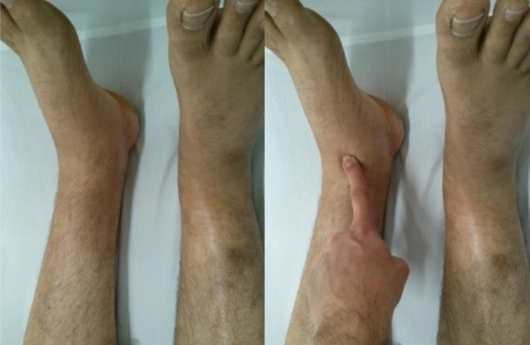
In the primary physical examination, we found Grade 2+ of lower limbs edema (Figure 1), and cardiac auscultation revealed soft Grade 2/6 systolic murmur at the both, right and left sternal border without any evidence of diastolic murmur and “tumor plop” in the apex. Laboratory findings shown a mild hypochromic microcytic anemia (hemoglobin = 10.9 g/dl, hematocrit = 34.9%, mean corpuscular volume = 71.96 fl, and mean corpuscular hemoglobin = 22.47 p.g). Chest X-ray (in posteroanterior view) revealed increased cardiothoracic ratio. Aortic arch was normal, main pulmonary artery was seen flat and right descending pulmonary artery was increased (Figure 2).
Figure 1.
2+ grade of lower limbs edema
Figure 2.

Antro-posterior chest X-ray
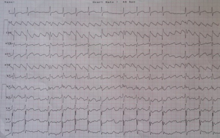
In electrocardiogram, there was no sign of abnormal conductive pathways. Cardiac rhythm was sinus tachycardia (Figure 3).
Figure 3.

An electrocardiogram (ECG) of the patient (before cardiac surgery) manifested that his cardiac rhythm was sinus tachycardia and precordial leads shown incomplete right bundle branch block. According to this ECG, the patient had a left atrium abnormality
The patient referred to echocardiographic study unit; transthoracic echocardiography showed a giant mobile mass in right side of the heart (11.4 × 4.2 cm) attached to the intratrial septum. Inferior vena cava was plethoric and tricuspid valve (TV), function was disturbed by this giant mass. RV function was reduced (Tricuspid annular plane systolic excursion = 0.85 cm) (Figure 4).
Figure 4.
(a and b) Apical four chambers view (an echocardiography before cardiac surgery), showing hypermobile huge mass in right side of the heart
The patient was scheduled for open heart surgery. General anesthesia was performed to median sternotomy. Mild hypothermia strategy (33.0° C) was stablished under cardiopulmonary bypass.
During an anoxic arrest, single aortic cross-clamping performed, and then, the tumor was completely excised through a right longitudinal atriotomy. The mass referred to histopathological analysis. Based on the microscopic evaluation, myxoid matrix rich in mucopolysaccharides, and polygonal cells appeared as a star and nest shape without atypical features, compatible with non-malignant myxoma.
During cardiac surgery and after tumor removal, saline test was performed by cardiac surgeon. According to this test, the surgeon concluded that inappropriate function of TV was due to mass.
After 1 month of discharge, our patient readmitted to the cardiac care unit complaining of palpitations. In electrocardiogram, cardiac rhythm was atrial flutter (Figure 5).
Figure 5.
An electrocardiogram after cardiac surgery demonstrate atrial flutter
Echocardiography of patient, as shown in figure 6, indicated that RV and RA, were severely dilated RV systolic function was severely impaired. TV, had leaflet malcoaptation (2 cm), with severe free low-pressure regurgitation [tricuspid regurgitation (TR)], and mild pulmonary insufficiency.
Figure 6.

Transthoracic echocardiography four chambers view, shows mal coaptation, with severe free low-pressure tricuspid regurgitation
Because of hemodynamic instability, cardiac rhythm converted to normal sinus rhythm by synchronized shock.
Because of severe low-pressure TR, the patient was candidate for TV repair (TVR), TVR surgery postponed to 6 months after myxoma excision. In this time, the patient has been closely monitored and controlled by medication.
Discussion
Myxoma is the most common primary cardiac mass with rare incidence and usually benign.5 More than 75% of myxomas are located in the left atrium, whereas right sided myxomas are scarce (15-20%). 3-4% of right-sided myxomas are located in RV or pulmonary artery being extremely rare.6
RA myxoma has a lower incidence that reported for a long time in several series of autopsy cases.1 Although only about 30% of affected patients are men,2,7 we reported a 45-year-old man suffering from a giant myxoma in right side of his heart.
In our patient like another case report,1 the myxoma originated in the left of RA and attached to the intratrial septum in some case reports that right atrial myxomas exude in the fossa ovalis or base of the interatrial septum.8,9
Nevertheless, transthoracic echocardiography cannot be recognized tumors smaller than 5 cm diameter, but still, it remains the gold standard method for diagnose the site and assessing the extent of myxomas and indicating their recurrence, with a sensitivity of up to 100%.7 In this report, our patient had a 13 × 9 × 3 cm, with an irregular and smooth surface tumor. In Samanidis et al. article, the mean diameter of the myxoma tumors that were evaluated in a 19 years’ period was 5.1 ± 1.8 cm.9 Therefore, our case had the largest myxoma in the right side of the heart, which heretofore reported.
In some case report, the most common manifestation of right side myxoma is dyspnea that occurring in 80% of affected patients.1,4,5 Our patient also compliant of dyspnea on exertion. dyspnea in our patient may not be due to huge myxoma itself, it may be caused by enlargement and increased size of RV due to hemodynamic changing in right ventricular function.10
Right side myxoma could be yielding other symptoms such as Raynaud’s phenomenon and syncope. Our patient had not any of these symptoms, despite the presence of huge myxoma in right side of the heart.
Conclusion
This case report suggests that in differential diagnosis of right-sided heart failure, the right-sided myxoma must be considered. Since surgical excision is reported to alleviate symptoms associated with cardiac myxomas, early identification and removal is preferable.
Acknowledgments
We would like to thank the patient participated in this study. We would also like to thank the all of nurses in Dey 9th Hospital, Torbat Heydariyeh, Iran, for patient management, and cardiac surgeon, cardiac anesthesiologist and perfusionist in Imam Reza Hospital, Mashhad, Iran.
Footnotes
Conflicts of Interest
Authors have no conflict of interests.
REFERENCES
- 1.Nina VJ, Silva NA, Gaspar SF, Raposo TL, Ferreira EC, Nina RV, et al. Atypical size and location of a right atrial myxoma: a case report. J Med Case Rep. 2012;6:26. doi: 10.1186/1752-1947-6-26. [DOI] [PMC free article] [PubMed] [Google Scholar]
- 2.Jang KH, Shin DH, Lee C, Jang JK, Cheong S, Yoo SY. Left atrial mass with stalk: Thrombus or myxoma? J Cardiovasc Ultrasound. 2010;18(4):154–6. doi: 10.4250/jcu.2010.18.4.154. [DOI] [PMC free article] [PubMed] [Google Scholar]
- 3.Vale Mde P, Freire Sobrinho A, Sales MV, Teixeira MM, Cabral KC. Giant myxoma in the left atrium: case report. Rev Bras Cir Cardiovasc. 2008;23(2):276–8. doi: 10.1590/s0102-76382008000200020. [DOI] [PubMed] [Google Scholar]
- 4.Diaz A, Di Salvo C, Lawrence D, Hayward M. Left atrial and right ventricular myxoma: an uncommon presentation of a rare tumour. Interact Cardiovasc Thorac Surg. 2011;12(4):622–3. doi: 10.1510/icvts.2010.255661. [DOI] [PubMed] [Google Scholar]
- 5.Gribaa R, Slim M, Kortas C, Kacem S, Ben Salem H, Ouali S, et al. Right ventricular myxoma obstructing the right ventricular outflow tract: a case report. J Med Case Rep. 2014;8:435. doi: 10.1186/1752-1947-8-435. [DOI] [PMC free article] [PubMed] [Google Scholar]
- 6.Huang SC, Lee ML, Chen SJ, Wu MZ, Chang CI. Pulmonary artery myxoma as a rare cause of dyspnea for a young female patient. J Thorac Cardiovasc Surg. 2006;131(5):1179–80. doi: 10.1016/j.jtcvs.2005.12.052. [DOI] [PubMed] [Google Scholar]
- 7.Manfroi W, Vieira SR, Saadi EK, Saadi J, Alboim C. Multiple recurrences of cardiac myxomas with acute tumoral pulmonary embolism. Arq Bras Cardiol. 2001;77(2):161–6. doi: 10.1590/s0066-782x2001000800007. [DOI] [PubMed] [Google Scholar]
- 8.Stolf NA, Benķcio A, Moreira L, Rossi E. Right atrium myxoma originating from the inferior vena cava: An unusual location with therapeutic and diagnostic implications. Rev Bras Cir Cardiovasc. 2000;15(3):255–8. [Google Scholar]
- 9.Samanidis G, Perreas K, Kalogris P, Dimitriou S, Balanika M, Amanatidis G, et al. Surgical treatment of primary intracardiac myxoma: 19 years of experience. Interact Cardiovasc Thorac Surg. 2011;13(6):597–600. doi: 10.1510/icvts.2011.278705. [DOI] [PubMed] [Google Scholar]
- 10.Mann DL, Zipes DP, Braunwald E, Bonow RO. Braunwald's heart disease: A textbook of cardiovascular medicine. 10th. Philadelphia, PA: Elsevier/Saunders; 2015. [Google Scholar]


