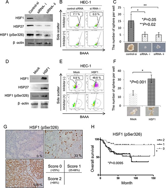
Figure 5. Phosphorylation of HSF1 at Ser326 is related to the maintenance of CSCs/CICs and poorer prognosis.

(A) Western blotting of HSF1 knockdown cells. HSF1 siRNA was transfected into HEC-1 cells. Twenty-four hours after transfection, total RNAs were purified and the expression of HSF1, HSF1 pSer326 and HSP27 were evaluated by Western blotting. β-actin was used as an internal control. (B) Aldefluor assay. ALDH activity of HSF1 knockdown cells was detected using the Aldefluor assay 48 hours after transfection. Percentage represents the proportion of ALDH1high cells. (C) Sphere formation assay. ALDH1high and ALDH1low cells derived from HSF1 siRNA-transfected HEC1 cells were cultured in serum-free medium. After 2 weeks of culture in vitro, a picture of a tumor sphere was taken. The counts were examined for statistical significance using Student's t-test. Data represent means ± SD. *P values. (D) Western blotting of HSF1-overexpressed cells. Cells stably transfected with HSF1 and control vector-transfected cells were used. The expression of HSF1, HSF1 pSer326 and HSP27 were evaluated by Western blotting. β-actin was used as an internal control. (E) Aldefluor assay. ALDH activity of HSF1-overexpressed cells was detected using the Aldefluor assay 48 hours after transfection. Percentage represents the proportion of ALDH1high cells. (F) Sphere formation assay. ALDH1high and ALDH1low cells derived from HSF1-overexpressed HEC1 cells were cultured in serum-free medium. After 2 weeks of culture in vitro, a picture of a tumor sphere was taken. The counts were examined for statistical significance using Student's t-test. Data represent means ± SD. *P values. (G) Immunohistological staining of phosphoHSF1 (pSer326). A total 122 of epithelial ovarian cancer tissues were immunohistochemically stained with anti-phopho-HSF1 pSer326 antibody and the expressions of HSF1 pSer326 were evaluated as (score 0: < 25%, n = 41; score 1: 25%–68%, n = 40; score 2: > 68%, n = 41). Representative pictures are shown. The HSF1 pSer326 positive rates are 0%, 33% and 100%, respectively. Magnification ×200. (H) HSF1 pSer326 positive cases are associated with poor prognosis. The differences of overall survival were examined for statistical significance using Fischer's test. *P values.
