
Figure 2. Invasive Fn in CRC tissues is associated with an activated β-catenin signaling pathway and TLR4/PAK1 protein abundance.
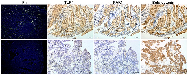
The upper panel shows that abundant invasive Fn in CRC tissue is associated with a high abundance of TLR4 and PAK1 and with β-catenin nuclear accumulation. The lower panel demonstrates that both TLR4 and PAK1 are absent in a Fn-negative CRC, and that cytoplasmic but not nuclear accumulation of β-catenin was seen in the tissue. Invasive Fn was detected by fluorescence in situ hybridization. 200× magnification.